Contrast agent and its use for imaging

a contrast agent and imaging technology, applied in the field of contrast agent enhanced medical imaging, can solve the problems of limited ultrasound imaging with respect to imaging depth, insufficient means to monitor cells, limited to target organs, etc., and achieve the effect of greatly improving the visibility of particles in ultrasound and photoacousti

Active Publication Date: 2015-09-10
STICHTING RADBOUD UNIVERSITAIR MEDISCH CENT
View PDF4 Cites 5 Cited by
  • Summary
  • Abstract
  • Description
  • Claims
  • Application Information

AI Technical Summary

Benefits of technology

[0011]We found that the ultrasound and photoacoustic visibility of particles greatly impro

Problems solved by technology

A key hurdle in the development and optimisation of cell therapy is that there are insufficient means to monitor the cells once they are in the patient, in a noninvasive manner.
Ultrasound imaging is potentially quantitative and it is not a whole body imaging modality, and is therefore limited to target organs.
Ultrasound imaging is limited with respect to depth of imaging.
They commonly have a relatively large size (1000-10000 nm diameter) which is generally unsuitable for cell labeling.
Moreover, they are also unsuitable to image smaller blood vessels e.g. in tumor imaging.
They also suffer from the additional disa

Method used


Image

  • Contrast agent and its use for imaging
  • Contrast agent and its use for imaging
  • Contrast agent and its use for imaging

Examples


example 1

Preparation of Particles Comprising Perfluoro-15-Crown-5-Ether

[0059]PLGA (0.09 gram) was dissolved in 3 ml dichloromethane in a glass tube. Liquid perfluoro-15-crown-5-ether (890 microliter) was added followed by 50 ml of a solution of Prohance® (a 3 mg / ml solution of gadoteridol) diluted in water. Optionally, additional agents, such as a fluorescent dye, may be added to the fluorocarbon at this stage. If a fluorescent particle was required, 1 mg of IcG or IC-Green (Indocyanine Green, Akorn Pharmaceuticals) was added to the solution.

[0060]As detailed herein below, we prepared particles with a high, medium and low content of Gadolinium. For that purpose, the above mentioned solution of Prohance® in water comprised 11.5, 5.75 and 2.85 ml respectively of Prohance® added up with water to 50 ml of solution. The entire mixture was then added dropwise into 25 ml of a solution of polyvinyl alcohol in water (20 gram / liter) under constant sonication (Branson Digital Sonifier 250; 3 minute cyc...

example 2

Characterisation of Particles

[0061]We found that particles as prepared above were stable for at least a year when kept at −20 degrees Celsius in the dry form. The particles were also stable in solution at working concentrations for at least 3 months at minus 4 degrees Celsius.

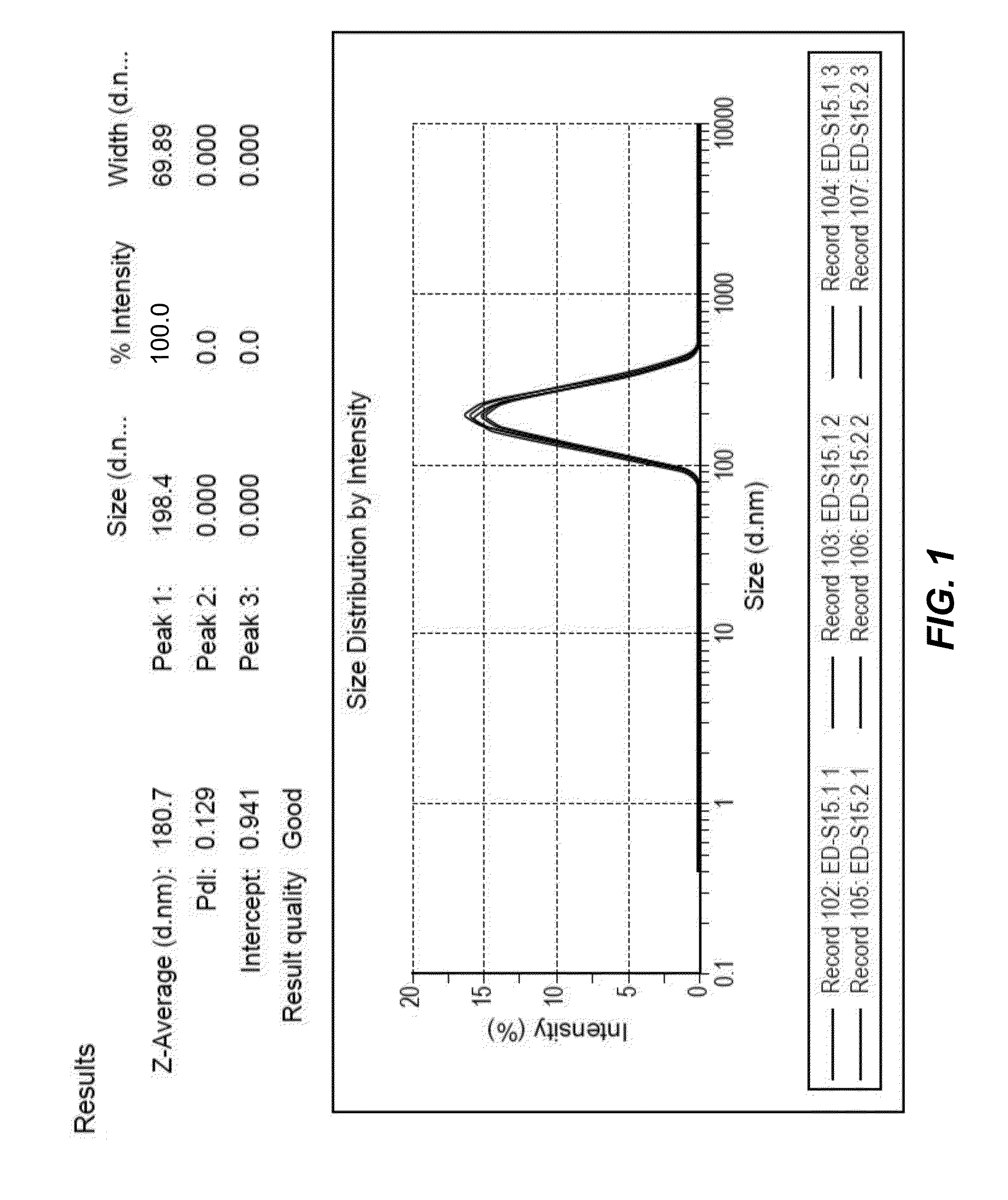
[0062]Diameter of particles prepared according to example 1 was determined using dynamic light scattering (DLS) as previously described (Biomaterials. 2010 September; 31(27):7070-7). FIG. 1 shows that the particle size ranged from 80 to 500 nm with a sharp peak at 181 nm. FIG. 1 shows the results of 6 individual and independent syntheses. The results are identical; all curves essentially overlap, indicating a high reproducibility. The particle diameter distribution remained stable for several months. The particles were lyophilised and frozen for storage. However, particles stored as aliquots in water (frozen) were also stable.

[0063]The particles prepared according to example 1 with high and medium gadolinium co...

example 3

Ultrasound Imaging in vitro

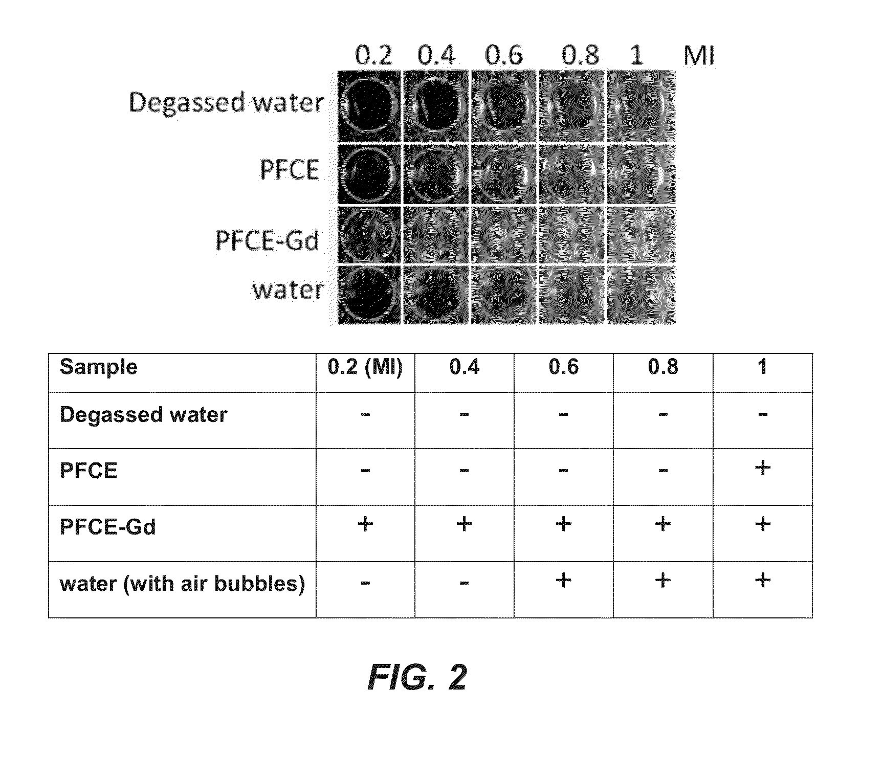
[0065]A linear array transducer (L11-3) with central frequency 7.5 MHz was used for all the ultrasound scans (SONOS 7500, Philips Medical Systems, Best, The Netherlands). The MI was variable, from 0.1-2.0, as indicated. Gain was typically set to 90%.

[0066]Gel phantoms consisted of 8% gelatin (Dr. Oetker, Ede, The Netherlands) and 2% agar (Agar Powder CMN, Boom, Meppel, The Netherlands) solution (these gels showed as bright in the ultrasound images).

[0067]Ultrasound exposure was performed at Mechanical Index (MI) ranging from 0.2 to 1. MI is a safety metric indicating how much energy is transferred to the subject or sample during imaging; clinical limits are 1.9 for diagnostic imaging and 1.0 for obstetric scans).


PUM


Abstract

The present invention relates to contrast agent enhanced medical ultrasound imaging. In particular, the contrast agents provided are useful for cell imaging and cell therapy, as well as in vivo targeting, drug delivery and perfusion or vascular imaging applications. More specifically, it provides a particle comprising a fluorinated organic compound and a metal. Such particles may be advantageously employed in qualitative or quantitative imaging such as acoustic imaging including photoacoustic and ultra-sound imaging, MRI imaging, such as 19F imaging, 1H imaging including T1 and T2 weighted imaging, SPECT, PET, scintigraphy, fluorescence imaging and optical coherence imaging and tomographic applications. This may then be employed in cell labeling, microscopy, histology or for imaging vasculature or perfusion in vivo and in vitro.

Description

FIELD OF THE INVENTION[0001]The present invention relates to contrast agent enhanced medical imaging. In particular, the contrast agents provided are useful for cell imaging, cell therapy and in vivo targeting and drug delivery applications.BACKGROUND OF THE INVENTION[0002]Cell therapy is an extremely promising field, which could potentially cure conditions such as diabetes, heart disease and cancer. There are currently over 15,000 ongoing clinical trials involving some form of cell therapy (www.clinicaltrials.gov). A key hurdle in the development and optimisation of cell therapy is that there are insufficient means to monitor the cells once they are in the patient, in a noninvasive manner.[0003]Imaging might provide a clinically applicable solution to monitor therapeutic cells once in the patient, in terms of their viability, localisation, numbers and functionality (see Srinivas M, Aarntzen E H, Bulte J W, Oyen W J, Heerschap A, de Vries I J, Figdor C G. Imaging of cellular therapi...

Claims


Application Information

Patent Timeline
IPC IPC(8): A61K49/22
CPCA61K49/221A61K49/225A61K49/0002A61K49/0034A61K49/0093A61K49/1818A61P35/00A61P37/00A61P37/02A61P37/06A61P9/00
Inventor FIGDOR, CARL GUSTAVDE VRIES, INGRID JOLANDA MONIQUESRINIVAS, MANGALACRUZ RICONDO, LUIS JAVIER
Owner STICHTING RADBOUD UNIVERSITAIR MEDISCH CENT
Why Eureka
Try Eureka