Nanoparticle formulations with enhanced mucosal penetration

a technology of nanoparticles and mucosal surface, which is applied in the direction of application, drug composition, biocide, etc., can solve the problems of limited control of drug delivery at mucosal surface, limited efficacy, and large amount of epithelium left susceptible and/or untreated, so as to improve the distribution over a mucosal surface, improve the residence time of mucus-penetrating particles, and improve the effect of diffusion

Active Publication Date: 2015-10-22
THE JOHN HOPKINS UNIV SCHOOL OF MEDICINE
View PDF4 Cites 41 Cited by
  • Summary
  • Abstract
  • Description
  • Claims
  • Application Information

AI Technical Summary

Benefits of technology

[0015]Osmosis can be used to cause rapid penetration of mucus-penetrating particles into deep recesses in highly-folded mucosal tissues. Absorption and penetrability into the deep recesses of mucosal tissues improves the distribution over a mucosal surface of otherwise poorly distributed entities. Rapid absorption and penetrability into deep recesses of mucosal tissues leads to increased residence time of mucus-penetrating particles. Rapid absorption facilitates user acceptability in addition to increasing the effectiveness of the treatment and minimizing the time between application and mucosal protection.
[0016]Hypotonic formulations were evaluated for delivering water-soluble drugs and for drug delivery with muco-inert (that is, non-adhesive) mucus-penetrating nanoparticles (MPP). Hypotonic formulations markedly increased the rate at which drugs and MPP reached the epithelial surface. Additionally, hypotonic formulations greatly enhanced drug and MPP delivery to the entire epithelial surface, including deep into the vaginal folds (rugae) that isotonic formulations failed to reach. Hypotonic formulations can cause free drugs not only to be drawn to the epithelium but also be drawn through the epithelium, reducing vaginal retention. In contrast, hypotonic formulations cause MPP to accumulate rapidly and uniformly on vaginal surfaces, but they do not pass through the epithelium and thus remain ideally positioned for sustained mucosal drug delivery. Minimally hypotonic formulations, preferably ranging from 20-220 mOsm / kg, provided rapid and uniform delivery of MPP to the entire vaginal surface, with minimal risk of epithelial toxicity. Hypotonic formulations for vaginal drug delivery via MPP should significantly improve prevention and treatment of reproductive tract diseases and disorders.
[0017]Data also show that there is a higher osmolality in the colon, such that vehicles with an osmolality above that of blood plasma (generally considered isotonic at ˜300 mOsm / kg), still lead to improvements in distribution in the colon due to rapid, osmotically-induced fluid absorption. The range for improved colon distribution with a hypotonic vehicle in the colon is ˜20 mOsm / kg-450 mOsm / kg. In the preferred embodiment, the formulation for application to the colon or rectum has an osmolality between about 20 mOsm / kg and 450 mOsm / kg, wherein sodium ions (Na+) cause at least 30% of the osmolality in excess of 220 mOsm / kg. (i.e., if the osmolality of the formulation is 450 mOsm / kg, Na+ ions must comprise at least 30% to 450-220=230 mOsm / kg, or 69 mOsm / kg). Improved distribution of hypotonically administered MPP (compared to CP) on rectal tissue with induced ulcerative colitis, including uptake of MPP into the ulcerated tissue, was also demonstrated. Hypotonic administration also leads to improved distribution of free drug (tenofovir labeled with FITC) in the colon.

Problems solved by technology

However, controlled drug delivery at mucosal surfaces has been limited by the presence of the protective mucus layer.
As a result of mucus turnover, most therapeutics delivered locally to mucosal surfaces suffer from poor retention and distribution, which limits their efficacy.
Without maximal distribution with penetration into these deep recesses, much of the epithelium is left susceptible and / or untreated.
Failure to achieve adequate distribution over the entire vaginal epithelium is a documented failure mode of vaginal microbicides.
Another significant barrier to effective drug delivery to the vagina is the viscoelastic layer of mucus secreted by the endocervix that coats the vaginal epithelium.
Although the use of mucoadhesive dosage forms has been proposed for increasing residence time in the vagina, mucus clearance occurs rapidly (on the order of minutes to hours), limiting the residence time of mucoadhesive systems.
Hypertonic formulations cause rapid, osmotically-driven secretion of fluid into the vagina, and this causes an immediate increase in fluid leakage from the vagina at a rate proportional to the hypertonicity of the formulation.
Moreover, recent investigations of candidate vaginal and rectal microbicides both in animal models and in humans have revealed that hypertonic formulations cause toxic effects that can increase susceptibility to infections.
Unfortunately, the gel formulation was highly hypertonic, leading investigators in the most recent clinical trial of tenofovir to reduce the concentration of glycerol to reduce toxicity.
However, the concentration was not reduced, and the formulation is still significantly hypertonic.
There appears to be no evidence to justify hypertonic formulations for vaginal drug delivery, since in addition to the documented toxic effects, hypertonic formulations cause rapid osmotically-driven secretion of vaginal fluid, fluid flow that opposes the delivery of drugs to the epithelium.

Method used


Image

  • Nanoparticle formulations with enhanced mucosal penetration
  • Nanoparticle formulations with enhanced mucosal penetration
  • Nanoparticle formulations with enhanced mucosal penetration

Examples


example 1

Preparation of Nanoparticles

[0188]Materials and Methods

[0189]Biodegradable nanoparticles were prepared by either o / w single emulsion or w / o / w double emulsion method as described in R. C. Mundargi et al, J Control. Release 125, 193 (2008), M. Li et al., Int. J Pharm. 363, 26 (2008), C. E. Astete and C. M. Sabliov, J. Biomater. Sci. Polymer Ed. 17, 247 (2006), and R. A. Jain, Biomaterials, 21, 2475 (2000).

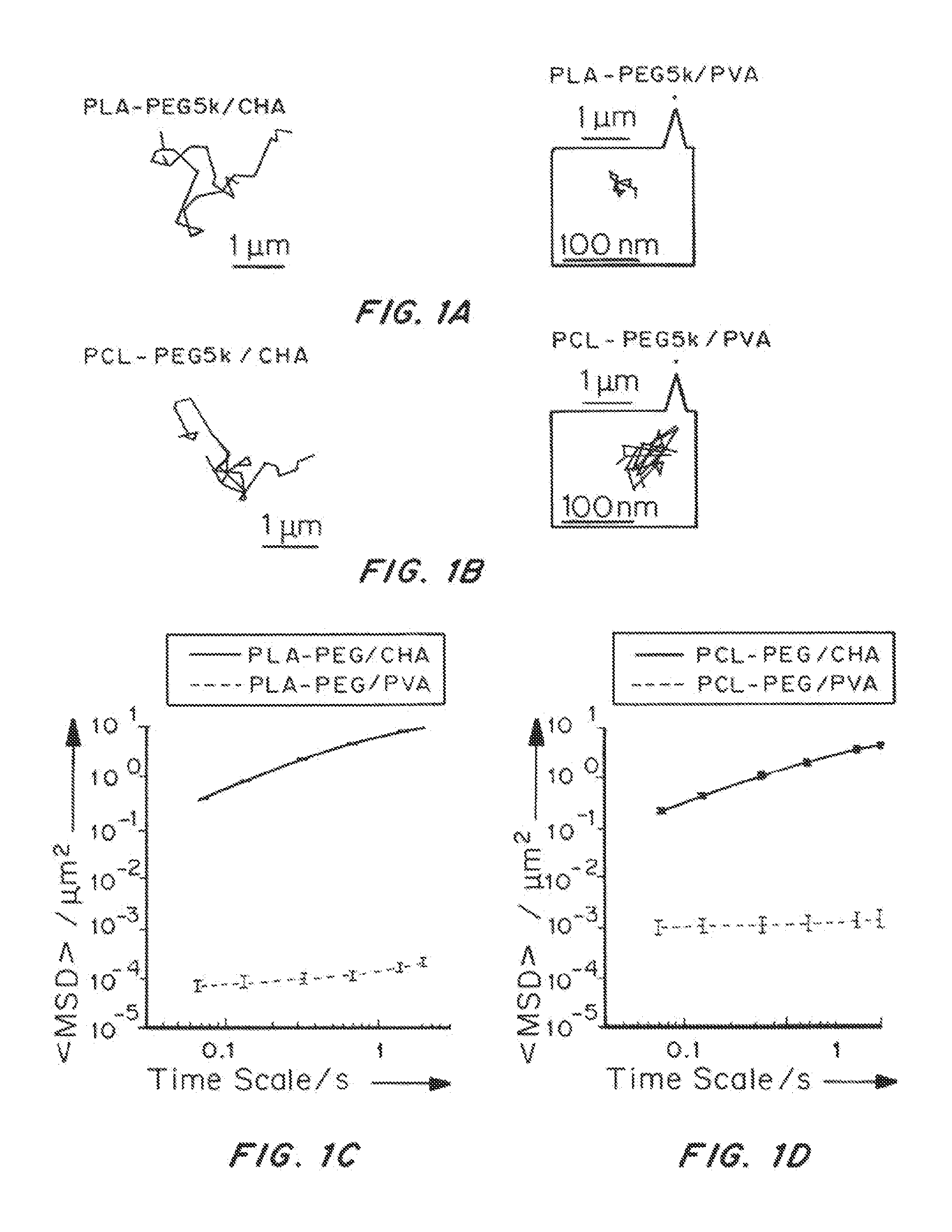
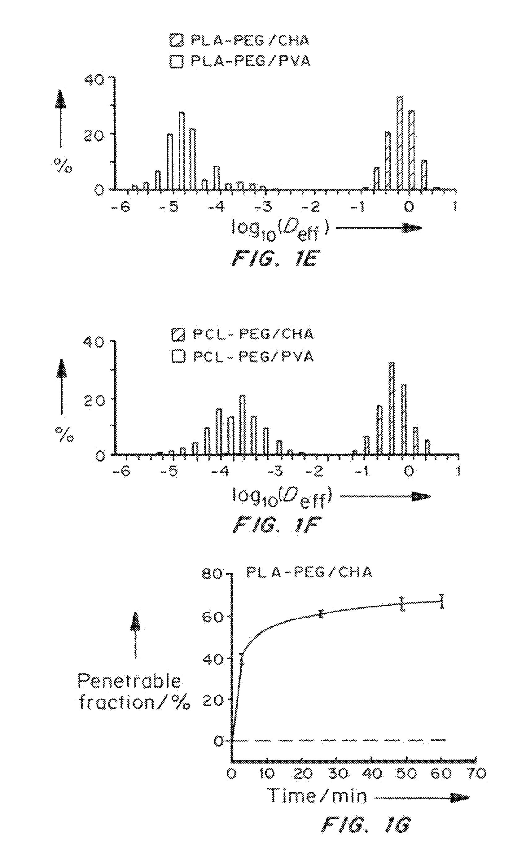
[0190]Nanoparticles were characterized for size, surface property and drug loading (for drug encapsulated nanoparticles). The displacements of nanoparticles were tracked in fresh, undiluted human CVM using multiple particle tracking.

[0191]Nanoparticles Prepared with Different Amounts of PEG

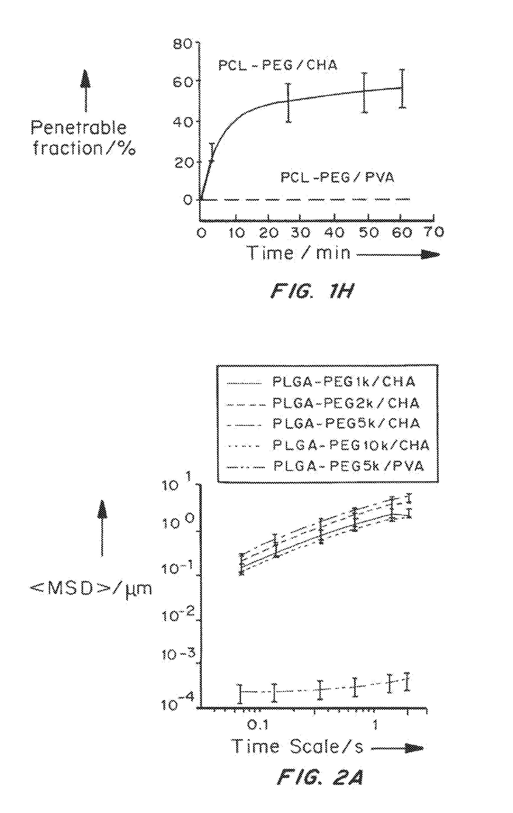
[0192]PLGA-PEG nanoparticles were prepared with varying target PEG contents (0, 2, 3, 5, 8, 10 and 25 wt %, referred to as PLGA, PLGA-PEG2%, PLGA-PEG3%, PLGA-PEG5%, PLGA-PEG8%, PLGA-PEG10% and PLGA-PEG25%) using emulsification. The PEG molecular weight 5 kDa was selected since at the same PEG conte...

example 2

Nanoparticles Prepared with Different Emulsifiers

[0197]Materials and Methods Alexa Fluor 555 cadaverine (AF555) was chemically conjugated to polymers. Nanoparticles were prepared using emulsification. Typically, a mixture (total 50 mg) of PLGA-PEG5k and AF555-labeled PLGA-PEG5k was dissolved in 1 mL dichloromethane (DCM). The oil phase was poured into 5 mL aqueous solution containing 1% emulsifier under sonication (VibraCell, Sonics & Materials Inc., Newtown, Conn.) at 30% amplitude for 2 mins in an ice-water bath to form the oil-in-water emulsion.

[0198]The emulsion was poured into another 40 mL aqueous phase of emulsifier solution under magnetic stirring at 700 rpm for at least 3 hours to allow the solvent to evaporate. The solvent was further evaporated by placing the solution in a vacuum chamber for 30 mins. The final nanoparticle suspensions were filtered through 1 μm syringe filter, centrifuged at 20,000 g for 25 mins and thoroughly washed with water.

[0199]Emulsifiers including...

example 3

Preparation of Drug Encapsulated Nanoparticles

[0205]Materials and Methods

[0206]Curcumin was selected as a model hydrophobic drug which was dissolved with polymer in DCM. The procedure was similar to that for preparation of unloaded nanoparticles. The prepared curcumin-nanoparticles can be visualized in mucus because of curcumin's intrinsic fluorescence.

[0207]BSA was used as a model hydrophilic drug because it is representative of large molecule biologics. BSA-FITC and BSA (10% ratio of BSA-FITC) were dissolved in 0.2 mL 16% w / v aqueous solution at 37° C. This solution was added to 1 mL of 100 mg / ml PLGA-PEG5k in DCM solution during probe sonication (30% amplitude, 1 min with 1 s pulse) in the ice-water bath. The resultant W / O primary emulsion was immediately added to a second water phase (5 mL 1% saponin solution) under sonication (20% amplitude for 2 min). The double emulsion was transferred to another 40 mL 1% saponin solution with magnetic stirring for 3 hours. Nanoparticles were...


PUM

PropertyMeasurementUnit
osmolalityaaaaaaaaaa

Abstract

Hypotonic formulations were evaluated for delivering water-soluble drugs and for drug delivery with muco-inert (that is, non-adhesive) mucus-penetrating nanoparticles (MPP). Hypotonic formulations markedly increased the rate at which drugs and MPP reached the epithelial surface, including deep into the vaginal folds. Minimally hypotonic formulations, preferably ranging from 20-220 mOsm/kg, provided rapid and uniform delivery of MPP to the entire vaginal surface, with minimal risk of epithelial toxicity. Data also show that there is a higher osmolality in the colon, such that vehicles with an osmolality above that of blood plasma (generally considered isotonic at ˜300 mOsm/kg), still lead to improvements in distribution in the colon due to rapid, osmotically-induced fluid absorption. The range for improved colon distribution with a hypotonic vehicle in the colon is ˜20 mOsm/kg-450 mOsm/kg.

Description

GOVERNMENT RIGHTS[0001]The United States government has certain rights in this invention. This work was supported by National Institutes of Health grants R01HD062844, R33AI079740, R01CA140746) (J.H. and R.C.), the National Science Foundation (L.M.E. and the National Institutes of Health / Microbicide Innovation Program 5R21AI079740.FIELD OF THE INVENTION[0002]This invention is in the field of nanoparticle formulations, particularly hypotonic nanoparticle formulations that rapidly deliver mucus-penetrating nanoparticles to mucosal covered epithelial surfaces, and methods of making and using thereof.PRIORITY CLAIMS[0003]U.S. Ser. No. 61 / 588,350 filed Jan. 19, 2012[0004]PCT / US2012 / 024344 filed Feb. 8, 2012[0005]PCT / US2012 / 069882 filed Dec. 14, 2012BACKGROUND OF THE INVENTION[0006]Localized delivery of therapeutics via biodegradable nanoparticles often provides advantages over systemic drug administration, including reduced systemic side effects and controlled drug levels at target sites....

Claims


Application Information

Patent Timeline
Patent Type & Authority Applications(United States)
IPC IPC(8): A61K9/51A61K31/704A61K9/00
CPCA61K9/5146A61K9/51A61K31/704A61K9/0031A61K9/5138A61K9/0034A61K9/0048A61K9/5153A61P11/00Y10S977/773Y10S977/906A61K9/0043A61K9/0053A61K9/0014
Inventor ENSIGN, LAURACONE, RICHARDHANES, JUSTIN
Owner THE JOHN HOPKINS UNIV SCHOOL OF MEDICINE
Why Eureka
Try Eureka