

METHOD FOR PRODUCING MONOCLONAL IgA ANTIBODY
a monoclonal antibody and antibody technology, applied in the field of monoclonal iga antibodies, can solve the problems of long-term administration, side effects, and disruption of the immune system of the intestinal tract, and achieve the effects of suppressing intestinal putrefaction, suppressing intestinal bacterial growth alternation, and improving or optimizing the intestinal environmen
- Summary
- Abstract
- Description
- Claims
- Application Information
AI Technical Summary
Benefits of technology
Problems solved by technology
Method used
Image

Examples
example 1
Preparation of Antibodies
1-1: Separation of Intestinal Lamina Propria Cells
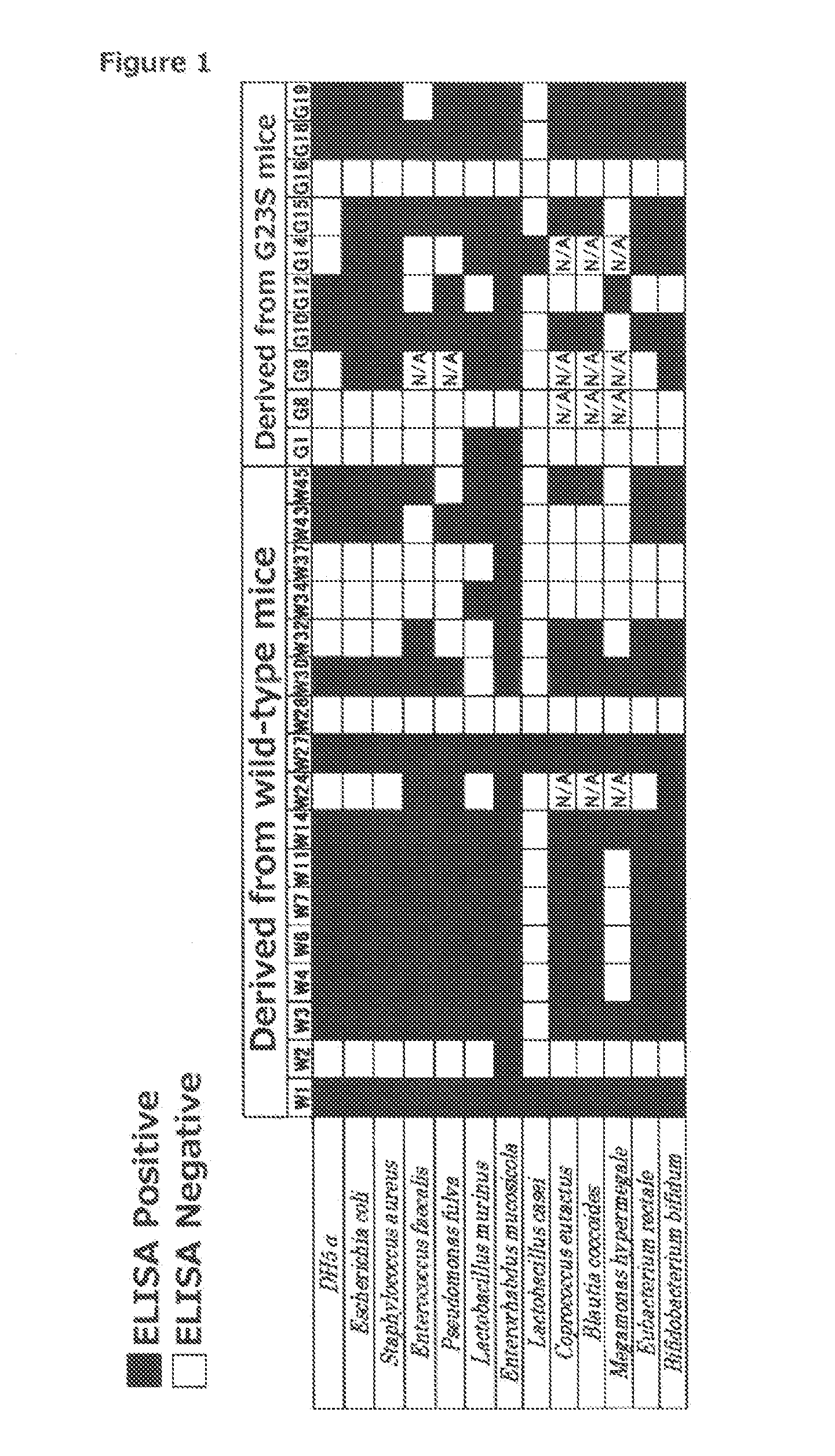
[0270]8-week-old wild-type CS7BL / 6 mice (CLEA Japan, Inc.) were fed until the age of 20 weeks with free access to food and water in an experiment facility of the Nagahama Institute of Bio-Science and Technology. Subsequently, the mice were euthanized with carbon dioxide and then subjected to laparotomy to remove the entire length of the small intestine. After the connective tissue and Peyer's patches were removed from the removed small-intestine sample, a longitudinal incision was made through the small intestine, and the intestinal contents were washed out in a 10-cm dish filled with PBS. The small intestine was washed well with three to four changes of PBS.
[0271]As with the wild-type C57BL / 6 mice, the small intestine was also collected from mice expressing AID G23S (AID G23S mice), in which a mutation is introduced at the N-terminal side of AID protein. Such AID G23S mice can be created using a known method...
example 2
Experiment to Identify Antigens for Monoclonal IgA Antibodies
2-1: Immunoprecipitation Experiment
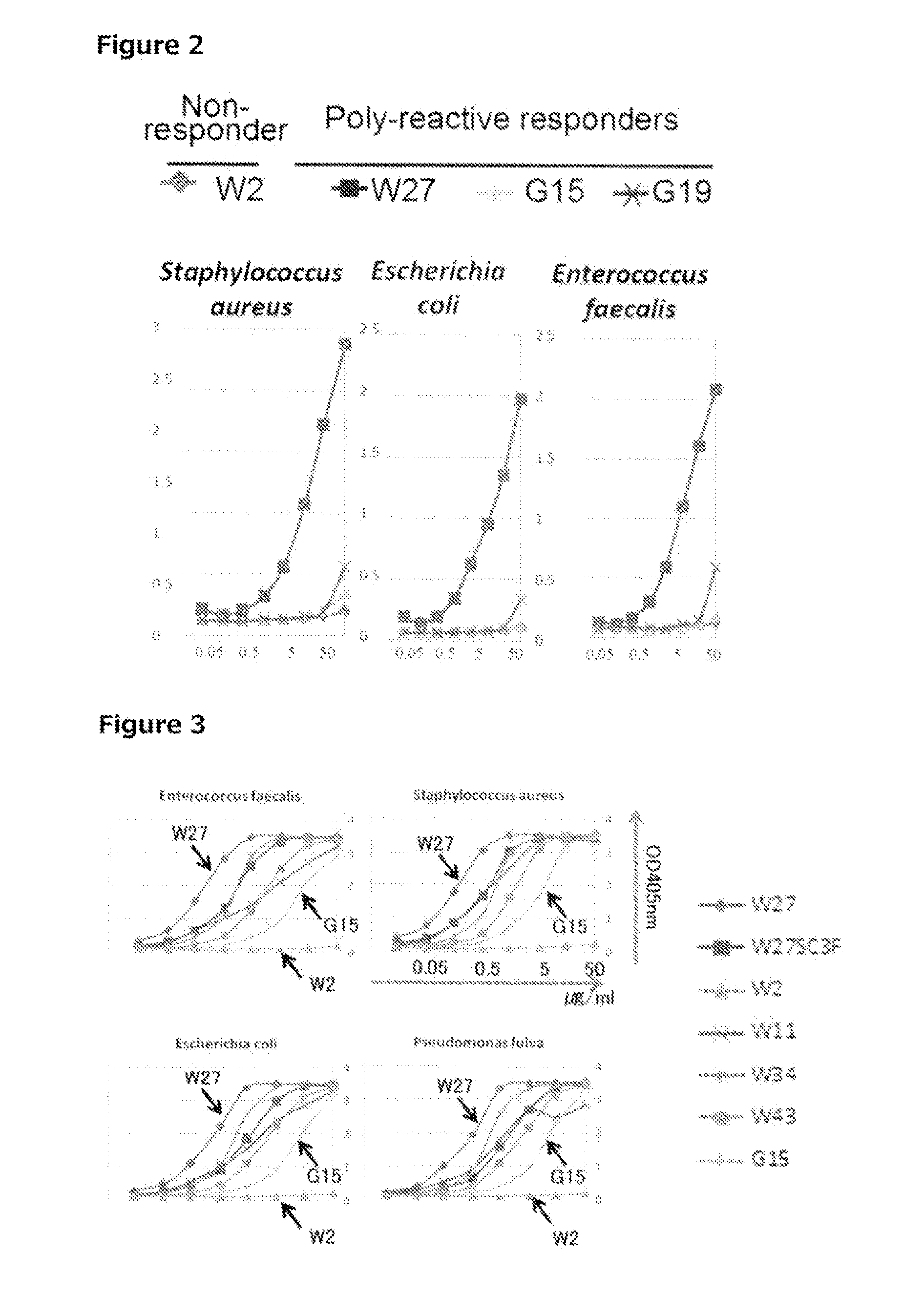
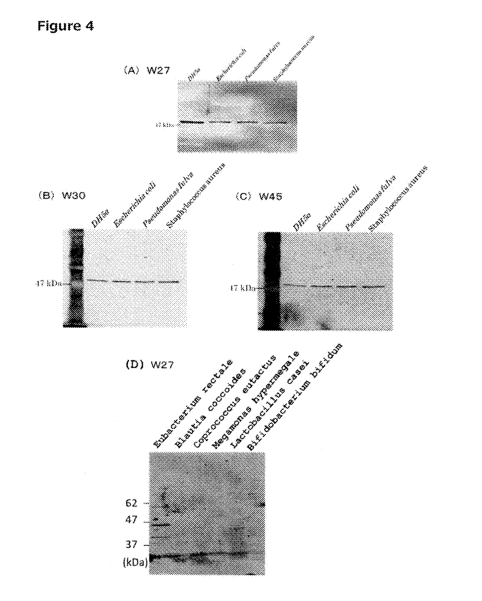
[0309]After cell extracts were prepared from three species of bacteria, i.e., Escherichia coli (DH5α), Pseudomonas fulva, and Staphylococcus aureus, by the method described in section 1-5 above, 2-ME (final concentration of 300 mM) and SDS (final concentration of 1%) were added, and heat denaturation was performed at 95° C. for 10 minutes. Thereafter, in each solution, the 2-ME concentration was adjusted to 60 mM, and the SDS concentration was adjusted to 0.2%. First, 100 μl of Protein G Sepharose 4 Fast Flow (GE) that had been washed was added to each cell extract, and the resulting mixtures were mixed by inversion at 4° C. for 30 minutes. Components that non-specifically bind to Protein G Sepharose were then eliminated by centrifugation. 5 μg of the W27 monoclonal IgA antibody was added to each of the cell extracts obtained after the pre-clear, and the resulting mixtures were allowed t...
example 3
Oral Administration Experiment for W27 Monoclonal IgA Antibody
3-1: Analysis of Disease Model of G23S Mice
[0320]Large intestinal tissue sections of AID G23S mice were prepared and stained with HE to investigate the degree of inflammation. As a result, significant reduction or atrophy in the crypts was observed even at 13-16 weeks of age compared with wild-type mice (FIG. 6). In particular, at 40 weeks or more of age, significant reduction or atrophy in the crypts and infiltration of inflammatory cells into mucosa of the large intestine were observed. Immunostaining showed that a larger number of CD11b+ cells, IL-6 positive cells, IL-6, IL-17, and like inflammatory cells were present than in the wild-type mice.
[0321]Further, the results of measuring the expression levels of TNFα, IFNγ, IL-1, IL-17, IL-6 and like inflammatory cytokines by a method using quantitative PCR similarly revealed that a larger number of inflammatory cells were present in the large intestinal tissues of AID G2...
PUM
 Abstract
Description
Claims
Application Information