
Self-assembled micro-and nanostructures

a micro-and nanostructure, self-assembling technology, applied in the direction of natural mineral layered products, drug compositions, peptides, etc., can solve the problems of low bond strength, toxic to living tissues, and associated risk of viral or prion contamination, and achieve antibacterial anti-fouling, superior adhesive and/or anti-oxidant properties

Inactive Publication Date: 2016-04-28
RAMOT AT TEL AVIV UNIV LTD +1
View PDF0 Cites 3 Cited by
  • Summary
  • Abstract
  • Description
  • Claims
  • Application Information

AI Technical Summary

Benefits of technology

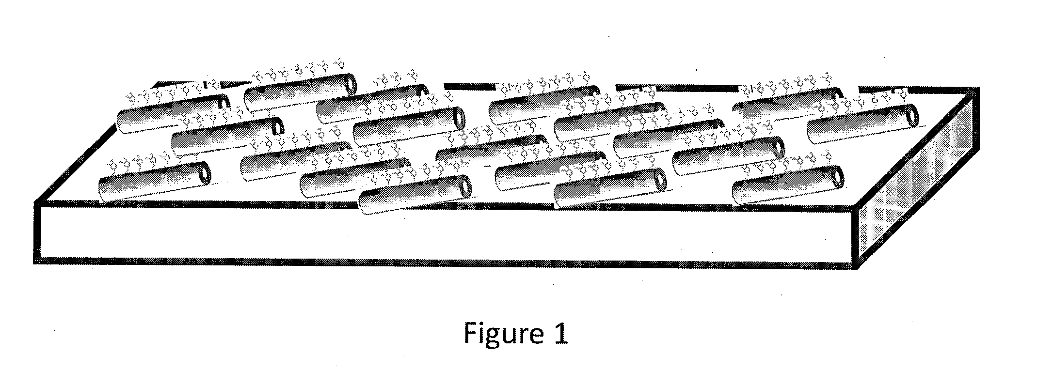
The present invention provides self-assembled micro- and nano-structures with controlled orientation of active moieties relative to the target surface, enhancing adhesive, anti-bacterial, anti-fouling and / or anti-oxidant properties. The micro- and nano-structures have superior properties compared to currently known products and are biocompatible, making them suitable for various pharmaceutical, cosmetic and medical devices applications. The invention is based on the use of amino acids comprising catecholic moieties or incorporation of such amino acids into self-assembled peptides. The resulting micro- and nano-structures provide a surface comprised of the controllably exposed catecholic groups, which display high mechanical stability and strength. The invention also provides a method for incorporating lysine residues into the amino acid / peptide assemblies, which may contribute to cohesion and adhesion.

Problems solved by technology

Current biological glues (fibrin, albumin, gelatin-resorcinol-formaldehyde, etc.) suffer from low bond strength and are in some cases derived from blood products, with associated risk of viral or prion contamination.
On the other hand, synthetic glues (e.g., cyanoacrylate adhesives) are very strong but they are also toxic to living tissues and form rigid, nonporous films that can hinder wound healing [1].
Extraction of MAPs from mussels is, however, not practical for commercial scale production.
The major disadvantage of presently known bioadhesive materials, mimicking mussel adhesive properties, is the uncontrollable presentation of the functional adhesive sites relatively to the surface.
Currently known DOPA-functional synthetic polymers expose the adhesive groups randomly, which reduces the adhesive properties of DOPA-functionalized materials.
Although this approach has been successfully used for various important applications, one major challenge remains: the ability to present a high density of catechol functional groups in a defined ultrastructural organization and architecture at the nano-scale.

Method used


Image

  • Self-assembled micro-and nanostructures
  • Self-assembled micro-and nanostructures
  • Self-assembled micro-and nanostructures

Examples


example 1

Experimental Methods

[0165]Nanostructures Self-Assembly

[0166]Material—Peptides (Fmoc-DOPA-DOPA, DOPA-DOPA, DOPA-Phe-Phe, Fmoc-DOPA-DOPA-Lys) were purchased from Peptron. Fmoc-DOPA was purchased from Ana Spec. A stock solution of Fmoc-DOPA was prepared by dissolving the building block with ethanol to a final concentration of 100 mg / ml. The stock solution was then diluted into water to the desired concentration (0.5 mg / ml, 0.75 mg / ml, 1 mg / ml, or 2 mg / ml). DOPA-Phe-Phe was prepared by dissolving lyophilized form of the peptide in 1,1,1,3,3,3,-hexafluoro-2-propanol (HFIP), at a concentration of 100 mg / ml or 50 mg / ml. The stock solution was either directly deposited on a cover slip glass slides or diluted into water to a final concentration of 2 mg / ml or 5 mg / ml.

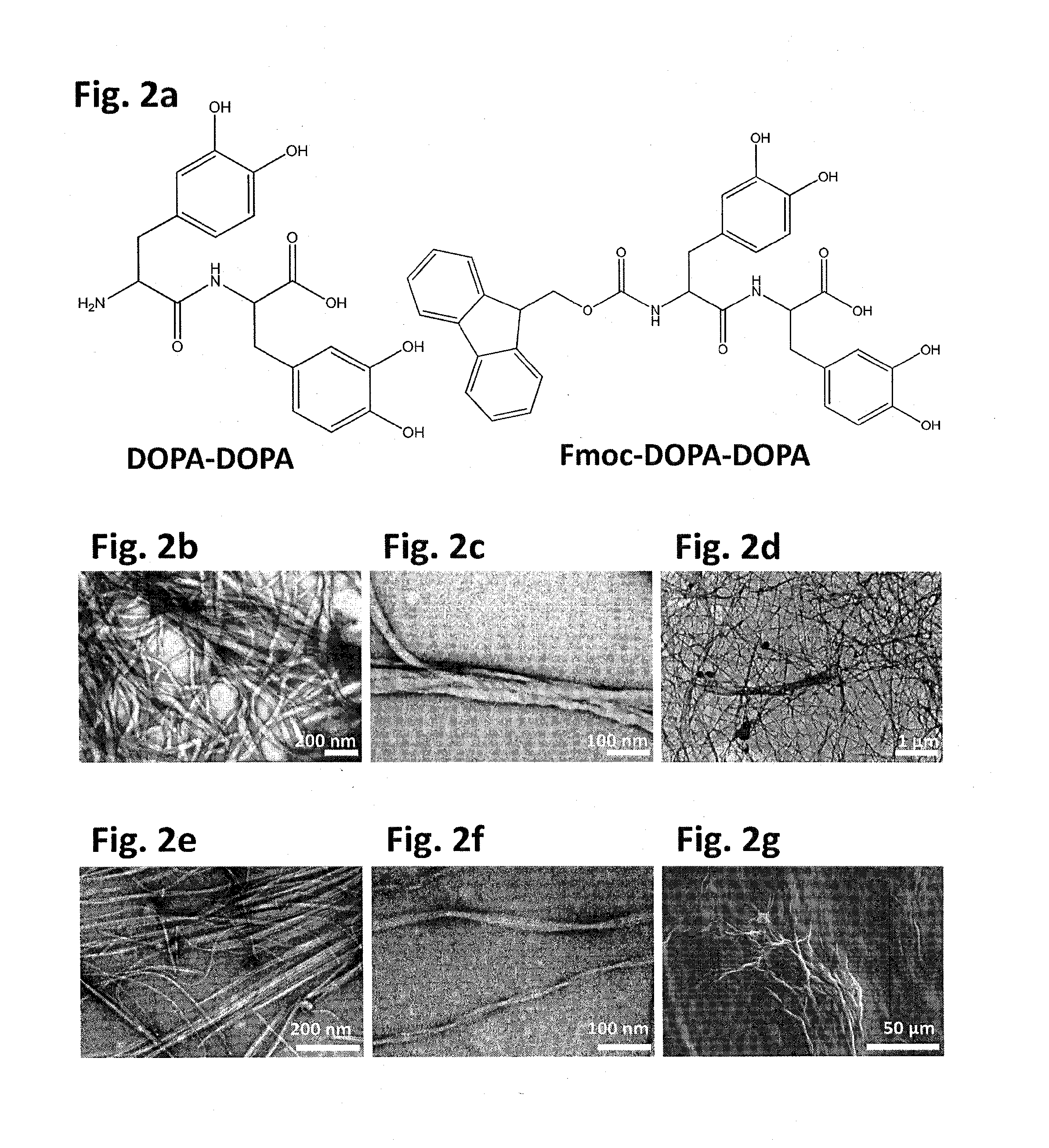
[0167]For the formation of the DOPA-DOPA dipeptide asssemblies (FIG. 2A-left panel), lyophilized peptide was dissolved in ethanol to a concentration of 33 mg / mL then diluted with Milli-Q water to a final concentration of 5 mg / mL....

embodiment 1

[0177]Atomic Force Microscopy (AFM) was used to assess the adhesion of the formed structures to a silicon oxide tip by employing “force / distance” measurements. This type of measurements allows deducing the attractive forces between the AFM tip and the contacted surface, when this force is represented by the minimum value of a force / distance curve. For AFM analysis, 10 μl aliquot of the peptide suspension was deposited on clean glass slide and dried at room temperature. Force / distance curves were determined at several points using NanoWizardIII of JPK instruments AG.

Adhesion Measurements

embodiment 2

[0178]In an alternative embodiment, AFM analysis was performed using an Asylum MFP-1D AFM instrument (Asylum Research, Santa Barbara, Calif., USA). To obtain force data the different peptides were prepared in ethanol: Fmoc-DOPA (1%) Fmoc-DOPA-DOPA (5%), Fmoc-DOPA-DOPA-Lys (12.5 mg / mL), samples were prepared in either DMSO or ethanol (12.5%) these results were compared to measurements performed on bare glass slide.

[0179]The AFM measurment was performed by emploting force mapping while simultaneously providing nanoscale topographical and mechanical information about the hydrogel. Force mapping involves generating individual force curves at discrete points on a material, which are then used to calculate stiffness and height values.

[0180]After overnight incubation, 10 μL of each sample were deposited on a glass slide and dried at room temperature. Force measurements of the samples were conducted using a SiO2 colloidal probe (tip velocity 1000 nm / s, compressive force 20 nN). To investiga...


PUM

PropertyMeasurementUnit
diameteraaaaaaaaaa
diameteraaaaaaaaaa
storage modulus G1aaaaaaaaaa

Abstract

The present invention discloses self-assembled bioadhesive anti-microbial, anti-fouling and / or anti-oxidant micro- and nano-structures comprising a plurality of amino acids or peptides, wherein each amino acid is an aromatic amino acid comprising a catecholic moiety, and / or each peptide comprises at least one aromatic amino acid comprising a catecholic moiety. Further disclosed are methods and kits for preparing these micro- and nano-structures. Further disclosed are uses of these micro- and nano-structures in pharmaceutical, cosmetic and medical devices applications.

Description

FIELD OF THE INVENTION[0001]The present invention relates to self-assembled bioadhesive, anti-microbial, anti-fouling and / or anti-oxidant micro- and nano-structures comprising a plurality of amino acids or peptides, the micro- or nano-structures comprising at least one aromatic amino acid comprising a catecholic moiety. The present invention further relates to methods of preparing the self-assembled micro- and nano-structures and to their use in a variety of biomedical and industrial applications, for example in pharmaceutical and cosmetic compositions and in medical devices.BACKGROUND OF THE INVENTION[0002]Bioadhesives are natural polymeric materials with adhesive properties. Bioadhesives may be comprised of a variety of substances, but proteins and carbohydrates feature prominently. Several types of bioadhesives offer adhesion in wet environments and under water, while others can stick to low surface energy—non-polar surfaces, such as plastic. However, the most prominent use of bi...

Claims


Application Information

Patent Timeline
Patent Type & Authority Applications(United States)
IPC IPC(8): C07K5/065C07K7/06A01N37/46C07K5/087
CPCC07K5/06078C07K5/0812A61K38/00A01N37/46C07K7/06C07K17/04
Inventor FICHMAN, GALITADLER-ABRAMOVICH, LIHIGAZIT, EHUDMESSERSMITH, PHILLIP B.
Owner RAMOT AT TEL AVIV UNIV LTD
Learn More