
Method and system for dual-band active thermal imaging using multi-frequency currents

a multi-frequency current and active thermal imaging technology, applied in the field of breast cancer diagnosis, can solve the problems of inaccurate results, inability to reliably sense the effect of this technique, and inability to comfort patients, so as to improve imaging performance, reduce individual drawbacks, and increase the sensitivity and specificity of eit

Active Publication Date: 2016-05-19
GENCER NEVZAT GUNERI +2
View PDF4 Cites 7 Cited by
  • Summary
  • Abstract
  • Description
  • Claims
  • Application Information

AI Technical Summary

Benefits of technology

This patent describes a new technique that uses both electric current injection and thermal imaging to detect cancer in the body. The technique involves measuring the conductivity distribution in the body and comparing it with data obtained from current injection at different frequencies. The system can be easily adjusted and can provide more reliable assessments by obtaining a larger amount of data. The technique combines the benefits of electrical impedance imaging and thermal infrared imaging, which improves sensitivity and specificity, and can detect cancer in a comfortable and fast examination.

Problems solved by technology

However, this method has accuracy problems especially for smaller tumors and it is not comfortable for patients since the breast needs to be squeezed to minimize the path length between source and detector.
If the dimension of a tumor is lower than 1.6 cm, it cannot be reliably sensed by this technique, since X-rays passing through the tumor are not detectable.
Another concern is the exposure of the patient to ionizing radiation (X-ray) which is potentially harmful for the human tissue.
However, this technique is not successful in detecting micro formations and monitoring deep regions of the breast.
Although it is more sensitive than mammography, it has a higher false-positive rate which may result in unnecessary biopsies.
At the same time, due to the relatively long data acquisition time, this technique is not the first choice by the patients.
Currently, there is no gold standard in detecting cancerous tissue in the breast.
However, these techniques are not also sufficient as a sole screening tool.
When the electric current is applied from the surface, the current cannot be focused homogeneously in the desired body location.
Therefore, desired amount of signal cannot be provided from the inner regions of the body.
In addition, the number of measurements are limited by the number of current drive electrodes.
This results in low-resolution images.
However, this requires a more demanding hardware, since the signal level decreases and the interaction between the electrodes increases.
In the early stages of the disease, diagnosis of smaller tumors is limited, especially for distant masses from the surface.
As a result, many studies were reported that abnormal breast thermogram shows a significant risk indicator for the breast cancer.
Although it has higher sensitivity and specificity, smaller cancerous tissues cannot be detected from deeper locations since the temperature contrast on the breast surface due to metabolic heat generation of the cancerous tissue may not be sufficient to be sensed.
Tumor tissue cannot produce adequate amount of temperature difference on the skin surface.
While, the noise equivalent temperature difference (NETD) of the state-of-the-art thermal infrared cameras is as low as several mK, detection may not be possible especially for smaller tumors located at deeper regions.
The thermal sensitivity can be increased by developments in the detector technology, but this requires a high cost and long-lasting R&D work.
Most of the clinicians and radiologists are well experienced on the structural anatomy and medical education but they are not experts on thermodynamics, thermokinetics and physical and biological basis of infrared imaging.
This may appear as a problem for the effective utilization of thermography for medical imaging.
This system has no clinical application.

Method used


Image

  • Method and system for dual-band active thermal imaging using multi-frequency currents
  • Method and system for dual-band active thermal imaging using multi-frequency currents
  • Method and system for dual-band active thermal imaging using multi-frequency currents

Examples


Embodiment Construction

[0116]Theory

[0117]We present below the theoretical analysis of thermal contrast enhancement with current injection. Electromagnetic problem of the method is modeled and the schematic of the electromagnetic problem is shown in FIG. 4. The electrical model of the body is represented using permeability μ=μ0, electrical conductivity κ and permittivity ε. Sinusoidal currents are applied using two electrodes attached on the body surface at points A and B. Applied currents generate an electric field in the conductive body. The steady-state electric field {right arrow over (E)}=−jw{right arrow over (A)}−∇Ø can be calculated using the following coupled partial differential equations,

∇2{right arrow over (A)}−jwμ(κ+jwε){right arrow over (A)}−μ(κ+jwε)∇Ø=0

∇.[(κ+jwε)∇Ø]+∇(κ+jwε).jw{right arrow over (A)}=0

[0118]and boundary conditions

κ∂∅∂n={IonA-IonB0otherwise

[0119]where {right arrow over (A)} is the magnetic vector potential, Ø is the scalar potential, and I is the current applied from the surfac...


PUM


Abstract

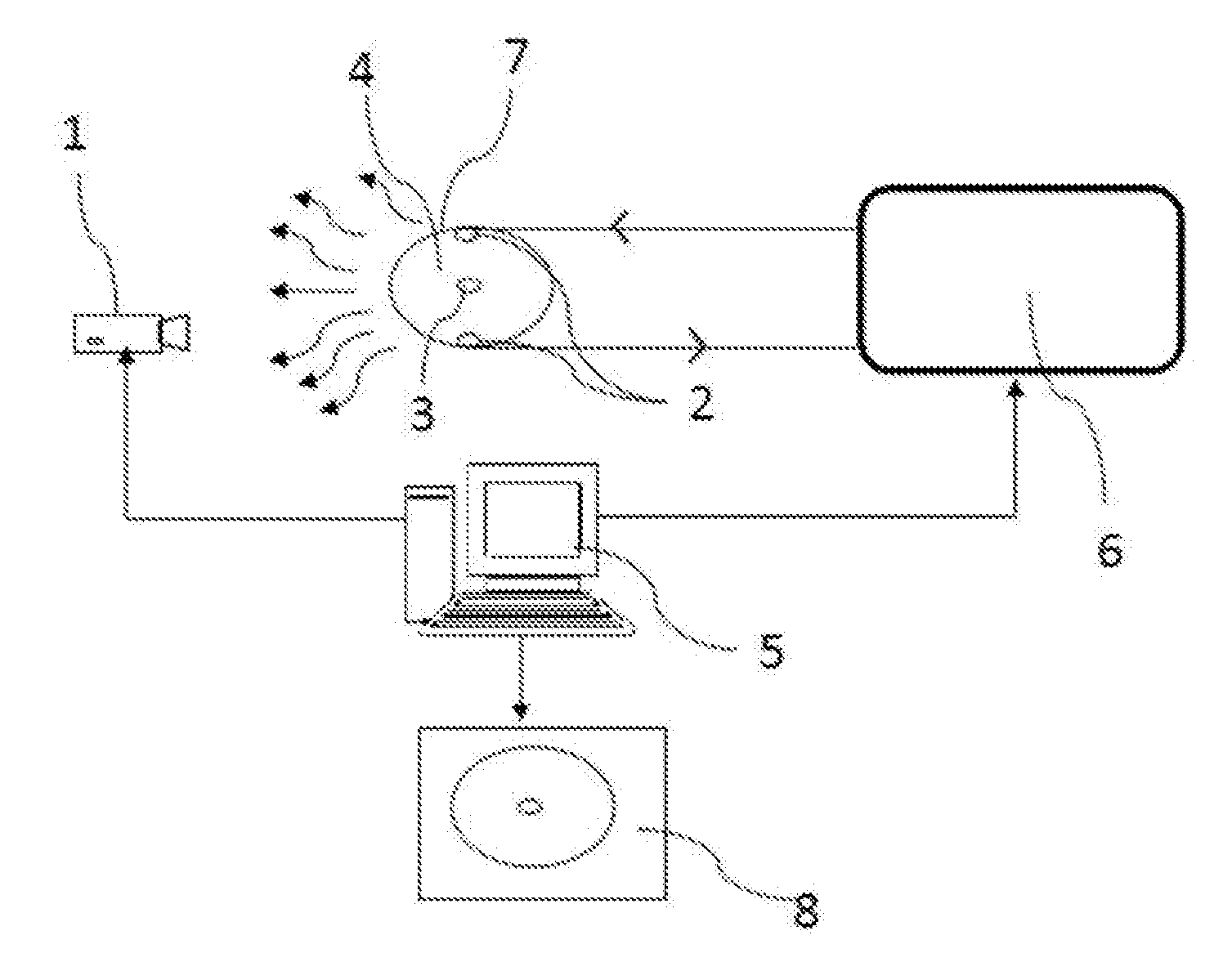
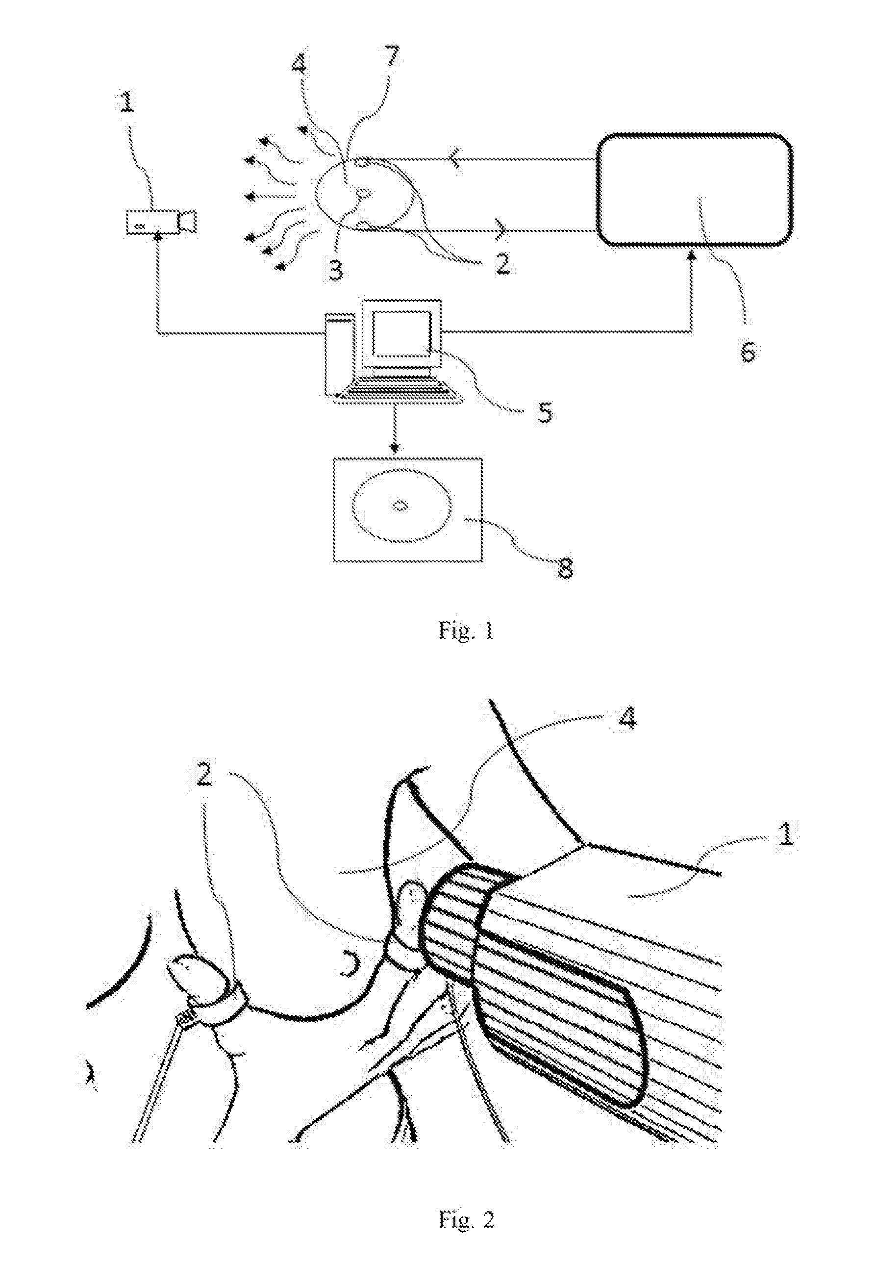
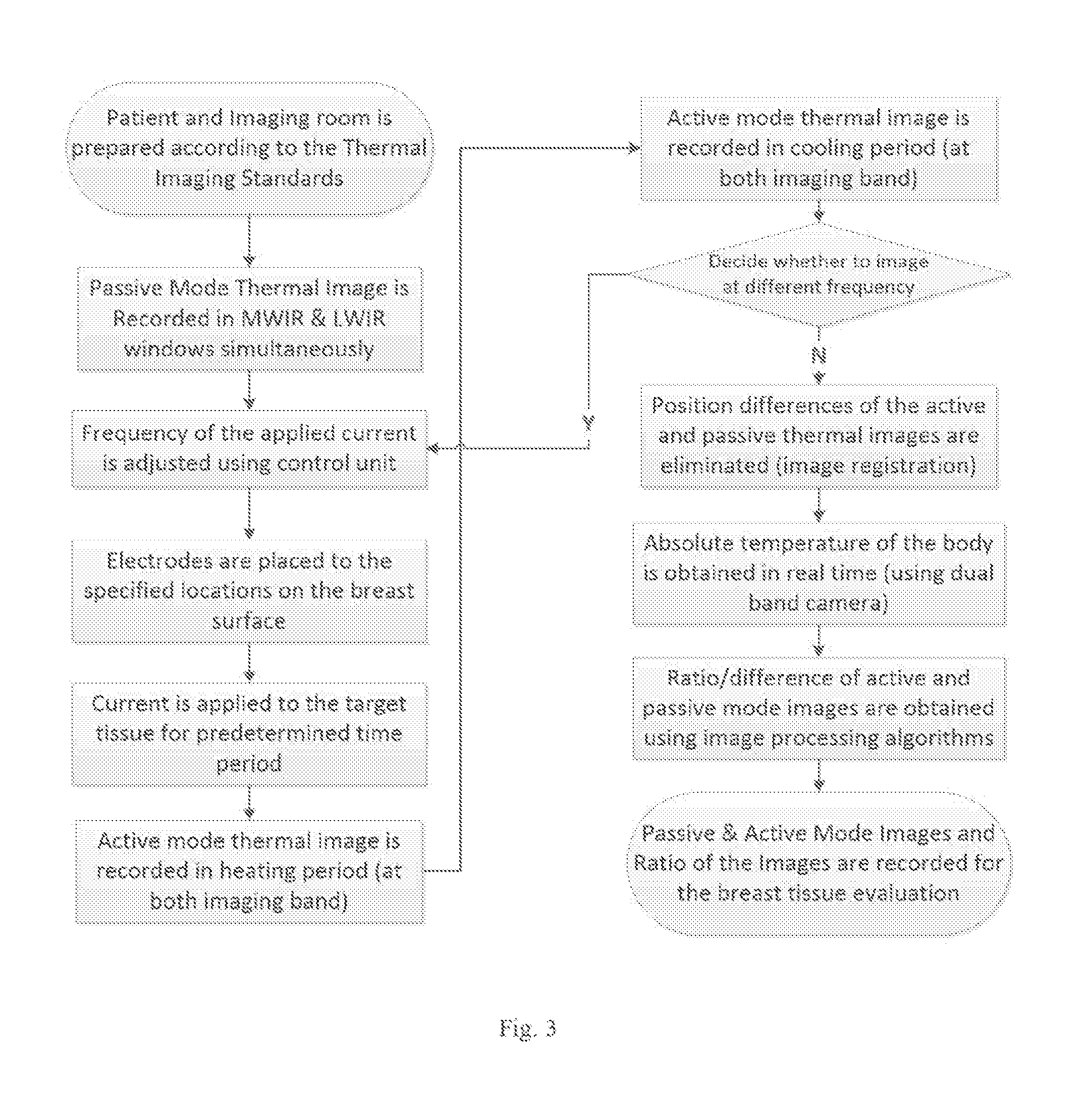
A hybrid system is developed using thermal and electrical impedance imaging methods together. The innovation of the approach relies on the frequency dependence of the tissue's electrical impedance which facilitates the acquisition of multiple thermal images with currents at different frequencies injected to the region of the body under inspection. Proposed method without current application (in passive mode of operation) provides images which are obtained by standard thermal imagers. On the other hand, the application of the electrical current (in active mode of operation) increases the temperature contrast on the body surface depending on the electrical property of tissue. Therefore, the technique while increasing the thermal contrast provides frequency dependent conductivity distribution data which can be used as a basis for the detection of the breast carcinoma. The sensitivity of the technique is increased by an infrared camera with dual band (MWIR / LWIR) imaging capability.

Description

BACKGROUND OF THE INVENTION[0001]The present invention is mainly concentrated on the diagnosis of the breast carcinoma using a hybrid technique incorporating both thermal and electrical impedance imaging methods. The innovation of the approach relies on the frequency dependence of the tissue's electrical impedance which facilitates the acquisition of multiple thermal images with currents at different frequencies injected to the region of the body under inspection. This new technique improves the sensitivity of medical imaging over the standard thermography as follows.[0002]Current injection at a single frequency increases the temperature gradients in the unhealthy regions enhancing the visibility (detection probability) of the cancerous tissues with a thermal camera,[0003]Current injection at multiple frequencies along with thermal imaging with a thermal camera results in generation of multiple thermal images according to the frequency dependencies of the tissue conductivities since...

Claims


Application Information

Patent Timeline
Patent Type & Authority Applications(United States)
IPC IPC(8): A61B5/00A61B5/053A61B5/01
CPCA61B5/0035A61B5/015A61B5/053A61B5/0093A61B5/6823A61B5/05A61B5/4312G06T7/0012G06T2207/10048G06T2207/30068
Inventor GENCER, NEVZAT GUNERICARLAK, HAMZA FEZABESIKCI, CENGIZ
Owner GENCER NEVZAT GUNERI
Learn More