ImmunoLipoplex Nanoparticle Biochip Containing Molecular Probes for Capture and Characterization of Extracellular Vesicles

a technology of micro-particles and probes, applied in the field of array biochips, can solve the problems of lack of sensitive, easy, fast, non-invasive and affordable screening tests for early disease detection, and inability to detect intra-ev contents, etc., and achieve the effect of eliminating most sample preparation steps

Active Publication Date: 2016-07-14
SPOT BIOSYST LTD
View PDF4 Cites 12 Cited by
  • Summary
  • Abstract
  • Description
  • Claims
  • Application Information

AI Technical Summary

Benefits of technology

[0006]The present invention is related to the design of new immunolipoplex nanoparticle (ILN) biochips for capture and characterization of extracellular vesicles (EVs) in blood and other body fluids using antibodies to identify membrane protein targets on EV surface and molecular probes (MPs) in lipoplex nanoparticles to detect intra-vesicular DNA/RNA/protein targets at the in

Problems solved by technology

Although significant progress has been made in disease diagnosis and treatment, mortality rates of diseases such as cancer, heart failure and AIDS have not changed much in the last several decades.
One reason is the lack of sensitive, easy, fast, non-invasive and affordable screening tests for early disease detection.
These methods, however, do not allow the detection of intra-EV contents and mainly serve as an EV isolation step in EV-based assays.
The entire sample preparation and target detection process requires more th

Method used


Image

  • ImmunoLipoplex Nanoparticle Biochip Containing Molecular Probes for Capture and Characterization of Extracellular Vesicles
  • ImmunoLipoplex Nanoparticle Biochip Containing Molecular Probes for Capture and Characterization of Extracellular Vesicles
  • ImmunoLipoplex Nanoparticle Biochip Containing Molecular Probes for Capture and Characterization of Extracellular Vesicles

Examples


example 1

Fabricate and Test Ab-ILN Biochip

[0096]We show a new biochip design which could simultaneously detect membrane protein targets and intra-vesicular RNA targets of individual EVs.

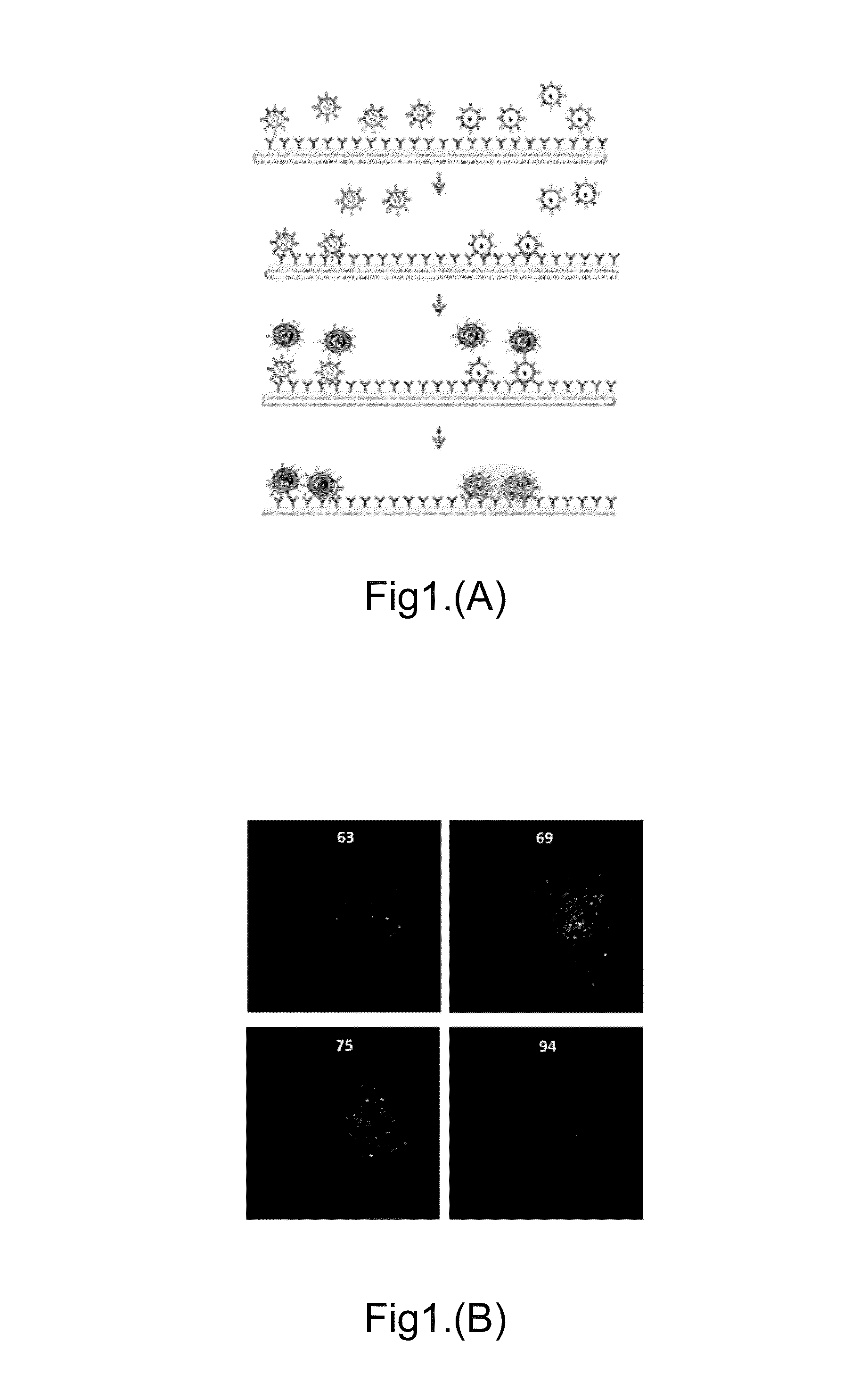
[0097]In one method, a mixture of 1-thiahexa(ethyleneoxide) lipidic anchor molecule WC14 [20-tetradecyloxy-3,6,9,12,15,18,22-heptaoxahexatricontane-1-thiol], biotin-PEG-SH and a lateral spacer β-mercaptoethanol (βME) in ethanol was added onto a gold coated glass substrate to form self-assembly monolayers (SAMs). Avidin was then added and un-reacted avidin was washed away using PBS. The biotin-conjugated Protein A was bound on the avidin-modified substrate surface through biotin-avidin interactions and the unbound Protein A was washed away with PBS. A target-specific antibody was then added and un-reacted antibody was washed away using PBS. The antibody-modified biochip can be used as an Ab-ILN biochip as shown in the first step of FIG. 1A.

[0098]We can tether antibodies on the chip surface to capture the EVs p...

example 2

Fabricate and Test Ab-TLN Biochip

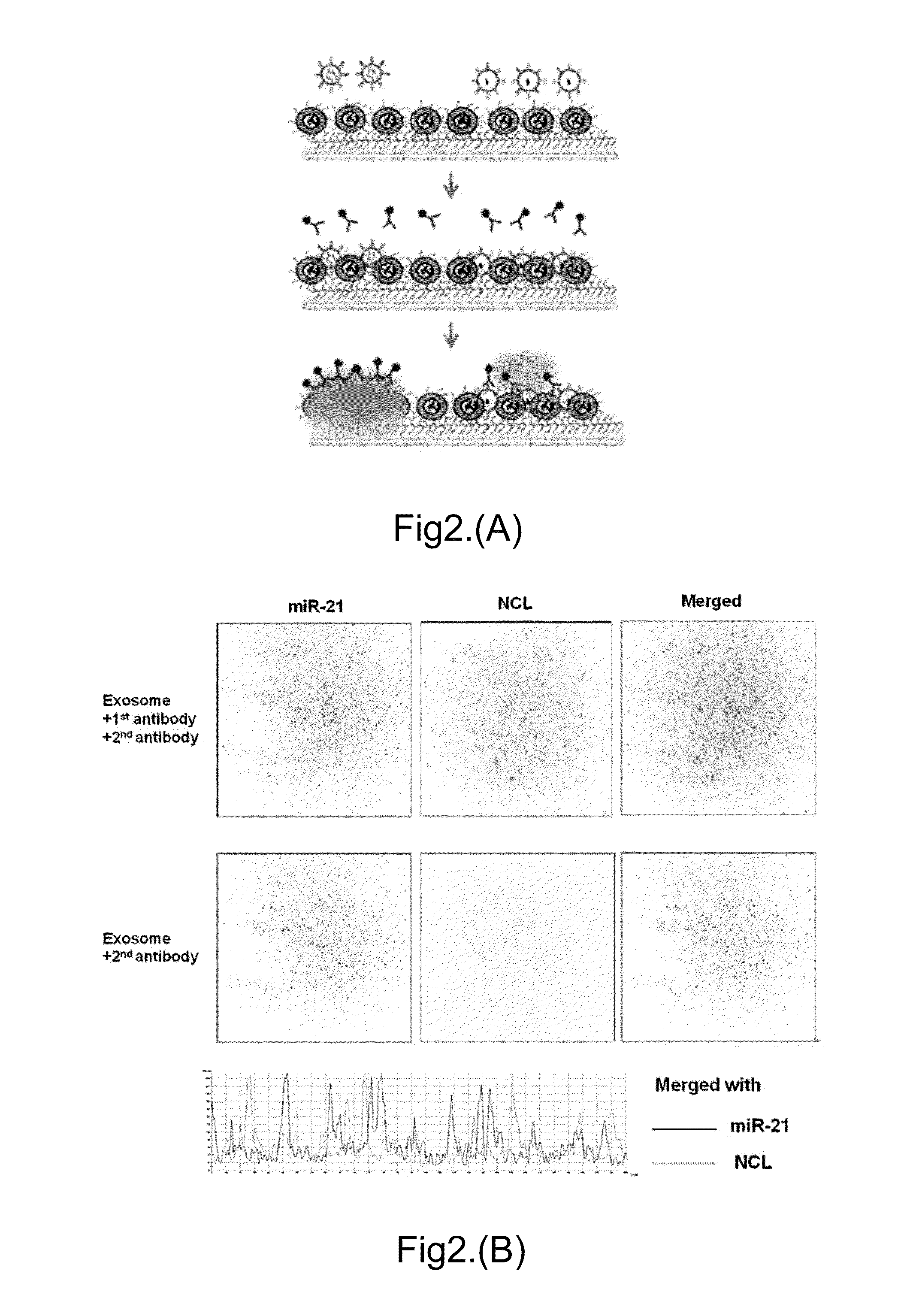
[0103]FIG. 2A shows the schematic of an Ab-TLN biochip design for EV capturing by tethered cationic lipoplex nanoparticles and detection of intra-vesicular RNAs by MPs in the lipoplex nanoparticles, followed with the detection of surface membrane proteins using first antibody and then fluorescence labeled second antibody.

[0104]EVs from culture supernatants of 2×10⁷ cells or 20 μL plasma samples were captured by an Ab-TLN biochip which consists of either empty liposomes or liposomes containing RNA-specific MBs (e.g. miR-21). After the fusion of negatively charged EVs with positively charged cationic LNs, a TIRF microscope was used to measure the surface receptors of captured EVs by fluorescence labelled antibody and RNA targets inside the captured EVs by the RNA-specific MBs. Using two different fluorescent molecules as shown in FIG. 2B, e.g. FAM (green) and Cy5 (red), the co-localization of both the RNA target and the surface receptor for individual ...

example 3

Ab-ILN Biochip Showing Single-EV Analysis of Co-localization of RNA and Membrane Protein Targets

[0106]We demonstrate single-EV analysis of co-localization of multiple RNA and membrane protein targets.

[0107]Our ILN biochip can be used to identify a membrane protein target and 1-2 RNA targets in a single EV.

[0108]FIG. 3A shows a biological atomic force microscopy (Bio-AFM) image of single EV secreted by A549 lung cancer cells captured by anti-CD63 antibody in aqueous solution. The diameter of this CD63-rich EV is ˜100 nm.

[0109]FIG. 3B shows a TIRF microscopy image of two miR-21-rich EVs with CD63 membrane proteins. The green fluorescent signal reports the fusion of LNs containing miR-21-specific MBs with the captured EVs.

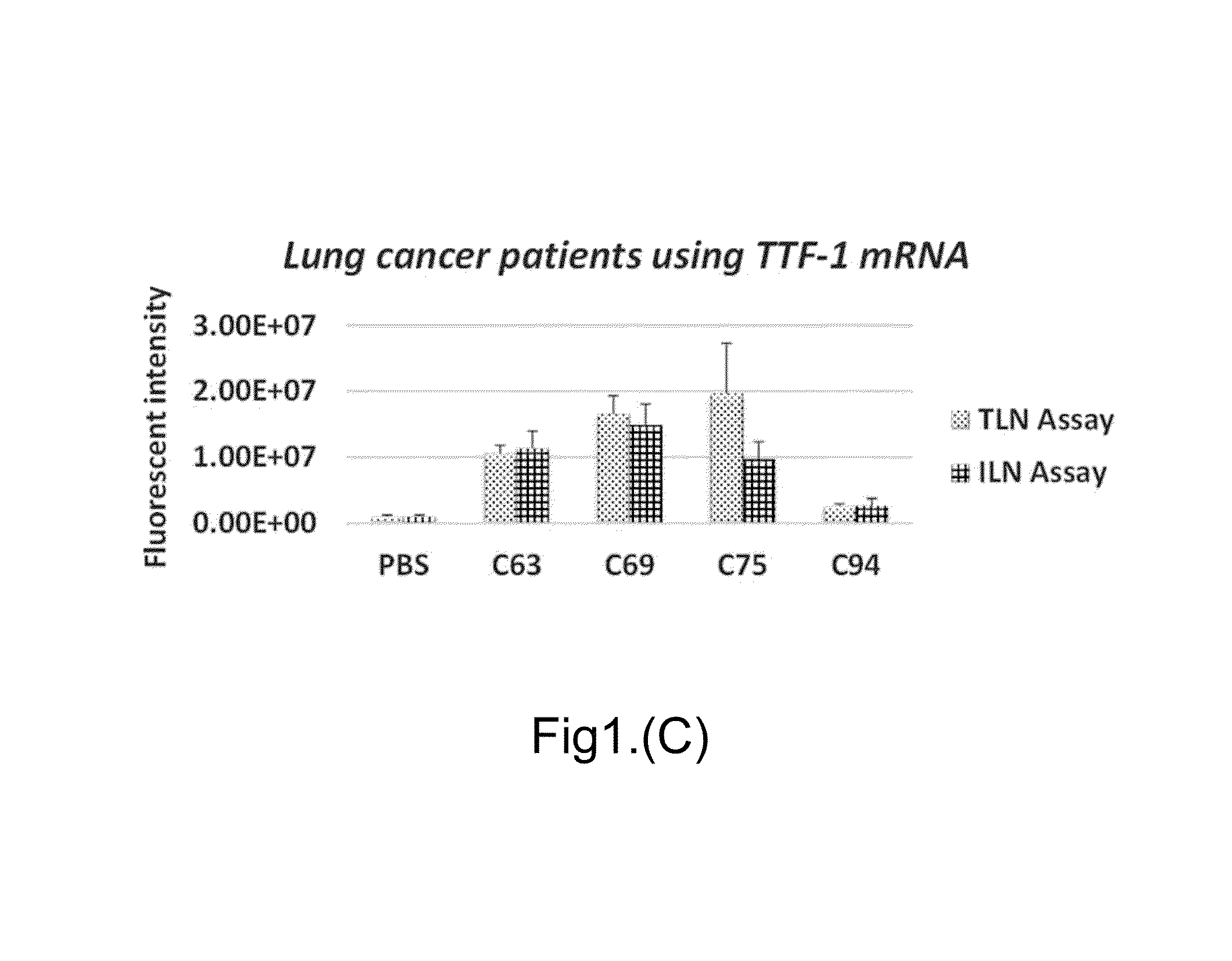
[0110]We compare CD63-rich and NCL-rich EVs with miR-21 and TTF-1 content from A549 cell secreted EVs as shown in FIG. 3C. There are more EVs containing CD63 than NCL surface receptor, and a small fraction (12.2 and 10.9% for CD63 and NCL EVs, respectively) of those E...


PUM


Abstract

The present invention disclosed a method of fabricating an antibody immunolipoplex nanoparticle (Ab-ILN) biochip and antibody tethered lipoplex nanoparticle (Ab-TLN) biochip. The aforementioned antibody-based lipoplex nanoparticle biochip or the related array contains molecular probes and is applied for detecting the presence of a disease or condition in a subject obtaining a body fluid sample by capturing and identifying both membrane protein and intra-vesicular DNA/RNA/proteins of extracellular vesicles (EVs).

Description

TECHNICAL FIELD OF THE INVENTION[0001]The present invention relates to array biochips consisted of an antibody based capture system for extracellular vesicles (EVs) and liposomal nanoparticles containing molecular probes (MPs), such as molecular beacons and aptamers, to identify nucleic acids and protein content inside the captured EVs. MPs inside the liposomal nanoparticles and antibody or other ligands on the biochip surface can simultaneously capture target EVs from body fluids based on EV surface receptors and identify target messenger RNAs, microRNAs, long non-coding RNAs (IncRNA), circulating genomic DNAs, circulating tumor DNA and / or proteins in the EVs based on MPs as biomarkers. The arrays can also be used to capture and identify viruses and cells.BACKGROUND OF THE INVENTION[0002]Early and convenient detection has become extremely important against various diseases including cancer, cardiovascular diseases and infectious diseases. The earlier a disease is diagnosed, the mor...

Claims


Application Information

Patent Timeline
IPC IPC(8): G01N33/531G01N33/543
CPCG01N33/54346G01N33/531
Inventor LEE, LYJAMESKWAK, KWANG JOOLEE, ANDREW
Owner SPOT BIOSYST LTD
Why Eureka
Try Eureka