Nanoparticles for magnetic resonance imaging tracking and methods of making and using thereof

a magnetic resonance imaging and nanoparticle technology, applied in the field of nanoparticles for magnetic resonance imaging tracking and methods of making and using thereof, can solve the problems of difficult to overcome metastases, difficult to form polymeric diacest, and difficult hydrophilic agent use, and achieve the effect of efficient incorporation of diacest agents

Active Publication Date: 2016-07-21
THE JOHN HOPKINS UNIV SCHOOL OF MEDICINE
View PDF2 Cites 3 Cited by
  • Summary
  • Abstract
  • Description
  • Claims
  • Application Information

AI Technical Summary

Benefits of technology

[0009]It is an object of the invention to provide micro- and/or nanoparticles, such as polymeric micro- and/or nanoparticles, which can efficiently incorporate ...

Problems solved by technology

While these nanocarriers have been shown to improve the efficacy of chemotherapy, there have been significant challenges.
In cancer therapy, for example, the balance between minimizing exposure of therapeutics to healthy tissues and concurrently target a high dose of chemotherapeutics to tumors and metastases are obstacles that have been difficult to overcome.
However, the use of hydrophilic agents can be problematic for formulating polymeric diaCEST particles.
The inte...

Method used


Image

  • Nanoparticles for magnetic resonance imaging tracking and methods of making and using thereof
  • Nanoparticles for magnetic resonance imaging tracking and methods of making and using thereof
  • Nanoparticles for magnetic resonance imaging tracking and methods of making and using thereof

Examples


example 1

Preparation of DiaCEST Liposomes

[0232]Drug-containing liposomes were prepared with the poly(ethylene) glycol (PEG) concentration varied systematically using the thin film hydration method. 25 mg of lipid dissolved in chloroform was dried, with the resultant thin film hydrated using 1 ml barbituric acid (BA) to form multilamellar vesicles. The mixture was then annealed at 55-65° C., sonicated, and subsequently extruded through stacked polycarbonate filters. Doxorubincin (DOX) was then loaded into the liposomes remotely.

example 2

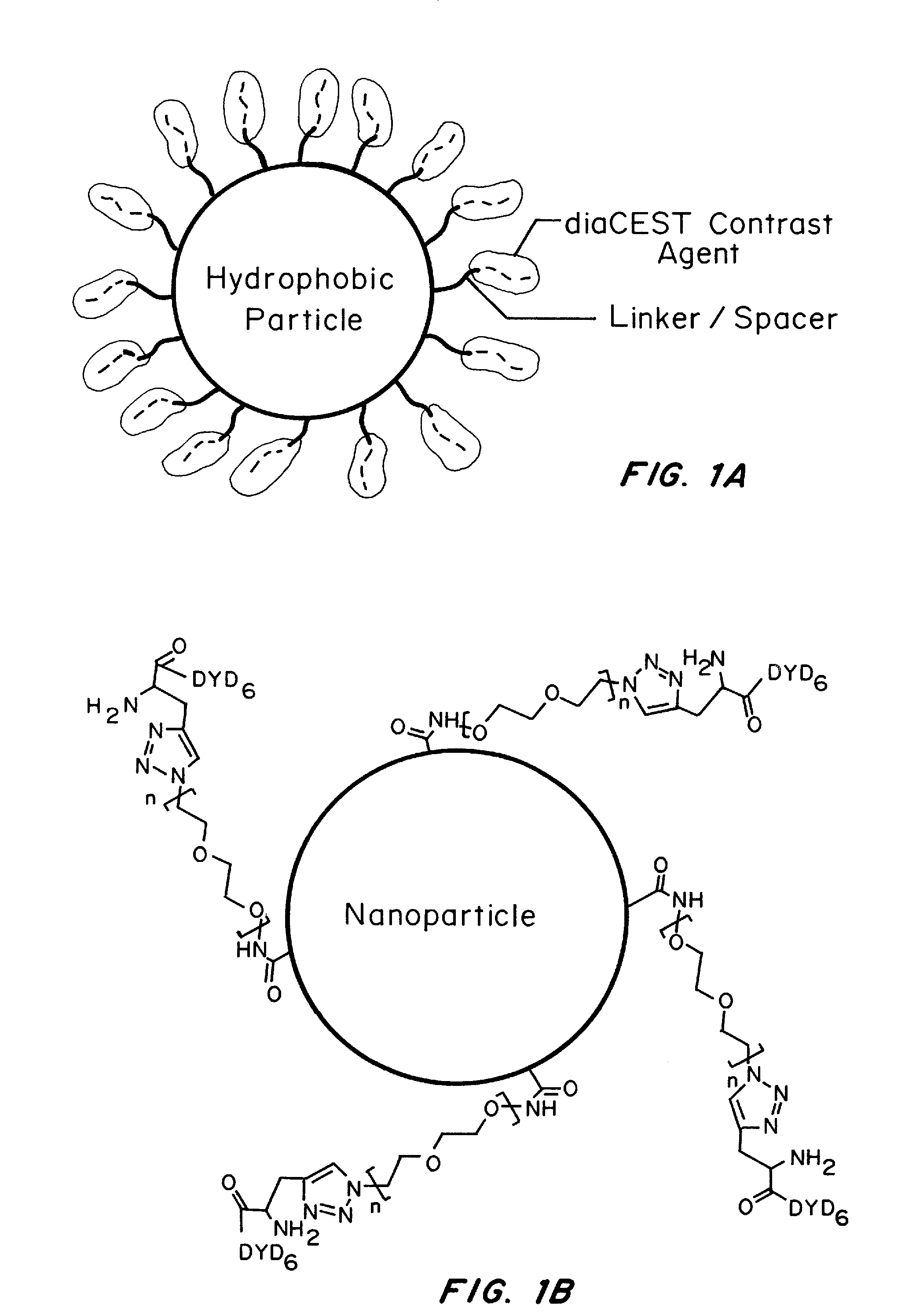
Preparation of DiaCEST Polymeric Particles

[0233]Polymeric particle preparation: The peptide contrast agent, propargylglycine-W-(DYD)6-NH2, was synthesized on a microwave-assisted peptide synthesizer Libertyl (CEM, USA). The CEST nanoparticles were then prepared by standard coupling chemistry. NH2PEG-N3 was coupled to carboxylates on the surface of polystyrene (PS) particles in the presence of EDC and NHS in 200 mM sodium borate buffer pH 7.8 at RT. Propargylglycine-W-(DYD)6-NH2 peptide was coupled with PS-PEG-N3 in the presence of catalytic amount of CuAcAc, TBTA, and excess ascorbic acid in sodium phosphate buffer pH 7, under nitrogen, using click chemistry. Biodegradable CEST nanoparticles based on the polymer of poly(lactic-co-glycolic) acid-co-PEG (PLGA-PEG) were synthesized using a similar procedure and formulated using a oil-in-water emulsion method.

example 3

Animal Preparation

[0234]Five million CT26 cells were injected subcutaneously into the right flank of a mouse and allowed to grow for ˜10 days prior to MRI.

[0235]CEST Imaging

[0236]Mice were anesthetized using isoflurane, positioned in a 11.7 T horizontal bore Bruker Biospec scanner, and imaged before and 24 h after intravenous administration of 100 ul of DOX / BA PEGylated liposomes. CEST images were acquired through collection of two sets of saturation images, a water saturation shift referencing (WASSR) set for B0 mapping and a CEST data set for characterizing contrast. For the WASSR images, the saturation parameters were tsat=500 ms, B1=0.5 uT, TR=1.5 sec with saturation offset incremented from −1 to +1 ppm with respect to water in 0.1 ppm steps.

[0237]For the CEST images, tsat=3 sec, B1=4.7 uT, TR=Ssec, with offset incremented from −6 to +6 ppm (0.3 ppm steps) with a fat suppression pulse. The acquisition parameters were TR=5.0 sec, effective TE=21.6 ms, RARE factor=8. The CEST imag...


PUM


Abstract

Surface conjugated diamagnetic Chemical Exchange Saturation Transfer (diaCEST) agent carriers and methods of making and using are described herein. The particles are safe alternatives to conventional paramagnetic or superparamagnetic metal-based MRI contrast agents that are often toxic and therefore not biocompatible. The carriers described herein can provide simultaneous monitoring of multiple particle types labeled with ‘multicolor’ diaCEST contrast agents. In some embodiments, the carriers are micro- and/or nanoparticles. In other embodiments, the carriers are liposomes. In some embodiments, the particles and/or liposomes are mucus penetrating. In other embodiments, the particles and/or liposomes are not mucus penetrating.

Description

CROSS-REFERENCE TO RELATED APPLICATIONS[0001]This application claims priority to U.S. Ser. No. 61 / 760,897 entitled “A Surface Conjugated Diamagnetic Chemical Exchange Saturation Transfer Nanoparticle and / or Nanocarrier for Magnetic Resonance Imaging (MRI) Tracking and Methods Therefor”, filed on Feb. 5, 2013. The contents of this application are incorporated herein by reference in their entirety.STATEMENT REGARDING FEDERALLY SPONSORED RESEARCH OR DEVELOPMENT[0002]This invention was made with Government Support under Agreement R01EB015031 awarded to Michael McMahon by the National Institutes of Health. The Government has certain rights in the invention.FIELD OF THE INVENTION[0003]This invention is in the field of nanocarriers for magnetic resonance imaging (MRI) tracking and methods of making and using thereof.BACKGROUND OF THE INVENTION[0004]In recent years there has been growing interest in the use of nanoparticles for drug delivery. While these nanocarriers have been shown to impr...

Claims


Application Information

Patent Timeline
IPC IPC(8): A61K49/18
CPCA61K49/1833A61K49/1878
Inventor YU, TAOPATEL, HIMATKUMARCHAN, KANNIE M.Y.OSKOLKOV, NIKITAMCMAHON, MICHAELHANES, JUSTIN
Owner THE JOHN HOPKINS UNIV SCHOOL OF MEDICINE
Why Eureka
Try Eureka