Eureka AIR delivers breakthrough ideas for toughest innovation challenges, trusted by R&D personnel around the world.

Method for assessment of neural function by establishing analysis module

a neural function and analysis module technology, applied in the field of neural function assessment by establishing an analysis module, can solve the problems of affecting the quality of life of patients, and requiring more time-consuming processes and molecular markers, and analyzing procedures and durations affecting the use of cell models for drug screening, etc., to achieve rapid and representative analysis, quick detection of drug neurotoxicity or protective effect on neurons

Active Publication Date: 2016-09-15
NAT CHENG KUNG UNIV +1
View PDF1 Cites 1 Cited by
  • Summary
  • Abstract
  • Description
  • Claims
  • Application Information

AI Technical Summary

Benefits of technology

The present invention provides a method for analyzing neural function quickly and representatively using important markers for nerve growth and optimized image analysis flow. This method can also be used for drug screening to detect neurotoxicity or protective effects on neurons.

Problems solved by technology

Among the side effects of chemotherapy, neuropathy significantly affects patient's quality of life.
However, there are some problems while using molecular markers for assessing neuronal function in high-throughput screening.
However, simultaneous multiparametric analysis for detecting neurite outgrowth and synaptogenesis requires more complicate, time-consuming processes and molecular markers.
Moreover, usage of cell model for drug screening also affects analyzing procedure and duration.
First, chemicals added for inducing differentiation may complicate the assessment of tested drug regarding to the unpredictable drug-drug interaction.
However, primarily cultured neurons are more difficult to obtain and maintain, to optimize the in vitro culture period is essential.

Method used

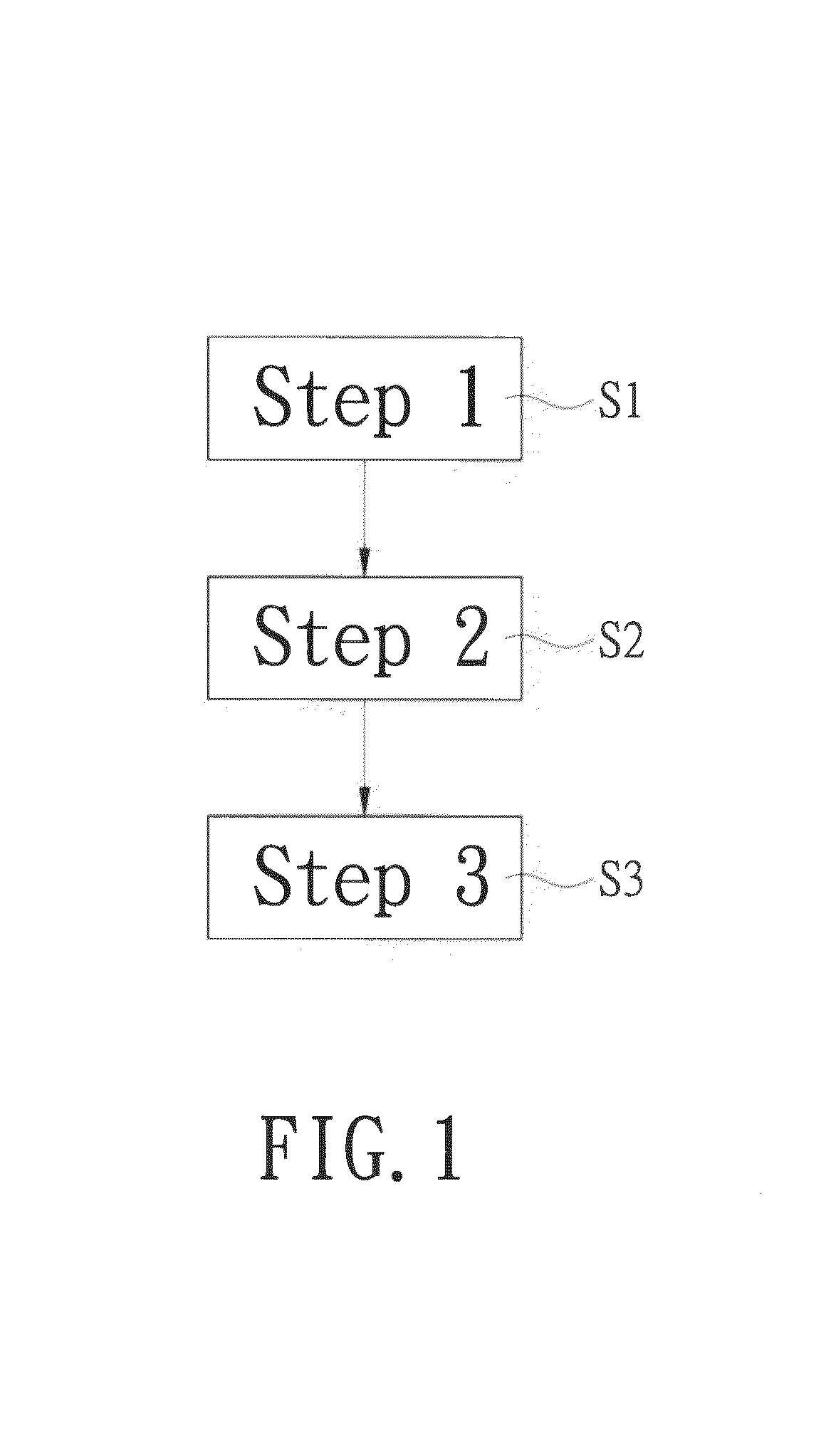

Image

  • Method for assessment of neural function by establishing analysis module
  • Method for assessment of neural function by establishing analysis module
  • Method for assessment of neural function by establishing analysis module

Examples


embodiment one

Preparing Primarily Cultured Cells and Performing Immunofluorescence Staining

[0026]Comparing with conventional method of inducing differentiation by treating cell lines with chemicals, the method uses primarily cultured cells for differentiating naturally, which shares more characteristic with normal neuron in vivo and is a better method in assessment of early formation of neural network. Moreover, the test results are not affected by drug-drug interaction. Thus the present invention uses primarily cultured cells for evaluating neural function.

[0027](1) Preparation of Primarily Cultured Cells

[0028]Cerebral cortex was obtained from 0˜1 day old B6 mice pup and meninges were removed. DMEM / F12 medium without serum was used to triturate the cortex tissue. Then, the DMEM / F12 medium without serum was refilled to 5 mL, mixed evenly with 0.5 mL trypsin, and placed at 37□ for 5 minutes. Then the mixture with 1 mL fetal bovine serum (FBS) was filtered through a cell strainer. After a centrifug...

embodiment two

Image Acquisition and Analysis

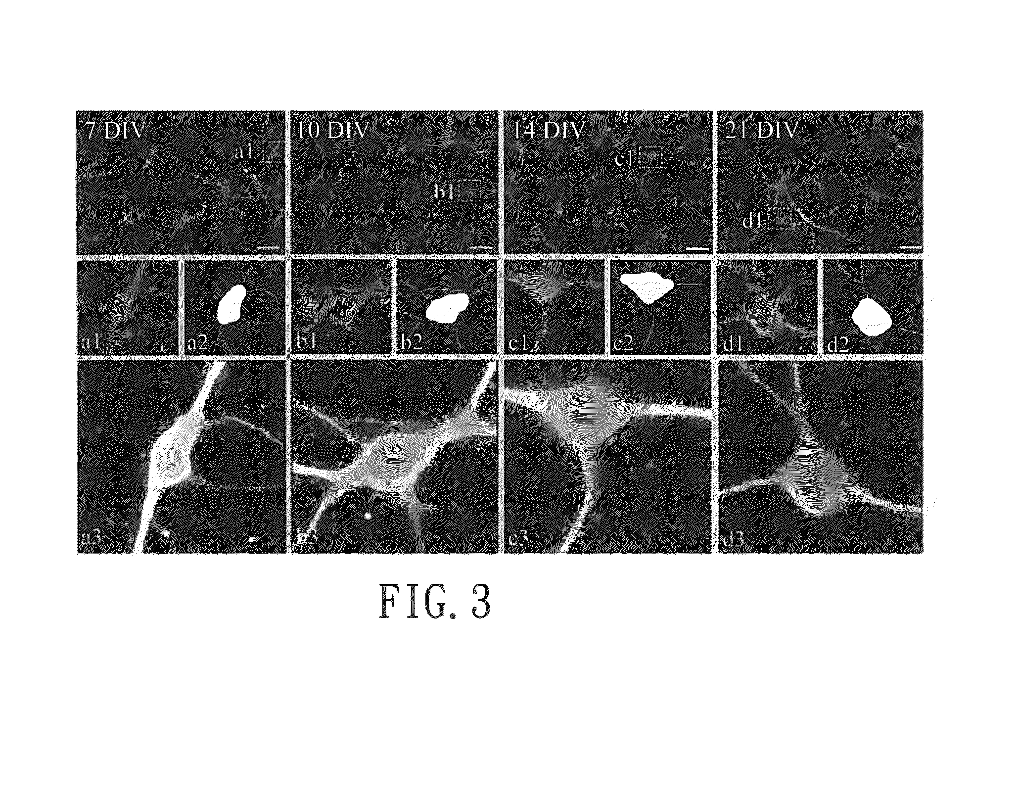
[0030]Fluorescence microscopy system was used to capture images at the wavelength of DAPI (4′-6-Diamidino-2-phenylindole), FITC (fluorescein isothiocyanate) and Cy5 for image analysis of the cultured cells mentioned above. The images were saved automatically for analysis.

[0031]To analyze neural function accurately, MetaXpress 3.1 software (“MetaXpresss Image Acquisition and Analysis Software (Analysis Guide)” was used on the website ftp: / / ftp.meta.moleculardevices.com / pub / uic / software / MX31R13 / HelpD ocs / MetaXpress / MetaXpress_3_1 Analysis_Guide.pdf) to design an analysis module. An automatic analysis was performed according to the following optimal indicators (a)˜(c) for image analysis with 32 steps.

[0032](a) Definition of Neurons

[0033]The cultured cells including neurons and glial cells were firstly separated according to the area and the fluorescence intensity of the nucleus stained by Hoechst dye together with MAP2 antibody. For example, built-in optio...

embodiment three

Detecting the Effect of Chemotherapy Drugs on Neurons

[0046](1) Preparation of Primarily Cultured Cell

[0047]Cerebral cortex was obtained from 0˜1 day old B6 mice pup and meninges were removed. DMEM / F12 medium without serum was used to triturate the cortex tissue. Then, the DMEM / F12 medium without serum was refilled to 5 mL, mixed evenly with 0.5 mL trypsin, and placed at 37□ for 5 minutes. Then the mixture with 1 mL FBS was filtered through a cell strainer. After a centrifugation at 3,000 rpm for 10 min, the supernatant was removed and precipitated cells were added to a medium required for nerve cell culture. The medium was prepared by adding B-27® Supplement, 0.5 mM L-glutamine, and 0.5% penicillin streptomycin to Neurobasal® A medium.

[0048](2) Black multi-well microplates with clear bottom were used for nerve cell culture. Each well was coated with 70 μL poly-D-lysine or poly-L-lysine. Cells were seeded in a density of 2.8×10⁴ / well. After cells were cultured for 10 days, chemothera...


PUM


Abstract

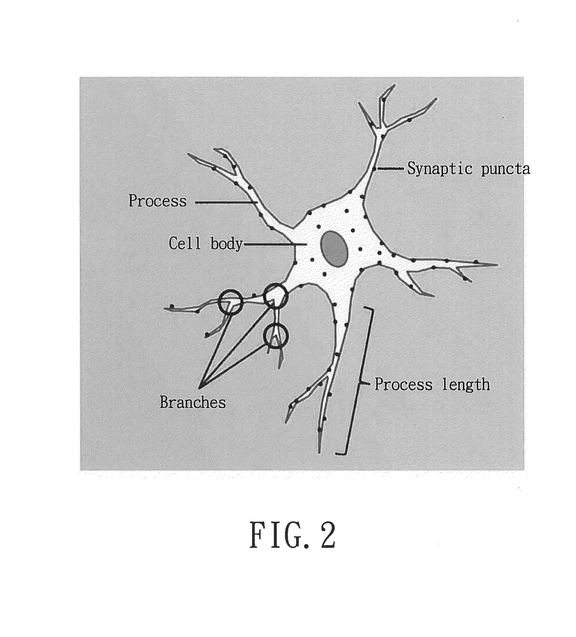
A method for assessment of neural function by establishing an analysis module is revealed. The first step of the method is to capture images of the cultured cells with a plurality of fluorescence labeling by a fluorescence microscopy system for image analysis. The cultured cells include neurons and non-neuronal cells. Then select neurons with neurites having fluorescence labeling and exclude non-neuronal cells according to an area and a fluorescence intensity of nucleus. Also calculate an area of the neuronal cell body, a length of the neurites and a number of processes and branches to verify outgrowth of the neurites of the neurons. Next calculate a number of synaptic puncta having fluorescence labeling on the area of the neurites having fluorescence labeling defined in Step 2 to verify formation of the synaptic puncta of the neurons for assessment of neural function.

Description

BACKGROUND OF THE INVENTION[0001]1. Fields of the Invention[0002]The present invention refers to a method for assessment of neural function by establishing an analysis module, especially to a method by which neurite outgrowth and function are analyzed fast and representatively. The method uses important markers related to neurite outgrowth in combination with optimized image analysis flow and program commands. Thereby the method can not only assess nerve growth and formation of neural network at early stage but also act as a high-content drug screening platform for quickly detecting the effect of drugs on neural function.[0003]2. Descriptions of Related Art[0004]With the progress of cancer therapies, survival rate of cancer patients is increased markedly. Recently, due to the extension of life span of cancer patients, side effects of chemotherapy drugs have drawn much more attention. Among the side effects of chemotherapy, neuropathy significantly affects patient's quality of life. ...

Claims


Application Information

Patent Timeline
IPC IPC(8): G01N33/53G01N33/50G01N33/58
CPCG01N33/5308G01N33/582G01N2500/10G01N2500/04G01N33/5058G01N33/5005C12N5/00
Inventor SHEN, MENT-RUCHANG, LIAN-YUNSUN, YUAN-TINGCHANG, JANG-YANG
Owner NAT CHENG KUNG UNIV
Why Eureka
Eureka Blog
Learn More