Anti-pacap antibodies and uses thereof

Active Publication Date: 2016-10-20
H LUNDBECK AS
View PDF0 Cites 28 Cited by
  • Summary
  • Abstract
  • Description
  • Claims
  • Application Information

AI Technical Summary

Problems solved by technology

There is a theoretical risk of coronary spasm in patients with established heart disease following administration, and cardiac events after taking triptans in rare instances may occur.
However, the administration of these treatme

Method used


Image

  • Anti-pacap antibodies and uses thereof
  • Anti-pacap antibodies and uses thereof
  • Anti-pacap antibodies and uses thereof

Examples


Example

Example 1

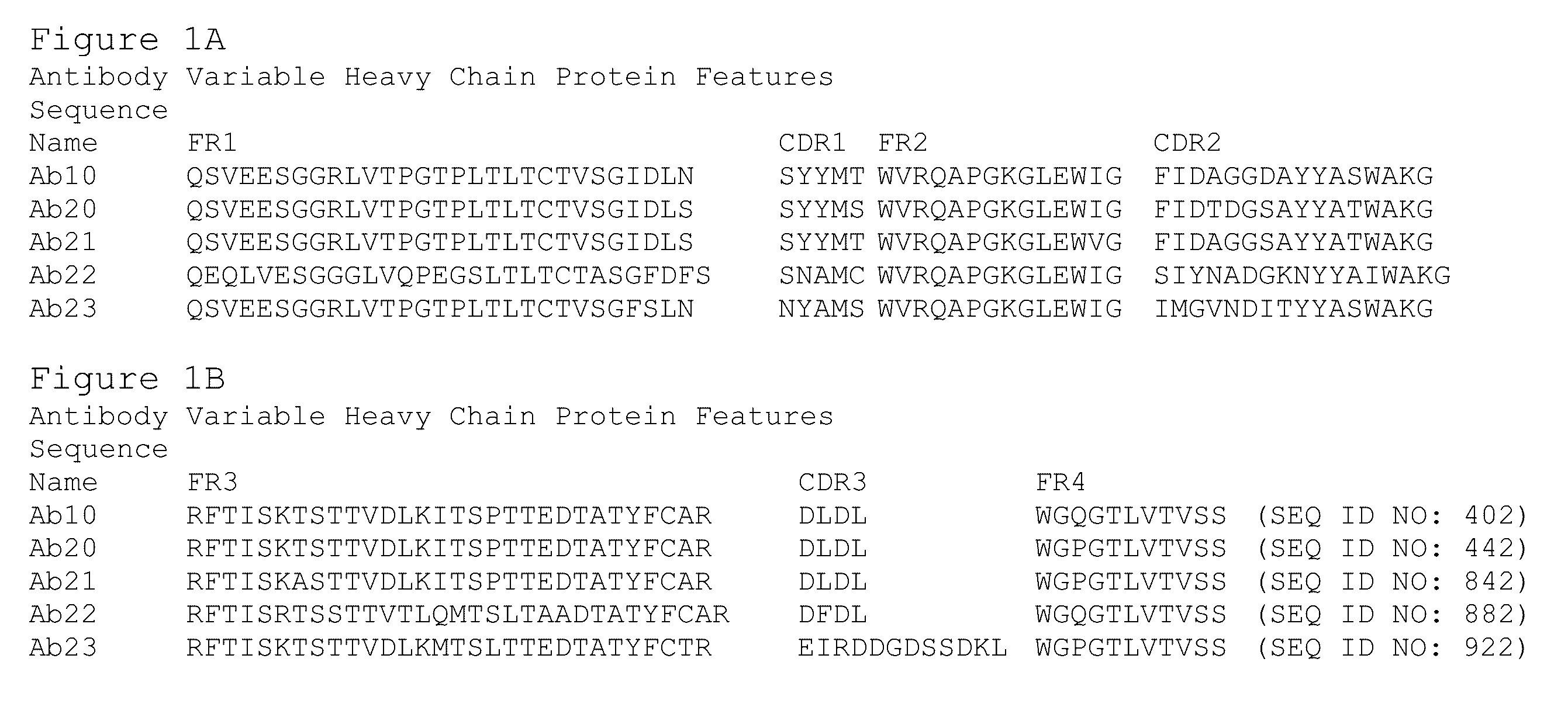
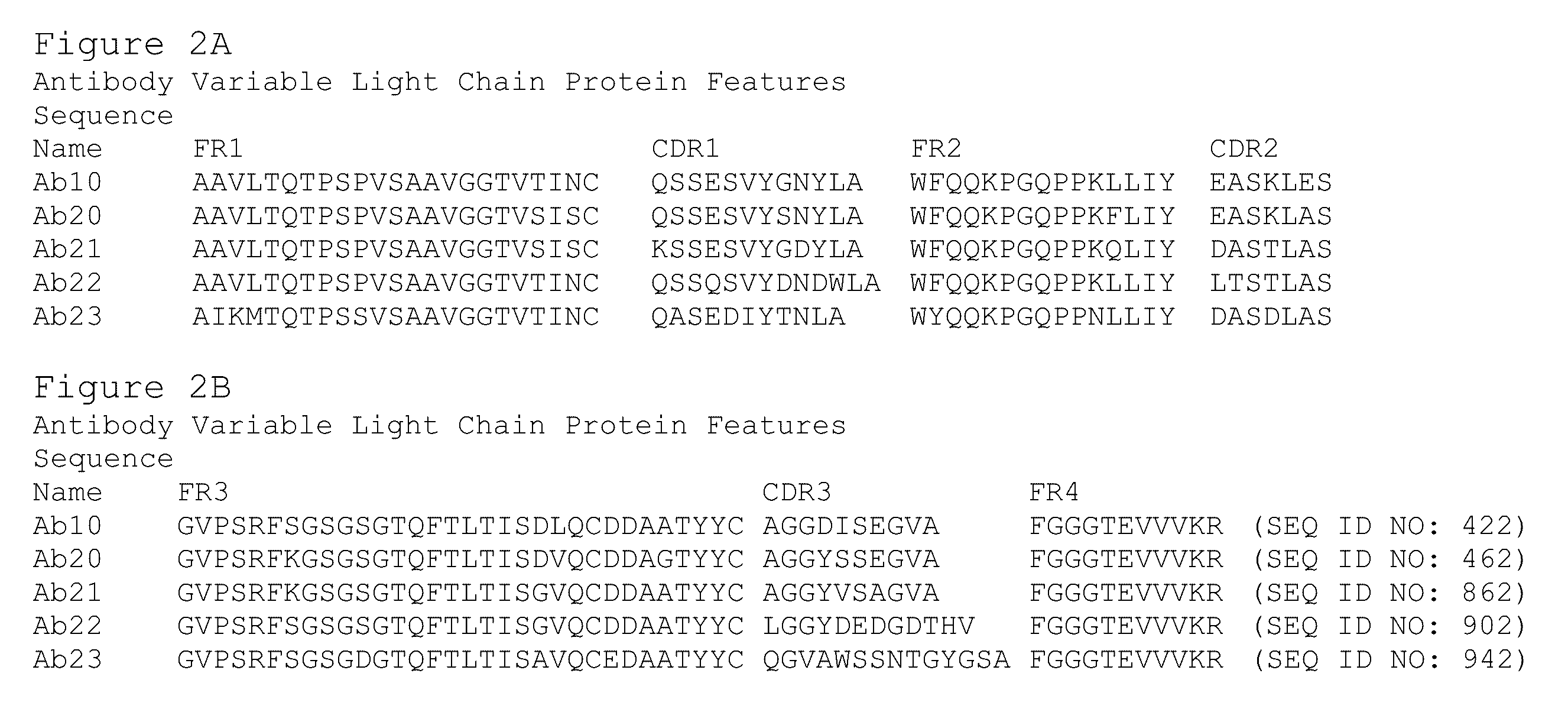
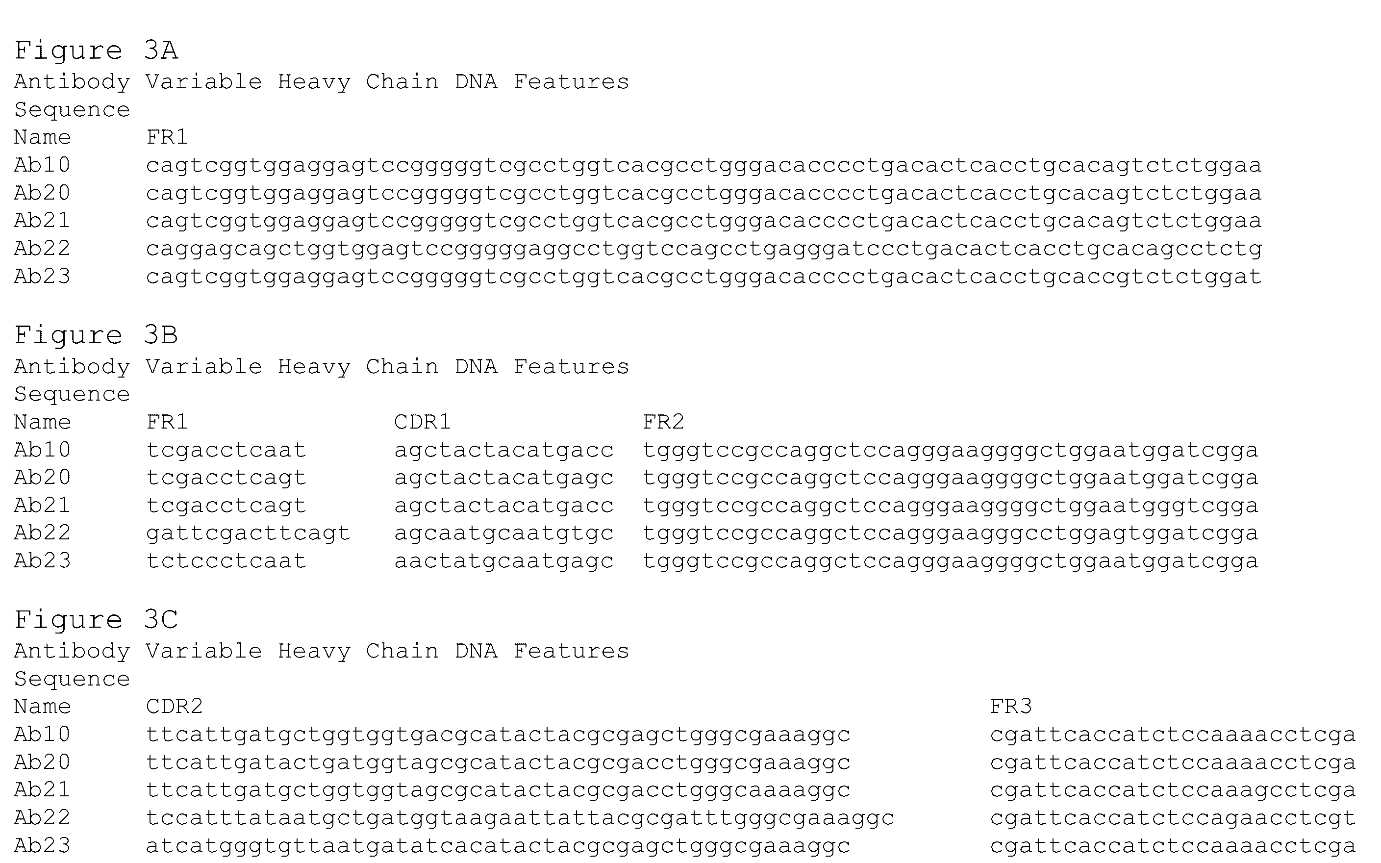
Preparation of Antibodies that Selectively Bind PACAP

[0450]By using an antibody selection protocol substantially as described herein, a panel of antibodies specific to PACAP38 and PACAP27, and a panel of antibodies specific to PACAP38 only, were produced.

Immunization Strategy

[0451]Rabbits were immunized with PACAP38 (American Peptide, Vista, Calif.) (SEQ ID NO: 1241). Peptides were prepared for immunization as follows. A 0.15 ml volume of 10 mg / ml keyhole limpet hemocyanin (“KLH”) dissolved in Dulbecco's phosphate buffered saline (“DPBS”) supplemented to 1M NaCl was combined with 1.0 ml of 1 mg / ml peptide (dissolved in deionized water). Then 1.0 ml of 40 mM carbodiimide was added prior to a 12-hour incubation at room temperature with gentle mixing. Excess carbodiimide and unconjugated peptide were removed by dialysis to DPBS prior to sterile filtration. Next unconjugated peptide equal to the initial mass of KLH was added prior to preparation for injection into rabbits. Alte...

Example

Example 2

Binding Affinities of Anti-PA CAP Antibodies

[0478]Binding affinities of monoclonal antibodies for human PACAP were estimated using SPR on the PROTEON™ XPR36 (Bio-Rad, Hercules, Calif.). Antibody was immobilized to the surface of general amine coupling (“GLC” or “GLM”) Chips (Bio-Rad, Hercules, Calif.). A dilution series of human PACAP38 (SEQ ID NO: 1241) prepared in 1×PBST Buffer (4.3 mM Na Phosphate, 1.4 mM K Phosphate, 135 mM NaCl, 2.7 mM KCl 0.05% Polysorbate-20) purchased from Teknova (Cat# P1192, Teknova, Hollister, Calif.) and supplemented with 0.25 M arginine (from J. T. BAKER®), 0.2 mg / ml BSA (Jackson Immuno Research Labs, West Grove, Pa.), and 0.005% sodium azide (VWR International, Radnor, Pa.) with the pH adjusted to 7 was used to query the antibodies. Antigen (ranging from 1.23 nM to 100 nM) was typically run sequentially with association times of 2-4 minutes and dissociation times of 3-120 minutes grouped with the PROTEON™ Manager Software (v3.1.0.6 (Bio-Rad, H...

Example

Example 3

Inhibition of PACAP38-Induced Signaling Via VPAC1-R

[0484]To identify antibodies that neutralize PACAP38-induced signaling via human VPAC1-R, CHO-K1 cells expressing human VPAC1-R were used in a cAMP HTRF cell-based assay. Antibody dilutions were incubated with PACAP38 at 4× the final concentration (5 nM) for 1 hour. While the antibody / antigen complexes were incubated for 1 hour, VPAC1-R expressing CHO-K1 cells (generated at Alder Biopharmaceuticals, by stable transfection of CHO-K1 cells (ATCC, catalog # CCL-61) with human VPAC1-R cDNA; selected clone 1 was used for in vitro cell based assays) were detached with 0.25% trypsin for 4 minutes. The cells were washed and re-suspended at 1×10⁶ cells per ml culture media. 20 μl of Ab / antigen mixture was mixed with 20 μl of cells in HTRF plates and incubated with shaking for 30 minutes. 20 μl of Eu3+ cryptate labeled anti-cAMP mAb (1:20 diluted) and 20 μl of (1:20 diluted) d2-labeled cAMP in lysis buffer were added to each well and...


PUM

PropertyMeasurementUnit
Temperatureaaaaaaaaaa
Fractionaaaaaaaaaa
Fractionaaaaaaaaaa

Abstract

The present invention is directed to antagonistic antibodies and antigen binding fragments thereof having binding specificity for PACAP. These antibodies inhibit, block or neutralize at least one biological effect associated with PACAP, e.g., vasodilation. In exemplary embodiments these antibodies and antigen binding fragments thereof may comprise specific VH, VL, and CDR polypeptides described herein. In some embodiments these antibodies and antigen binding fragments thereof bind to and/or compete for binding to specific epitope(s) on human PACAP. The invention is further directed to using these antagonistic anti-PACAP antibodies, and binding fragments thereof, for the diagnosis, assessment, and treatment of diseases and disorders associated with PACAP and conditions where antagonism of PACAP-related activities, such as vasodilation, mast cell degranulation, and/or neuronal activation, are therapeutically beneficial, e.g., headache and migraine indications.

Description

CROSS-REFERENCE TO RELATED APPLICATIONS[0001]This application claims the benefit of U.S. Provisional Application Ser. No. 62 / 148,550, filed Apr. 16, 2015, U.S. Provisional Application Ser. No. 62 / 148,557, filed Apr. 16, 2015, U.S. Provisional Application Ser. No. 62 / 148,562, filed Apr. 16, 2015, U.S. Provisional Application Ser. No. 62 / 148,596, filed Apr. 16, 2015, U.S. Provisional Application Ser. No. 62 / 148,643, filed Apr. 16, 2015, U.S. Provisional Application Ser. No. 62 / 148,583, filed Apr. 16, 2015, U.S. Provisional Application Ser. No. 62 / 148,640, filed Apr. 16, 2015, each of which is hereby incorporated by reference in its entirety.SEQUENCE DISCLOSURE[0002]This application includes as part of its disclosure an electronic sequence listing text file named “43257o5809.txt”, having a size of 165,209 bytes and created on Feb. 29, 2016, which is hereby incorporated in its entirety.FIELD OF THE INVENTION[0003]This invention generally pertains to antibodies and antigen binding fragme...

Claims


Application Information

Patent Timeline
IPC IPC(8): C07K16/26A61K39/395G01N33/74A61K45/06
CPCC07K16/26A61K45/06A61K39/3955C07K2317/24C07K2317/76C07K2317/92G01N2333/5757G01N33/74G01N33/5088A61P25/06A61K2039/505C07K2317/34C07K2317/33Y02A50/30A61K39/39566A61K49/0004A61K49/0008C07K14/57536C07K14/57563C07K16/4241C07K2317/21C07K2317/40C07K2317/50C07K2317/54C07K2317/55C07K2317/622C07K2317/56C07K2317/565
Inventor LOOMIS, MARIA-CRISTINAGARCIA-MARTINEZ, LEON F.DUTZAR, BENJAMIN H.ALLISON, DANIEL S.HENDRICKS, LEEOJALA, ETHAN W.FAN, PEISMITH, JEFFREY T.L.LATHAM, JOHN A.KARASEK, CHARLIEMULLIGAN, JENNYSCALLEY-KIM, MICHELLESTEWART, ERICARUBIN, VANESSA LISBETHBILLGREN, JENS J.
Owner H LUNDBECK AS
Why Eureka
Try Eureka