Small molecule binding pockets in nucleic acids

a nucleic acid and small molecule technology, applied in the field of small molecule binding pockets in nucleic acids, can solve the problems of inability to interrogate the binding interaction between rnas and small molecules, inability to rationally design small molecule modifications to improve mirna binding affinity and activity, and inability to meet the requirements of asos delivery, etc., to achieve rapid identification, efficient and cost-effective

Active Publication Date: 2017-02-02
NYMIRUM
View PDF0 Cites 16 Cited by
  • Summary
  • Abstract
  • Description
  • Claims
  • Application Information

AI Technical Summary

Benefits of technology

[0091]In some embodiments, a significant advantage of the present invention is the use of lower magnetic fields (for example, 300 MHz or less), which can significantly shorten the repetition delay and the total experimental time can be reduced to ¼-⅕ of that of high fields because the repetition delay depends on Ti relaxation time which is significantly shorter at low magnetic field (i.e., Ti relaxation time at 100 MHz is more than 6 times shorter than that of 600 MHz for molecules of correlation time of 4-8 ns (oligonucleotides of 25-50 bases)). This Ti relaxation time difference at between high and low magnetic fields becomes larger as molecular weight or size of a molecule increases. Within given time, 4-5 times more measurements can be repeated and added at low magnetic fields to yield signal-to-noise gain of factor of 2.
[0092]In some embodiments a number of small molecule-bound bimolecular structures can be determined for uses comprising computer aided drug discovery efforts, which commonly rely on biomolecular structures determined when bound to a small molecule. Representative small molecules include aminoglycosides, flavin / flavonoids, intercelators (e.g., acridine orange, proflavine and the like), and tetracylines (e.g., tetracycline, doxycycline and the like). An exemplary set of small molecules that can be used to determine a small molecule-bound biomolecular structure is shown below in Table 1.TABLE 1Non-limiting examples of small molecules.NameNameNetilmicin9-AminoacridineParomomycinLincomycinNeomycin BKanamycin BMitoxantroneSisomicin5-(N-N-dimethyl)-amilorideThiazole orangeDiminazeneEthidium BromideKanamycin A4′,6-diamidino-2-phenylindoleStreptomycinQuinacridineGentamicinAcridine orangeAmikacinErythromycinPentamidineCycloheximideHoechst 33258Blasticidine STobramycinChloramphenicolHoechst 33342SpectinomycinChlortetracyclinePuromycinMinocyclineAcetylpromazineOxytetrtacyclineYohimbineTetracyclineUsnic AcidDoxycyclineEnamine T0503-4982RiboflavinGossypinProflavineQuercitrinApramycinL-arg-p-nitroanilideKasugamycinRutinHygromycin BChembridge 5484617GeneticinChembridge 5566364RibostomycinLife Chemicals F3246-0011AmilorideEnamine 4864450SpiramycinPrinceton BioResearch OSSK-789168TylosinMaybridge JFD02062LinezolidEnamine AR-471 / 431186792-(2,3-diamino-3H-benzimidazol-1-yl)-1-(4-methoxyphenyl)ethanone2-amino-3-hydroxy-N-[(2,3,4-trihydroxyphenyl)methyleneamino]propanamide4-(2-hydroxyethylamino)-7-methyl-2-oxo-1,5-diazabicyclo[4.4.0]deca-3,5,7,9-tetraene-3-carbaldehyde3-chloro-2-(2,3,4,5,6-pentahydroxyhexylideneamino)naphthalene-1,4-dioneSmiles: c12c(c(═O)n3c(n2)c(ccc3)C)cc(c(n1Cc1ccccc1)═N)C(═O)NCCN1CCOCC15-((3-Aminopropyl)amino)-7,10-dihydroxy-2-(2-((2-hydroxyethyl)amino)ethyl)anthra(1,9-cd)pyrazol-6(2H)-one[3-(diethylamino)isoindol-1-ylidene]-diethyl-ammonium1-[6-amino-8-(2-hydroxyethylamino)purin-3-yl]-3-morpholin-4-ylpropan-2-ol
[0093]In some embodiments, one or more small molecules described herein are salts. In some embodiments, one or more small molecules described herein are not salts but are in free form. In some embodiments, one or more small molecules described herein are complexed with another molecule. In some embodiments, one or more small molecules described herein are mixed and / or in the presence of one or more molecules, atoms, ions, and / or other matter.
[0094]In order to identify which small molecules interact with the biomolecule, in some embodiments, one synthesizes a uniformly isotopically labeled biomolecular sample, individually or in a combinatorial manner mix each small molecule at a ratio that one would expect to see changes in NMR signals for relatively tight binding small molecules (for a low μM Kd, a ratio of 2:1 or 4:1 could be used), collect the NMR data such as chemical shifts, resonance intensities, and / or NOEs, compare the NMR data of the biomolecule in the presence of the small molecule to the NMR data of the biomolecule in the absence of the small molecule, and select small molecules that cause significant changes in the NMR data. In some embodiments, changes in NMR data comprise a portion of a chemical shift linewidth, for example a one linewidth. In some embodiments, changes in NMR data comprise a significant reduction in an NOE and / or a resonance intensity when comparing the biomolecule NMR data in the absence and presence of the small molecule is significant). In various embodiments, NMR data of the small molecule could be monitored and similar perturbations observed on addition of the biomolecule of interest, where, in some embodiments, the biomolecule is non-isotopically labeled. In various embodiments, the same solution conditions (e.g., buffer or solubilization solution) for each sample are used to minimize random noise due to differences in solution environments.
[0095]In some embodiments, the present invention provides a method for determining the 3-D atomic resolution structure of a polynucleotide when bound to a small molecule, ligand or other chemical entity for purposes comprising computer-aided drug design. The method includes providing a polynucleotide sample comprising a plurality of polynucleotides, the plurality of polynucleotides having an identical nucleotide sequence, wherein each polynucleotide comprises at least one nucleotide isotopically labeled with one or more atomic labels selected from the group consisting of 2H, 13C, 15N, 19F and 31P; generating an NMR sample comprising one or more polynucleotides admixed with one or more small molecules that form one or more bound complexes; obtaining an NMR spectrum of the NMR sample using an NMR device; determining one or more chemical shifts of the one or more atomic labels; and determining the 3-D atomic resolution structure of the polynucleotides from the chemical shifts determined in step (d).
[0096]In some embodiments, the present invention provides a method for determining the 3-D atomic resolution binding pocket of a polynucleotide purposes comprising computer-aided drug design. The method includes identifying one or more small molecule binders that bind the polynucleotide; providing a polynucleotide sample comprising a plurality of the polynucleotide, wherein each polynucleotide comprises at least one nucleotide isotopically labeled with one or more atomic labels selected from the group consisting of 2H, 13C, 15N, 19F and 31P; generating an NMR sample comprising the polynucleotide sample admixed with one or more of the small molecule binders; obtaining an NMR spectrum of the NMR sample using an NMR device; determining one or more chemical shifts of the one or more atomic labels; and determining the 3-D atomic resolution structure of the polynucleotides from the chemical shifts determined in step (d); identifying the binding pocket of the one or more small molecule binders, wherein the binding pocket is identified using methods comprising, computational docking, molecular dynamics, quantum mechanics, and / or any other computer-aided methods useful to determine a small molecule binding pocket.

Problems solved by technology
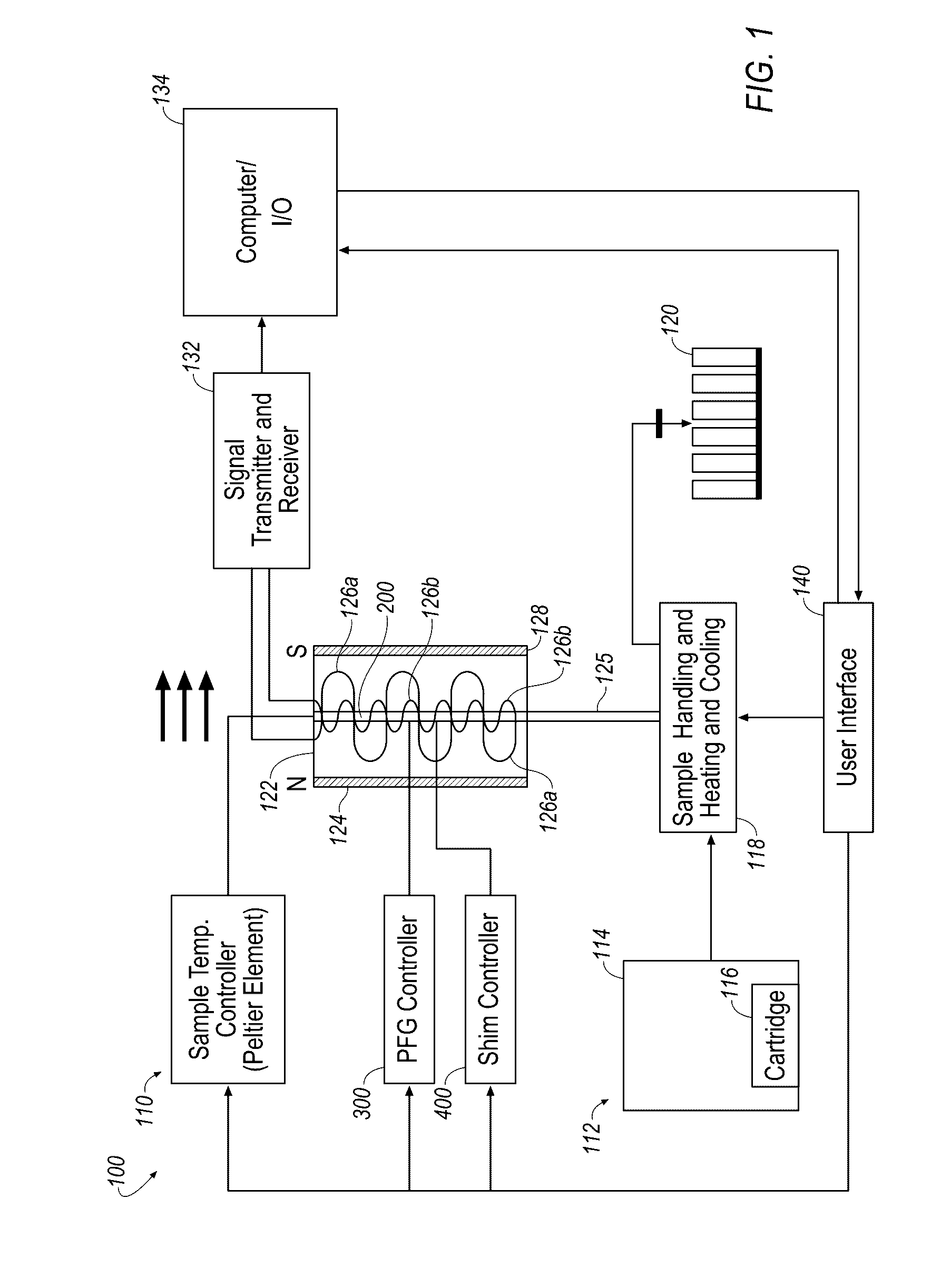
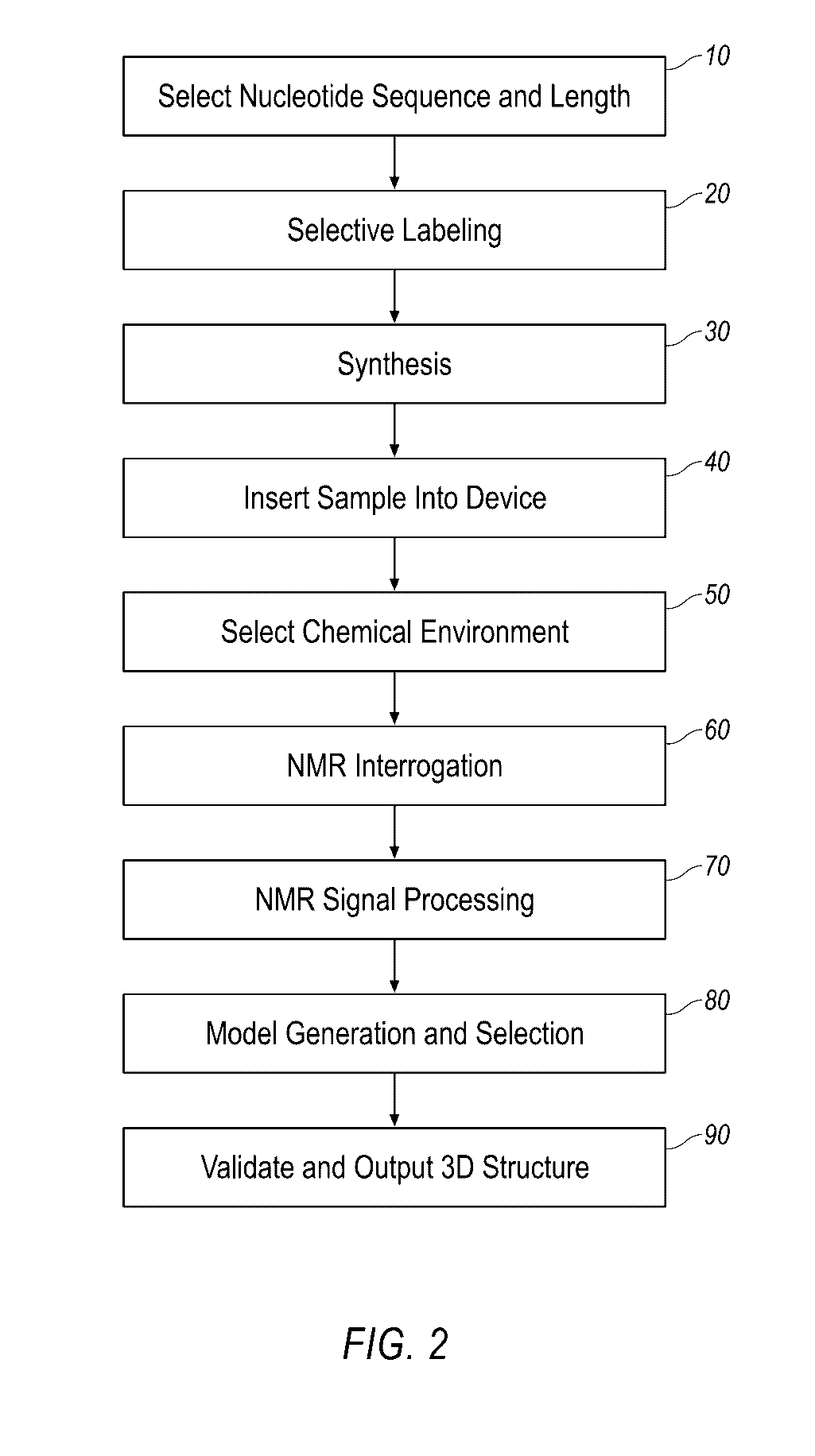
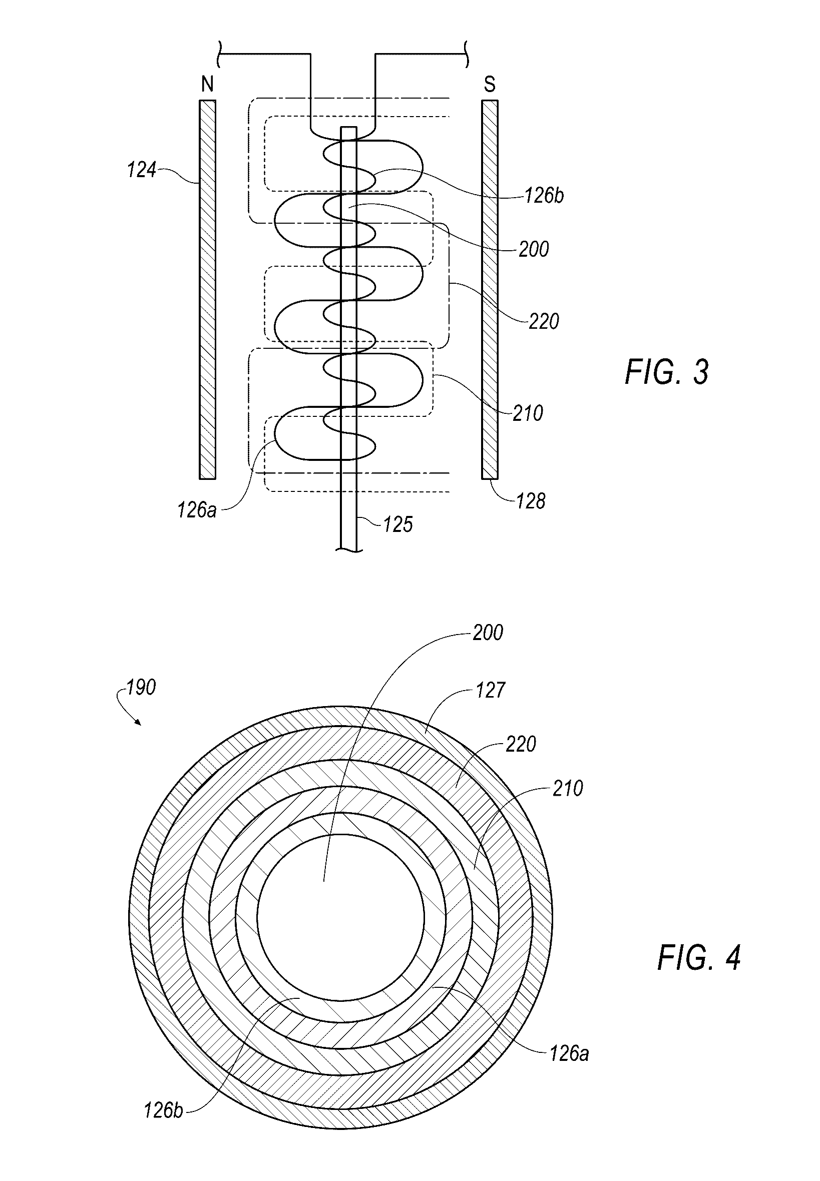
However, delivery of ASOs has been limited to accessible tissues such as the liver, kidney, spleen, and to a certain extent the lungs.
Moreover, due to poor understanding of miRNA structural biology and the lack of miRNA-Dicer or RISC complex structure, rational design of small molecule modifications to improve miRNA binding affinity and activity remains a significant challenge.
In some embodiments, small molecule drug discovery programs utilize structure-based drug design methods, however, current approaches to interrogate binding interactions between RNAs and small molecules have been unsuccessful.
Traditional approaches such as x-ray crystallography and classic Nuclear Magnetic Resonance methods are prohibitively time consuming and expensive.
Further, in some embodiments, these approaches do not account for the inherent plasticity of RNA, which is prevalent in miRNAs, thus preventing accurate atomic-level interpretation the data.
While methods relying solely on molecular modeling have been reported, none have proven useful in the drug discovery process.

Method used


Image

  • Small molecule binding pockets in nucleic acids
  • Small molecule binding pockets in nucleic acids
  • Small molecule binding pockets in nucleic acids

Examples


example 1

Structure Determination of an Apical Loop Sequence of Human Pre-miR 122 RNA

[0326]The apical loop sequence of human pre-miR 122 RNA sequence was investigated as an example. Two base pairs at the 5′- and 3′-ends of the sequence were modified from the native pre-miR 122 sequence in order to stabilize the 24-mer stem loop construct for NMR studies (5′-GGCUUGUGUCUAAACUAUCAAGCC-3′) (SEQ ID NO: 2).

[0327]The secondary structures predicted by the software program MFold suggest the possibility of a long stretch (up to 12 bases) of an apical loop which combined together with redundant adenine bases in the loop sequence prohibits conventional sequential assignment using uniformly non-selectively labeled RNA sample and thus requires selective labeling for unambiguous resonance assignment and 3-D structure determination. Four RNA oligonucleotides sequences are designed for selective labeling. Each has two selectively 13C / 15N isotope labeled residues. Labeling a pair of purine (A or G) and pyrimid...

example 2

Methods of Using 1H NMR Chemical Shifts in Determining RNA Structure and Dynamics

[0340]Methods and Materials

[0341]Predicting RNA 1H Chemical Shifts. A panel of 18 RNA structures (1ZC5, 2KOC, 1Z2J, 1XHP, 2QH2, 2KYD, 1JU7, 2JTP, 2FDT, 1N8X, 2L3E, 1LW, 2JYM, 2L5Z, 1NC0, 1IDV, 2L1V, 1OW9) was used to evaluate 1H chemical shift predictions using SHIFT and NUCHEMICS. This panel represents RNA structures that have been determined by NMR following the introduction of SHIFTS and NUCHEMICS (2002-2011) for which 1H chemical shift assignments were also available in the Biological Magnetic Resonance Bank (BMRB)29 (http: / / www.bmrb.wisc.edu / ). Four additional structures (2QH3, 2QH4, 1YMO, 2JWV) were not included due to undocumented or incomplete chemical shift referencing. However, including these structures had little to no impact on the results presented here.

[0342]Molecular dynamics (MD) simulations. MD simulations of an RNA duplex (PBID:2KYD)30, UUCG tetra-loop (PBID:2KOC)31, and pre-quenosine...

example 3

Solving the A-Site Structure

[0375]The technology described herein was established using the apramycin-bound bacterial ribosomal A-site structure. A-site is a well-studied RNA that undergoes an induced-fit structural change on binding small molecule antibiotics. Specifically, A1492 and A1493 loop from inside to outside the helix on small molecule binding, which in the ribosome signals acceptance of a codon-anticodon pair effectively eroding translational fidelity leading to bacterial death. In order to test whether the chemical shifts can be used to solve the A-site bound structure, we attempted to solve the 2.7 Å resolution apramycin-bound A-site x-ray structure (PDB ID#1YRJ) using only NMR chemical shifts and a newly developed method adapted from our recent publication. Briefly, apramycin-bound A-site chemical shifts were downloaded from the Biomagnetic Resonance Databank and re-referenced using the Aeschbacher method. The A-site secondary structure was input into CONSTRUCTOR and 1...


PUM

PropertyMeasurementUnit
frequencyaaaaaaaaaa
frequencyaaaaaaaaaa
frequencyaaaaaaaaaa

Abstract

Described herein is technology for determining the 2-D or 3-D atomic resolution structure of a polynucleotide bound to and / or interacting with another molecule, for example a small molecule. In some aspects of the technology, NMR and isotopic labeling strategies are used. The technology described herein is useful for a plurality of applications including but not limited to drug discovery and chemical biology probe discovery.

Description

CROSS REFERENCE TO RELATED APPLICATIONS[0001]This application is a continuation-in-part of U.S. patent application Ser. No. 14 / 650,087, filed Jun. 5, 2015, currently pending, which is a 35 U.S.C. §371(c) U.S. National Phase filing of International Patent Application Serial No. PCT / U52013 / 073330, filed Dec. 5, 2013, which claims the benefit of priority to U.S. Provisional Application No. 61 / 733,784, filed Dec. 5, 2012. This application also claims the benefit of priority to U.S. Provisional Application No. 62 / 214,040, filed Sep. 3, 2015. The entire contents of each of the aforementioned disclosures are incorporated herein by reference in their entirety.BACKGROUND[0002]Nucleic acids are being targeted to treat various diseases. Tools to rapidly identify molecules that bind nucleic acids are beneficial to the drug discovery process.SUMMARY OF THE INVENTION[0003]The central dogma of molecular biology, DNA→RNA→Protein, has undergone a significant revision. Rather than being merely a mess...

Claims


Application Information

Patent Timeline
Patent Type & Authority Applications(United States)
IPC IPC(8): G06F19/16G01N33/53C40B30/02G16B15/00G16B15/10G16B35/20
CPCG06F19/16G01N33/5308C40B30/02G01N33/6845G16B15/00G16B35/00G16C20/60G16B35/20G16B15/10G01N24/087
Inventor STELZER, ANDREW CORBETMCBRAIRTY, MITCHELL ALANAL-HASHIMI, HASHIM M.BAE, SUNG-HUN
Owner NYMIRUM
Why Eureka
Try Eureka