Method for preparation of retinal ganglion cells

a retinal ganglion and ganglion cell technology, applied in the field of preparation of retinal ganglion cells, can solve the problems of low possibility of successfully inducing the cellular components of the retina, insufficient lowering of intraocular pressure to stop the progression of glaucoma, and insufficient regenerative of the retinal nerve fiber layer with elongated axons, etc., to achieve no risk of retinal detachment, easy supply, and efficient production

Active Publication Date: 2017-07-06
SENJU PHARMA CO LTD +1
View PDF0 Cites 4 Cited by
  • Summary
  • Abstract
  • Description
  • Claims
  • Application Information

AI Technical Summary

Benefits of technology

[0017]According to the method of the present invention, retinal ganglion cells with elongated axons that are applicable to actual clinical practice can be efficiently produced from pluripotent stem cells within a short period of time. In addition, such retinal ganglion cells can be produced in a simple manner without inserting a gene from the outside. The retinal ganglion cells produced by the method of the present invention can be used for cell replacement therapy that involves implantation of cells into the damaged site of a retinal nerve. The cell replacement therapy involving the use of the retinal ganglion cells is advantageous over conventional cell replacement therapy involving the use of retinal pigment epithelial cells, the clinical application of which to humans has already been initiated, or cell replacement therapy involving the use of visual cells, the application of which is expected in the future, for the following reasons. That is, retinal ganglion cells are present in the inner layer of the retina and in the vicinity of the vitreous cavity, and, accordingly, cells can be easily supplied. In addition, there is no risk of retinal detachment. With the use of such retinal ganglion cells, also, screening of a drug, such as an agent for glaucoma treatment (e.g., a “protective agent for the retinal nerve” that delays retinal nerve cell death or a “regenerative agent for the retinal nerve” that promotes plasticity or regeneration of the retinal nerve), can be carried out readily and rapidly in vitro. Accordingly, the time, labor, cost, and the like required for drug screening involving the use of experimental animals comprising breeding of experimental animals, preparation of pharmacological samples, and immunostaining can be reduced to a significant extent.

Problems solved by technology

In Japan, about 20% of visual impairment cases are caused by glaucoma, which is the leading cause of blindness.
While eye drops are effective for the lowering of intraocular pressure to a certain extent, the lowering of intraocular pressure is often insufficient to stop the progression of glaucoma.
According to the method described in Non-Patent Document 2, however, a retinal layer structure is not formed, the possibility of successfully inducing cellular components constituting the retina is low, and axons of retinal ganglion cells have not resulted in elongation.
That is, clinically applicable retinal nerve fiber layers with elongated axons have not yet been regenerated according to conventional techniques.
In addition, such techniques are not practically satisfactory in terms of the rate of induction or the number of days until induction.
However, such disease animal models are disadvantageous since breeding and management thereof are laborious, animal cells are significantly different from human cells, and evaluation of therapeutic effects of drugs may not be definite.

Method used


Image

  • Method for preparation of retinal ganglion cells
  • Method for preparation of retinal ganglion cells
  • Method for preparation of retinal ganglion cells

Examples


examples

[0108]Hereafter, the present invention is described in greater detail with reference to the examples, although the technical scope of the present invention is not limited to these examples.

(Example 1) Preparation of Retinal Ganglion Cells from Human iPS Cells

1. Method

[0109](1) Induction of Differentiation from Human iPS Cells into Retinal Progenitor Cells

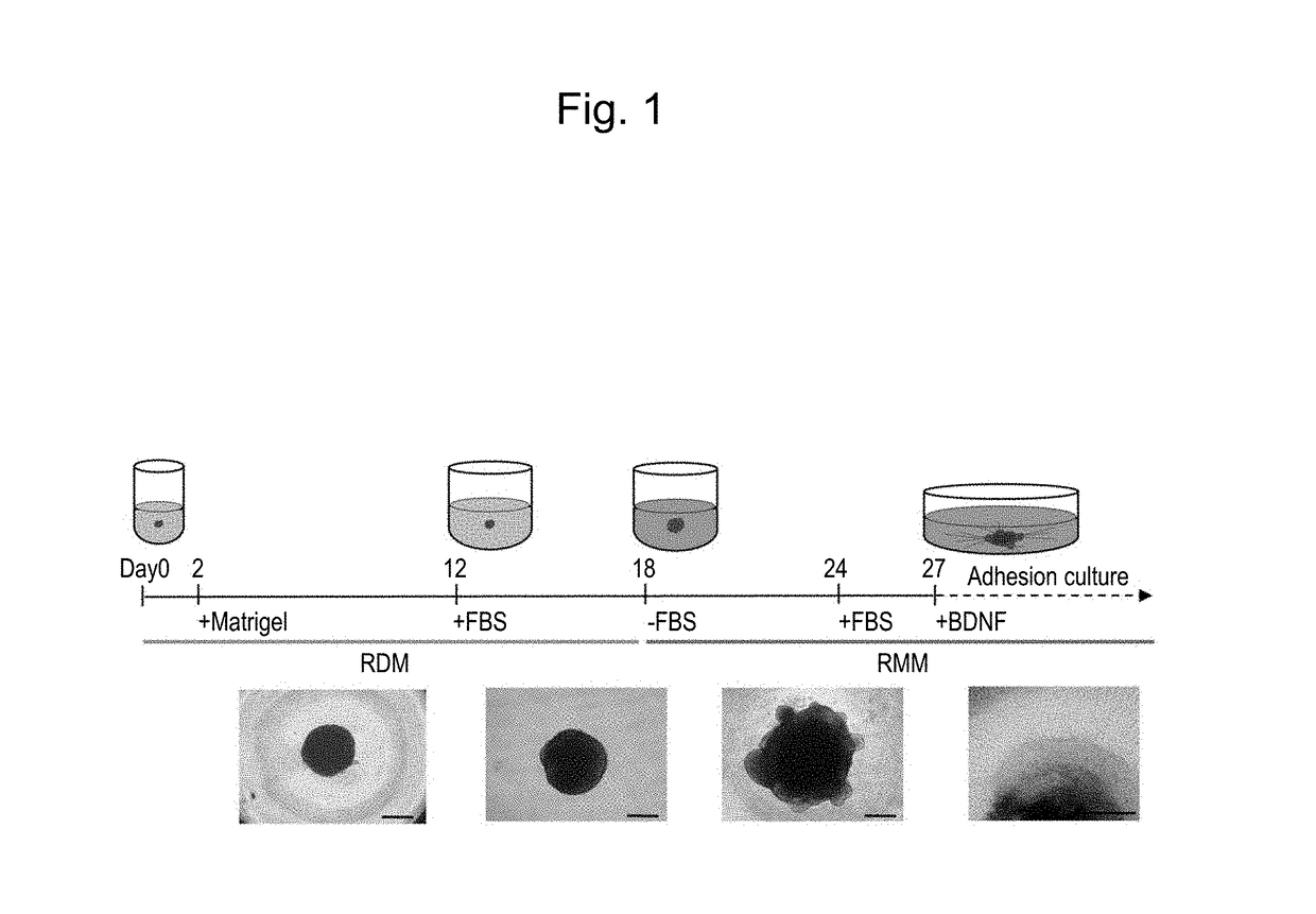
[0110]Human skin-derived iPS cells 409B2 provided by the Riken BioResource Center (RIKEN BRC) were used as human iPS cells (Okita et al., 2011, A more efficient method to generate integration-free human iPS cells, Nat. Methods). Basically, human iPS cells were induced to differentiate into retinal progenitor cells in the manner described below in accordance with the serum-free floating culture of embryoid body-like aggregates with the quick reaggregation (SFEBq) method described in the literature (Nakano, T. et al., Self-formation of optic cups and storable stratified neural retina from human ESCs, Cell Stem Cell 10, 771-785, 2012),...

example 3

(Example 3) Controlled-Release Test of Neural Inhibitor on Retinal Ganglion Cells

[0139]Floating culture was terminated 27 days after the initiation of differentiation induction that was conducted in Example 1, embryoid bodies were transferred and allowed to adhere to a 6-well plate coated with poly-D-lysine and laminin, and adhesion culture was then initiated using a neuronal maintenance medium supplemented with BDNF (final concentration: 100 ng / ml) (GlutaMAX-containing D-MEM / F12, N2 Supplements (Life Technologies), 1% FBS). The nerve growth inhibitor (Semaphorin 3A (SemaA)) was subjected to a controlled-release test 2 days after the initiation of adhesion culture. At the outset, MedGEL SP P15 (MedGEL Corporation) was soaked in 30 μl of 100 μg / μl Sema3A, and it was allowed to stand at 4° C. for 60 minutes. MedGEL was placed on Kimwipes, an excess solution was removed therefrom, and MedGEL was placed in a region approximately 5 mm away from the embryoid bodies. Thereafter, 10 μl of 1...

example 8

(Example 8) Functional Analysis of Axons

(1) Observation of Axonal Flow

(Method)

[0159]RGCs and their axons are known to be rich in mitochondria and to maintain homeostasis by axonal transport, as in other neurons. Neurotrophic tyrosine kinase receptor 1 (NTRK1), a member of the neurotrophic tyrosine kinase receptor family, is a nerve growth factor receptor. It is known to be expressed in RGCs, especially when the cells are damaged.

[0160]Using these two factors; i.e., NTRK1 and mitochondria, as tracers, a plasmid vector composed of GFP-NTRK1 with a CMV promoter and a plasmid vector composed of mCherry-mitochondria with a CMV promoter were directly introduced into the RGC cell body 34 days after the initiation of differentiation induction from human iPS cells (Day 34) via electroporation, and the anterograde rapid axonal transport was analyzed.

[0161]A specific test procedure is described below. The NTRK1 expression vector (RG213091; OriGene Technologies, Inc.), the pPAmCherry-Mito Vecto...


PUM

PropertyMeasurementUnit
culture temperatureaaaaaaaaaa
concentrationaaaaaaaaaa

Abstract

An object of the present invention is to provide a method of inducing pluripotent stem cells to differentiate into clinically applicable retinal ganglion cells.The present invention provides a method for inducing pluripotent stem cells to differentiate into retinal ganglion cells that can be used for clinical application. Such method is a method for producing retinal ganglion cells with elongated axons comprising the following steps: (a) a step of inducing pluripotent stem cells to differentiate into retinal progenitor cells via floating culture; (b) a step of inducing the retinal progenitor cells obtained in step (a) to differentiate into retinal ganglion cells via floating culture; and (c) a step of allowing axons to elongate via adhesion culture of the retinal ganglion cells obtained in step (b).

Description

TECHNICAL FIELD[0001]The present invention relates to a method for preparation of retinal ganglion cells, more particularly, a method for preparation of retinal ganglion cells comprising inducing pluripotent stem cells to differentiate into retinal ganglion cells with elongated axons.BACKGROUND ART[0002]At present, glaucoma is acknowledged as a major cause of blindness in developed countries. According to World Health Statistics 2002, the number of patients with blindness was 37,000,000 worldwide on the basis of the WHO standard, and 12% of such patients (i.e., 4,500,000 patients) suffer from glaucoma-induced blindness. In Japan, about 20% of visual impairment cases are caused by glaucoma, which is the leading cause of blindness. According to survey results, in addition, about 5% of the population aged 40 and older has been afflicted with glaucoma in Japan. It is also known that the prevalence rate of glaucoma increases with age. In the years to come after the arrival of an aging so...

Claims


Application Information

Patent Timeline
Patent Type & Authority Applications(United States)
IPC IPC(8): C12N5/079A61K35/30G01N33/50C12Q1/68
CPCC12N5/0621C12Q1/6876A61K35/30G01N33/5058C12Q2600/158C12N2506/08C12N2506/02C12N2501/13C12Q2600/136C12N2506/45A61L27/00C12Q1/02C12N15/09A61P27/06
Inventor AZUMA, NORIYUKITANAKA, TAKUYOKOI, TADASHI
Owner SENJU PHARMA CO LTD
Why Eureka
Try Eureka