
Prostatic liquid biopsy for the detection of prostate cancer and benign prostatic hyperplasia

a prostate cancer and liquid biopsy technology, applied in the field of prostate cancer and benign prostatic hyperplasia detection, can solve the problems of limited management and treatment of prostate cancer, inconvenient use of instruments, and inability to detect cancer at such a stage, so as to facilitate identification of individuals, accelerate exfoliation (shedding) of prostate cells, and increase the number of prostate epithelial cells available for detection

Active Publication Date: 2017-09-14
WAVESENSE INC
View PDF3 Cites 0 Cited by
  • Summary
  • Abstract
  • Description
  • Claims
  • Application Information

AI Technical Summary

Benefits of technology

The present invention provides minimally invasive methods for collecting target prostate cells or other biomarkers for testing and treating prostate cancer. The methods involve administering an agent that promotes the detachment and migration of cells from the prostate through the ureteral epithelial cells and collecting them for testing. The collected cells can be tested for a variety of analytes of interest using conventional means. The methods are less painful, cost-effective, and result in better samples with greater sensitivity and accuracy compared to prior art methods. The invention also includes the use of a therapeutic agent to accelerate the shedding of prostate cells for increased detection.

Problems solved by technology

Although highly ideal, the detection of cancer at such stage is considered almost accidental and is typically incidental to a separate transurethral resection of the prostate (TURP).
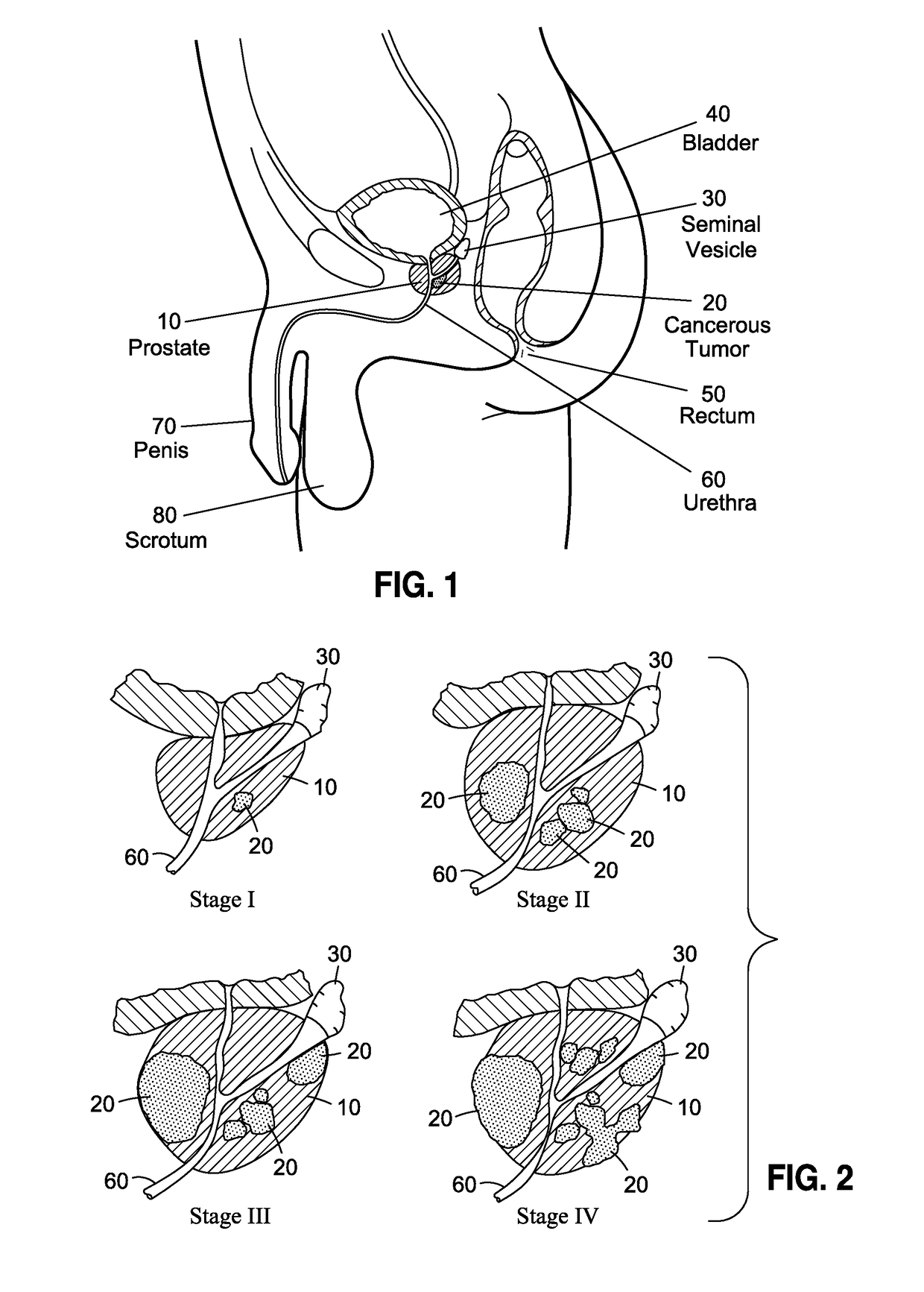
Given the anatomical positioning of the prostate, however, coupled with numerous drawbacks associated with the accurate diagnosis of prostate cancer, the ability to detect prostate cancer at its earliest stages is elusive and well-known to result in the implementation of harmful, sub-optimal care, as discussed below.
Management and treatment of prostate cancer is limited by access to an adequate sample of prostate tissue.
In this regard, the definitive diagnosis of prostate cancer is hampered by the limitations of acquiring prostate gland tissue using an invasive surgical procedure known as a prostate core needle biopsy.
Due to physical limitations of the prostate core biopsy, there are more that 3 million false negative biopsies reported presumably resulting from failure of the needle to pass through cancer tissue.
As is recognized, a key cause for this excessive false negative rate is the limited needle path inherent to core needle biopsy procedures.
When the prostate core biopsy procedure is examined closely, it is apparent that small tumors are easily missed and will continue to flourish undetected and untreated.
Also contributing to this failure to detect tumors is the small percentage of prostate tissue sampled by the biopsy procedure.
As a consequence, even to the extent needle biopsies could access and detect the presence of cancerous tissue, such approach is only as effective as the entirety of the random sampling made about prostate organ, which needs to be sufficiently sampled completely thereabout rather than just not in the more easily accessed areas.
There can likewise be significant discomfort and medical risk associated with prostate core needle biopsies.
Bleeding, antibiotic resistant infection / sepsis, urine retention, and tumor seeding (i.e., dislodging of tumor cells into tissue fluid or the circulation) are all well-known drawbacks associated with such procedure.
Taken together, the medical risk, poor sensitivity, and patient discomfort, the prostate core biopsy procedure is a less than ideal active surveillance tool that is limited to annual or semi-annual use.
Unfortunately, despite the availability of multiple biomarkers relating to prostate cancer, none can match the unequivocal specificity of skilled examination of prostate tissue by a pathologist.
Moreover, biomarker indications of prostate cancer are always confirmed by tissue biopsy prior to treatment and management of the patient and the data supporting this standard of care are overwhelming.
As such, biomarkers cannot achieve specificity equivalent to interrogation of the physical prostate cell.
In fact, despite the belief that biomarkers may offer a less invasive method of patient management, data shows that a great deal of patient harm is associated with the use of biomarkers in patient management for prostate cancer.
Along these lines, convincing evidence demonstrates that the PSA test often produces false-positive results, with reports that approximately 80% of positive PSA test results are false-positive when cutoffs between 2.5 and 4.0 μg / L are used.
There is also adequate evidence that false-positive PSA test results are associated with negative psychological effects, including persistent worry about prostate cancer.
Radiotherapy and surgery result in long-term adverse effects, including urinary incontinence and erectile dysfunction in at least 200 to 300 of 1000 men treated with these therapies.
Some clinicians have used androgen deprivation therapy as the primary therapy for early-stage prostate cancer, particularly in older men, despite the fact such an approach is not a U.S. Food and Drug Administration (FDA)-approved indication and has not been shown to improve survival in localized prostate cancer.
As discussed above, there is convincing evidence that PSA-based screening leads to substantial over-diagnosis of prostate tumors.
Thus, many men are being subjected to the harms of treatment of prostate cancer that will never become symptomatic.
Even for men whose screen-detected cancer would otherwise have been later identified without screening, most experience the same outcome and are, therefore, unnecessarily subjected to the harms of treatment for a much longer period of time.
Such PSA-based screening for prostate cancer has resulted in considerable overtreatment and its associated harms, and the United States Preventative Services Task Force (USPSTF) has considered the magnitude of these treatment-associated harms to be at least moderate.
Several researchers have reported, however, that attempts to detect prostate tumor cells in these specimens routinely is thwarted by unacceptably low sensitivities due to the rare numbers of prostate cells found in the urine.
However, using these cell concentration techniques also failed to provide adequate cell numbers and diagnostic sensitivity.
Although exfoliated prostatic epithelial cells can be acquired by non-invasive or minimally invasive sample collection methods, the potential for use as a reliable diagnostic method for detection of prostate cancer has not been realized due to the low numbers of prostate cells available even following prostatic massage.
Indeed, despite efforts to develop devices operative to facilitate collection of samples that seek to improve the probabilities that target cells of interest can be isolated and detected, such devices have proven ineffective.
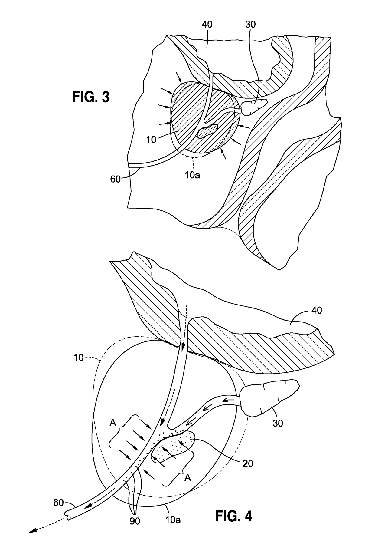
Among the drawbacks associated with all such devices and collection techniques using the same include the inability to selectively deploy such devices in a manner that maximizes the potential to capture the target cells of interest at the target prostatic urethral site.
Indeed, the use of such devices is completely random and there is no way to determine, and much less selectively deploy such devices at a time when the probability of collecting target cancer cells of interest is greatly enhanced.
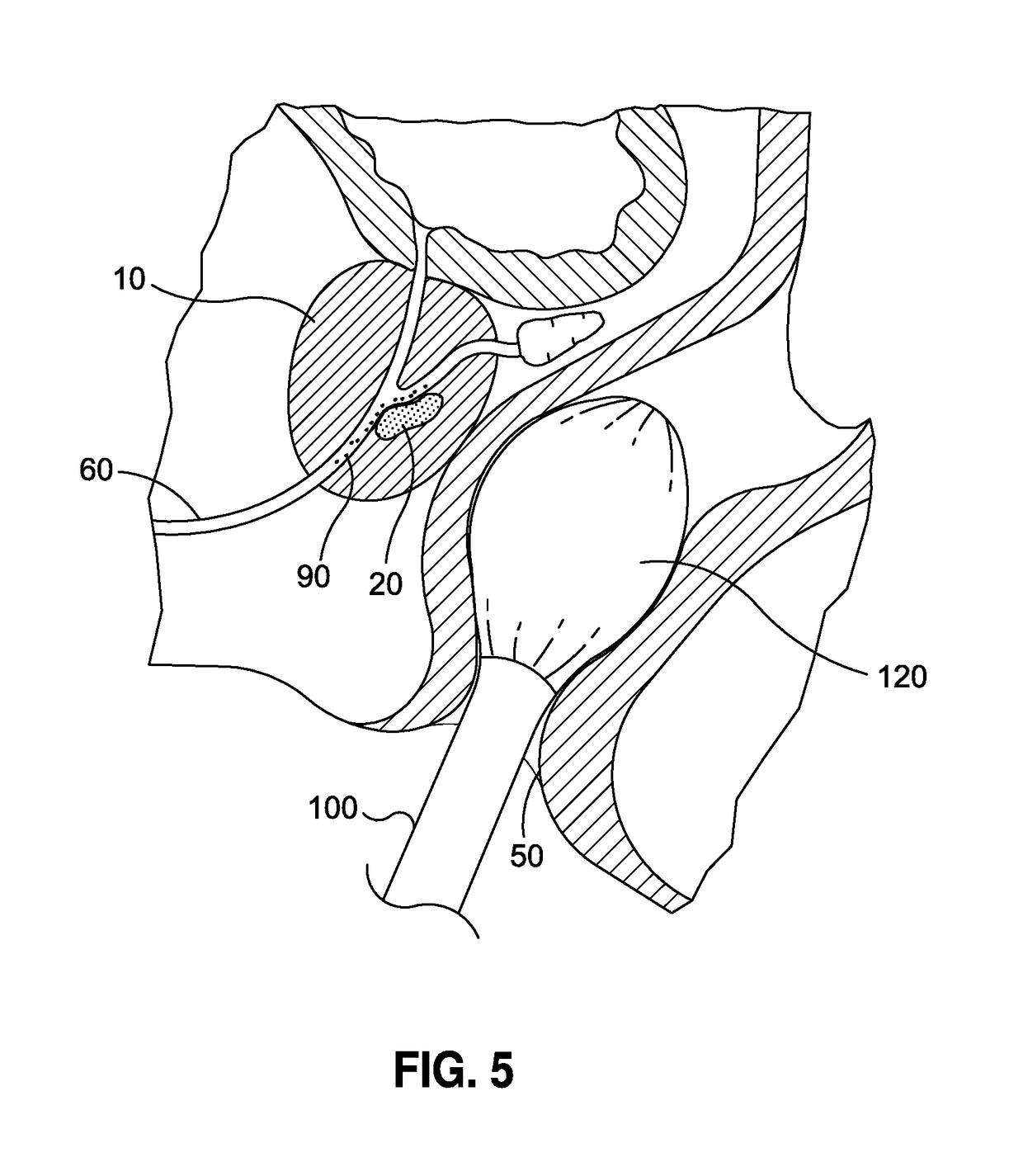
There is likewise no type of means for maximizing the probability that the target cancer cells will be in the prostatic urethra, as opposed to being located deep within the prostate, as illustrated in FIG. 5, and thus incapable of being accessed by such devices.
Accordingly, despite being slightly less invasive, such devices at best only provide a moderately increased chance of detecting the target cancer cells of interest.
Smith likewise does not anticipate the use of an exfoliating agent to increase the sensitivity of the biomarker assay.
The prior art, however, is completely deficient in any sort of structured methodology that can substantially increase the probability that the sought after biomarker, genetic material, or prostate cells can be increased in population and density so as to more easily, readily and accurately assess a patient's condition.

Method used


Image

  • Prostatic liquid biopsy for the detection of prostate cancer and benign prostatic hyperplasia
  • Prostatic liquid biopsy for the detection of prostate cancer and benign prostatic hyperplasia
  • Prostatic liquid biopsy for the detection of prostate cancer and benign prostatic hyperplasia

Examples


Embodiment Construction

[0049]The detailed description set forth below is intended as a description of the presently preferred embodiment of the invention, and is not intended to represent the only form in which the present invention may be implemented or performed. The description sets forth the functions and sequences of steps for practicing the invention. It is to be understood, however, that the same or equivalent functions and sequences may be accomplished by different embodiments and that they are also intended to be encompassed within the scope of the invention.

[0050]In this regard, the present invention is directed to methods for facilitating the ability of prostatic epithelial cells situated in the prostate to become dislodged from the prostate and migrate into the urethra for subsequent collection. The methods of the present invention are thus operative to generate a greater population of prostatic epithelial cells to be more readily collected and detected for any of a wide variety of uses, inclu...


PUM

PropertyMeasurementUnit
timeaaaaaaaaaa
timeaaaaaaaaaa
lengthaaaaaaaaaa

Abstract

Disclosed are methods to induce dislodgement of target prostatic cells from the prostate organ, collecting said cells, and subsequently examining the cell population. Such methods comprise the administration of an agent that facilitates the dislodgement of the target cells from within the prostate, which then migrate into the urethra. Exemplary agents include 5 alpha-reductase inhibitors. The cells induced to pass into the urethra are then collected non-invasively, such as through urine or semen samples. Such collection is further strategically calculated relative the administration of the agent so as to maximize the sample collection of the target cells of interest. The exfoliated prostatic epithelial cells are subsequently utilized for purposes such as detecting prostate cancer, predicting / measuring prostate tumor susceptibility to drug regimes, active surveillance of patients whose prostate biopsy results are negative, but continue to exhibit symptoms consistent with prostate cancer, and identifying false positive results associated with biomarker assays.

Description

CROSS-REFERENCE TO RELATED APPLICATIONS[0001]Not ApplicableSTATEMENT RE: FEDERALLY SPONSORED RESEARCH / DEVELOPMENT[0002]Not ApplicableBACKGROUND[0003]Prostate cancer is the most common cancer found in men and it is the second leading cause of death among men who die from cancer. By the age of 50, very few men have symptoms of prostate cancer, yet some precancerous or cancer cells may be present. More than half of all American men have some cancer in their prostate glands by the age of 80. Most of these cancers never pose a problem. See, National Cancer Institute Understanding Prostate Changes: A Health Guide for Men (http: / / wwwdotcancerdotgov / types / prostate / understanding-prostate-changes) current website guide 1 / 26 / 16. The American Cancer Society's estimates for prostate cancer in the United States for 2015 are approximately 220,800 new cases of prostate cancer; and 27,540 deaths from prostate cancer. See, American Cancer Society Prostate Cancer Guide (Ref: http: / / wwwdotcancerdotorg / ...

Claims


Application Information

Patent Timeline
Patent Type & Authority Applications(United States)
IPC IPC(8): G01N33/574A61K31/58C12Q1/68
CPCG01N33/57434A61K31/58C12Q1/6886A61B10/0045G01N33/57438G01N33/57488A61B2017/00274A61B2018/00547
Inventor FEISTEL, CHRISTOPHER
Owner WAVESENSE INC
Learn More