Three-dimensional structure for cardiac muscular tissue regeneration and manufacturing method therefor

a cardiac muscle tissue and three-dimensional technology, applied in the field of three-dimensional construct preparation, can solve the problems of limited material choice, minimal cell-material interaction, inferior tissue formation, etc., and achieve the effect of effective implementation of the micro-environment of cardiac muscle tissue and significant improvement of cell transfer efficiency into the myocardium

Inactive Publication Date: 2018-02-08
T&R BIOFAB
View PDF1 Cites 2 Cited by
  • Summary
  • Abstract
  • Description
  • Claims
  • Application Information

AI Technical Summary

Benefits of technology

The present invention is about a method for 3D printing using certain cells and a specific growth factor to create a micro-environment that mimics cardiac muscle tissue. This method improves the viability of cells and increases their transfer efficiency into the heart tissue, which can be useful for treating heart diseases like ischemic heart disease. The method also results in a vascular network within the 3D construct, which further enhances cell viability and transfer efficiency.

Problems solved by technology

This limitation reduces the choice of materials, due to the need to perform in an aqueous or aqueous gel environment.
Thus, although there are some success reports on bioprinting of cell-print constructs, minimal cell-material interactions and inferior tissue formation are the most important problems.
Indeed, these materials cannot exhibit complexity of natural extracellular matrices (ECMs), and thus they are not sufficient to reproduce the microenvironment having typical cell-cell connections and three-dimensional (3D) cell configurations of biological tissues.
Thus, cells in the hydrogel cannot exhibit unique morphology and function of living tissues in vivo.
Furthermore, in severe end-stage heart failure patient, there is no cure method other than cardiac transplantation or mechanical left ventricular assist device.
However, there are problems such as shortage of donor organs, difficulty in securing organs, high mortality rate and expensive treatment cost, etc.

Method used


Image

  • Three-dimensional structure for cardiac muscular tissue regeneration and manufacturing method therefor
  • Three-dimensional structure for cardiac muscular tissue regeneration and manufacturing method therefor
  • Three-dimensional structure for cardiac muscular tissue regeneration and manufacturing method therefor

Examples


preparation example 1

rst Bioprinting Composition

[0091]1-1: Preparation of Decellularized Extracellular Matrix

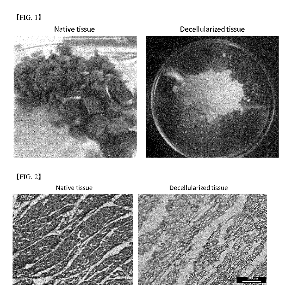
[0092]Decellularized extracellular matrix was prepared according to a method disclosed in Falguni Pati, et al., Nat Commun. 5, 3935 (2014) using porcine cardiac tissues (hereinafter, ‘hdECM’). The prepared hdECM was finally lyophilized and kept frozen before used. The optical microscope photograph and tissue staining photograph were shown in FIG. 2.

[0093]1-2: Preparation of Pre-Gel Form of a Tissue Engineering Construct Forming Solution

[0094]Liquid nitrogen was poured to the obtained lyophilized hdECM and it was crushed with mortar and a pestle. After the obtained hdECM powder (330 mg) was added to 0.5M acetic acid aqueous solution (10 ml), and pepsin (33 mg) (P7125, Sigma-Aldrich) was added, it was stirred for 48 hrs at a room temperature. Maintaining the temperature of the obtained solution below 10° C., riboflavin (2 mg) was added and 10 NaOH solution which was cooled below 10° C. was added, t...

preparation example 2

cond Bioprinting Composition

[0097]2-1: Preparation of Decellularized Extracellular Matrix

[0098]Decellularized extracellular matrix was prepared according to a method disclosed in Falguni Pati, et al., Nat Commun. 5, 3935 (2014) using porcine cardiac tissues (hereinafter, ‘hdECM’). The prepared hdECM was finally lyophilized and kept frozen before used. The optical microscope photograph and tissue staining photograph were shown in FIG. 2.

[0099]2-2: Preparation of Pre-Gel Form of a Tissue Engineering Construct Forming Solution

[0100]Liquid nitrogen was poured to the obtained lyophilized hdECM and it was crushed with mortar and a pestle. After the obtained hdECM powder (330 mg) was added to 0.5M acetic acid aqueous solution (10 ml), and pepsin (33 mg) (P7125, Sigma-Aldrich) was added, it was stirred for 48 hrs at a room temperature. Maintaining the temperature of the obtained solution below 10° C., riboflavin (2 mg) was added and 10 NaOH solution which was cooled below 10° C. was added, ...

example 1

on of a Three-Dimensional Construct for Tissue Engineering

[0105]A three-dimensional construct was fabricated using the first bioprinting composition and the second bioprinting composition obtained from the preparation examples 1 and 2.

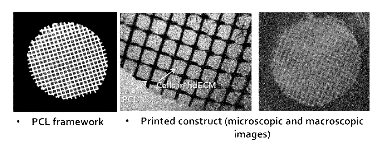
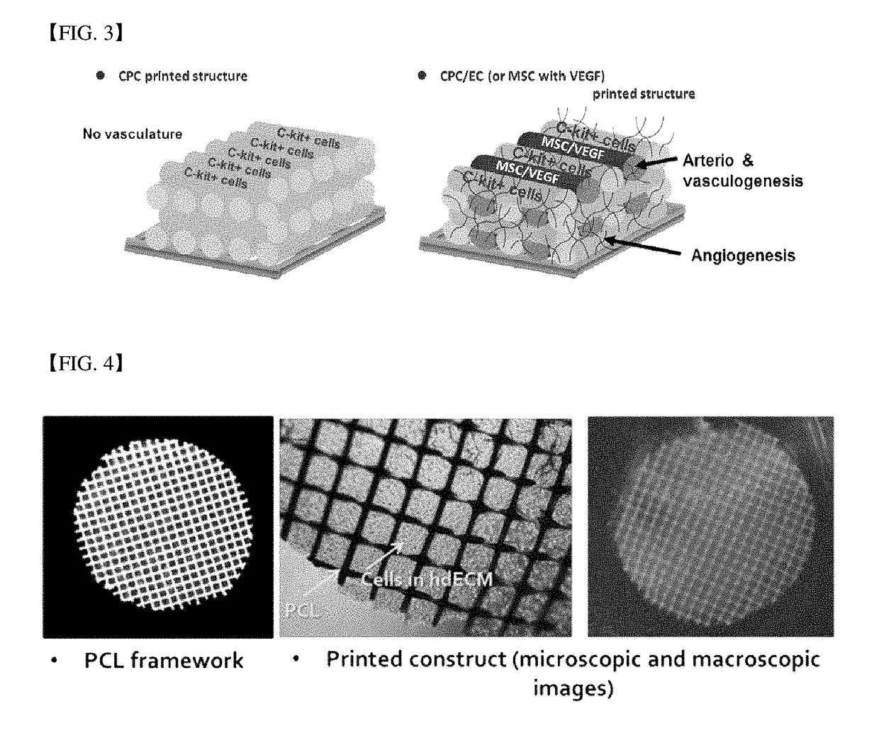
[0106]Specifically, polycaprolactone (PCL) framework was loaded on the syringe (the first syringe) of multi-head tissue and organ printing system (Jin-Hyung Shim et al., J. Micromech. Microeng. 22 085014 (2012)), and it was heated at approximately 80° C. to melt a polymer. The pre-gel form of the first bioprinting composition obtained from the preparation example 1 and the second bioprinting composition obtained from the preparation example 2 were loaded on another syringe (the second and third syringes, respectively), and the temperature was maintained below approximately 10° C. A thin PCL framework having below approximately 100 um line width, approximately 300 um of gap and 120 um of thickness by putting approximately 600 kPa of pneumatic pressure t...


PUM


Abstract

The present invention provides a preparation method of a three-dimensional construct for regenerating a cardiac muscle tissue comprising; a step of forming a three-dimensional construct by printing and crosslinking the first bioprinting composition comprising a tissue engineering construct forming solution containing decellularized extracellular matrix and a crosslinking agent, and cardiac progenitor cells, and the second bioprinting composition comprising the tissue engineering construct forming solution, mesenchymal stem cells and a vascular endothelial growth factor, to arrange the first bioprint layer and the second bioprint layer alternately; and a step of obtaining a crosslink-gelated three-dimensional construct by thermally gelating the crosslinked three-dimensional construct, and a three-dimensional construct for regenerating a cardiac muscle tissue, and the preparation method according to the present invention not only equally positions the cardiac progenitor cells in the construct but also implements a vascular network composed of vascular cells in the construct, so that the viability of cells can be maintained for a long time and the cell transfer efficiency into the myocardium can be significantly improved.

Description

TECHNICAL FIELD[0001]The present invention relates to a three-dimensional construct for regenerating a cardiac muscle tissue and a method for preparing thereof. More specifically, the present invention relates to a preparation method of a three-dimensional construct for regenerating a cardiac muscle tissue comprising; a step of forming a three-dimensional construct by printing and crosslinking the first bioprinting composition comprising a tissue engineering construct forming solution containing decellularized extracellular matrix and a crosslinking agent, and cardiac progenitor cells, and the second bioprinting composition comprising the tissue engineering construct forming solution, mesenchymal stem cells and a vascular endothelial growth factor, to arrange the first bioprint layer and the second bioprint layer alternately; and a step of obtaining a crosslink-gelated three-dimensional construct by thermally gelating the crosslinked three-dimensional construct, and a three-dimensio...

Claims


Application Information

Patent Timeline
IPC IPC(8): C12N5/071A61L27/36A61F2/24B33Y70/00B33Y80/00A61L27/38B33Y10/00
CPCC12N5/0697A61L27/3891A61L27/3826A61L27/3834A61L27/3895A61L27/367A61L27/3633B33Y10/00B33Y70/00B33Y80/00A61F2/24C12N2513/00C12N2502/1329C12N2502/1352C12N2501/165C12N2533/90C12N2537/10A61L2430/20A61F2240/001C07K14/475C12N5/0657A61L27/36
Inventor CHO, DONG-WOOJANG, JINAHPARK, HUN-JUN
Owner T&R BIOFAB
Why Eureka
Try Eureka