Medical implant

a technology of medical implants and implants, applied in the field of medical implants, can solve the problems of not providing the individual with the full range of natural movement, damage to skeletal tissues, and damage to skeletal tissues, and achieve the effects of stimulating stem cell homing, promoting tissue formation, and promoting regeneration of articular cartilag

Active Publication Date: 2018-08-02
UNIV OF SHEFFIELD
View PDF4 Cites 0 Cited by
  • Summary
  • Abstract
  • Description
  • Claims
  • Application Information

AI Technical Summary

Benefits of technology

[0011]The invention is an acellular, multifunctional, biomimetic medical device designed to be used as a medical implant which, depending on its selected bioactive factors, can be used to regenerate articular cartilage, bone, meniscal cartilage, and ligament or tendon tissues in the body. The novelty lies in the custom application of more than one biological factor, each bound to GAG oligosaccharides in a biomimetic way, at low levels (nanogram/picomole amounts) to a known scaffold material. These factors can be chosen to st...

Problems solved by technology

Damage to skeletal tissues can be caused through trauma, for example, but not exclusively, torn ligaments and tendons and bone fractures.
Damage to skeletal tissues can also be caused by degenerative diseases.
However, problems associated with joint prostheses include a finite working life14 and may not provide the individual with the full range of natural movement as compared to a natural healthy joint.
In addition, device loosening is a longer-term problem requiring invasive revision surgery15 and is particularly problematic in younger more active patients (16-17. who tend to have an active lifestyle and so put more demands on their prostheses.
However, while giving valuable pain relief, a fibrocartilage tissue is formed which is biomechanically inferior to native hyaline cartilage and degenerates after several years.
However, these procedures require the patient to have two separate surge...

Method used


Image

  • Medical implant
  • Medical implant
  • Medical implant

Examples


example 1

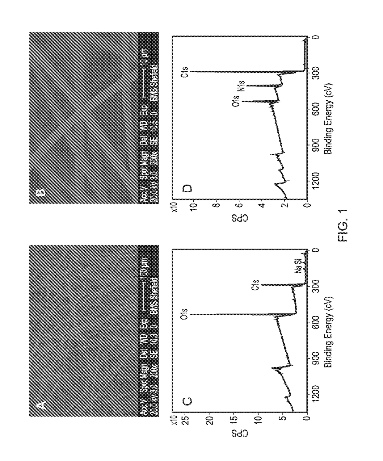
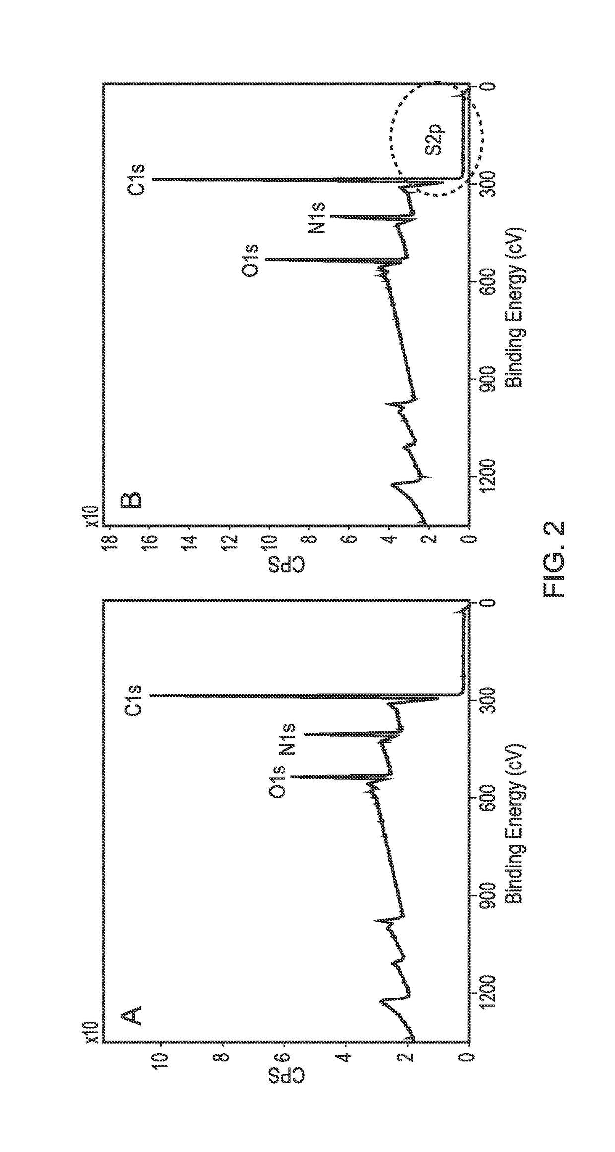
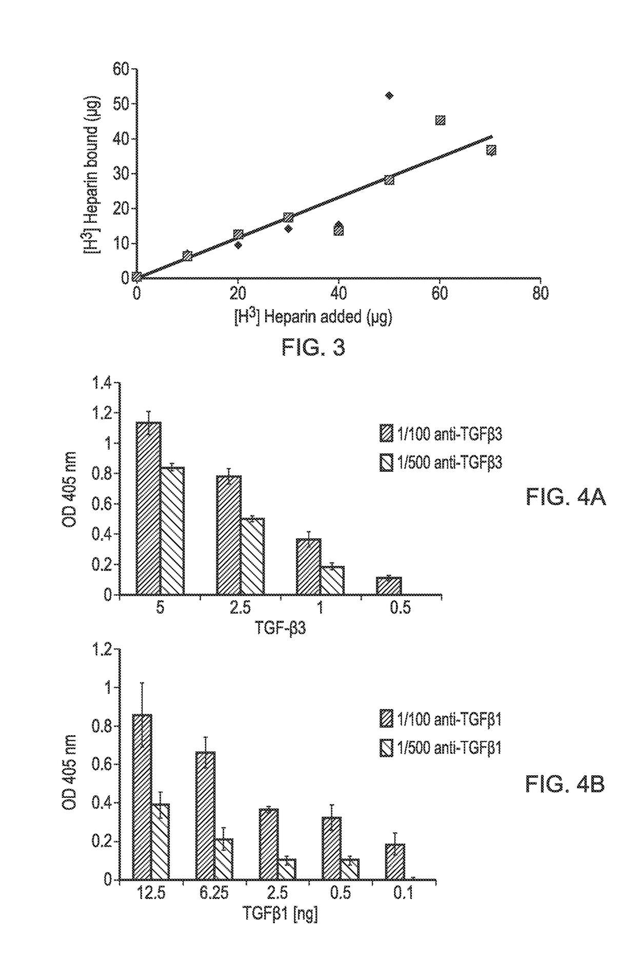
[0090]A PLLA scaffold was subjected to cold plasma treatment with allylamine as hereinbefore described and a coating of allylamine was confirmed by the presence of a nitrogen peak (FIG. 1D). FIG. 2B shows the X-ray photon spectroscopy image of the heparin-treated, allylamine coated PLLA scaffold. The small peak occurring at 168.1 ev shows the presence of sulphur groups confirming binding of heparin to the allylamine-treated PLLA. FIG. 3 shows a linear relationship of the amount of [H3]-radiolabelled heparin bound to the allylamine-treated scaffold vs the amount added The scaffold thus provides a positive surface coating of allylamine to which heparin binds non-covalently. Once the heparin has bound further experiments were conducted to show that heparin-binding bioactive factors such as for example TGFβ1, TGFβ3 and CXCl12 applied singularly or in combination will bind directly to the heparin layer by a tight non-covalent interaction. FIG. 4 shows the binding of TGFβ1 and TGFβ3 to an...

example 2

[0091]FIG. 5 shows the binding of several example heparin-binding bioactive factors to various allylamine and glycosaminoglycan functionalised surface. The results show the means+ / −standard error of the mean. We have demonstrated that TGFβ3 and TGFβ1, CXCL12, SDF1β, MIA, Wnt 3a and BMP2 bind to a selection of oligosaccharides directly. MIA is considered to bind to heparin via binding to fibronectin which will bind to heparin41. Here we have shown that MIA will bind directly to immobilised sulphated oligosaccharides as well as via binding to sulphated oligosaccharides via binding to fibronectin which then binds to the oligosaccharides. There are literature reports which state that TGFβ3 does not bind to sulphated oligosaccharides56,58. However, we have clearly demonstrated that TGFβ3 does bind directly to various sulphated glycosaminoglycans.

[0092]Binding of bioactive factors (BFs) which bind to GAG-binding proteins such as fibronectin or laminin, can be bound to the oligosaccharide-...

example 3

[0093]Studies were undertaken with bovine synovial fluid mesenchymal stem cells (SF-MSCs) or chondrocytes to investigate the attachment of the cells to an example of PLLA scaffolds functionalised with an example chondrogenic BF. Results for the SF-MSCs are shown in FIG. 6 and results for chondrocytes in FIG. 7. Cells were seeded onto the scaffolds as described herewithin and 48 hr after seeding the cell-scaffold constructs were fixed and examined by scanning electron microscopy. FIGS. 6 and 7 show scanning electron micrographs of SF-MSCs and chondrocytes attached to PLLA. (FIGS. 6A7A) and PLLA scaffolds functionalised with allylamine (FIGS. 6B and 7B), heparin (FIGS. 6C and 7C) and TGFβ3 (FIGS. 6D and 7D). For both cell types, TGFβ3-functionalised PLLA scaffolds showed a greater density of cells bound than allylamine or heparin functionalised PLLA scaffolds or unmodified PLLA. In summary, this data shows that MScs and chondrocytes can attach to PLLA scaffolds in higher cell densitie...


PUM

PropertyMeasurementUnit
Areaaaaaaaaaaa
Levelaaaaaaaaaa
Bioactiveaaaaaaaaaa

Abstract

The present invention relates to a cell-free, multi-layered medical device having bespoke, multifunctional bioactivity for the purpose of regeneration of skeletal tissues. The medical device may actively promote homing of stem cells into the medical device and promote their differentiation into the required cell type and promote de-novo tissue formation. The invention includes methods of making the medical device, uses of the medical device in promoting regeneration of the articular cartilage of a joint surface and in promoting healing and regeneration of skeletal tissues, for example, meniscal cartilage, tendon and ligament tissues and also healing of bone tissue indications such as fractures.

Description

[0001]The present invention relates to an acellular, multi-layered medical device having bespoke, multifunctional bioactivity, for the purpose of regeneration of skeletal tissues. The invention includes inter alia uses of the medical device, especially but not exclusively, in promoting regeneration of articular cartilage in lesions of the joint surface and in promoting healing of joint tissues, for example, meniscal cartilage and tendon and ligament injuries and also healing of bone injury such as fractures or subchondral bone degeneration in joints. For completeness of understanding, this application includes inter alia preferred methods of fabrication of the medical device.BACKGROUND[0002]Biological / bioactive factors (BFs), including growth factors, differentiation factors, cytokines and chemokines are crucial in maintaining normal tissue homeostasis, and wound healing and tissue regeneration. This is also the case for the skeletal tissues including the cartilage covering the surf...

Claims


Application Information

Patent Timeline
IPC IPC(8): A61L27/54A61L27/34A61L27/18A61L27/56A61F2/30
CPCA61L27/54A61L27/34A61L27/18A61L27/56A61F2/30756A61L2430/06A61L2430/02A61L2430/24A61L2300/608A61F2002/30766A61F2002/30677A61L27/20A61L2300/414C08L67/04C08L5/10A61K47/642A61K38/1841
Inventor CRAWFORD-CORRIE, AILEENBUTTLE, DAVID JOHNHAYCOCK, JOHN WILLIAM
Owner UNIV OF SHEFFIELD
Why Eureka
Try Eureka