Biodegrading implantable ocular sustained release drug delivery system

a drug delivery and biodegradable technology, applied in the field of biodegradable ocular sustained release drug delivery implant system and formulation, can solve the problems of biologic therapeutic agents being associated with undesired side effects, significant inconvenience and expense, and several difficulties in use of biologic therapeutic agents, and achieves high localization of delivery

Inactive Publication Date: 2018-09-20
BIOHEALTHWAYS INC
View PDF4 Cites 5 Cited by
  • Summary
  • Abstract
  • Description
  • Claims
  • Application Information

AI Technical Summary

Benefits of technology

[0045]In an embodiment, the implant may have a generally cylindrical shape, in which a bioerodable core is encapsulated by a tubular shell. The implant device may have end caps of the shell material at one or both ends of the cylinder, or both ends may be open. The open ends expose the bioerodable core to bodily fluids at the site of the implant. As the core erodes, the active agent is released into the biological fluids around the implant, thereby providing highly localized delivery of the drug to the specific site where the implant is placed.

Problems solved by technology

However, biologic therapeutics (biopharmaceuticals) have several difficulties associated with their use.
Biologic therapeutic agents may be associated with undesired side effects at high systemic concentrations.
Biologic therapeutic agents generally must be injected (oral delivery is not normally an option), so repeated painful and expensive administration of biologics may be required, causing significant inconvenience and expense.
Biologics also tend to be chemically unstable compared to small molecules, making it a challenge to deliver sustained required doses over time.
In general, topical eye drop application of drugs do not efficiently reach anterior or posterior anatomy of the eye.
Thus, it is difficult to provide and maintain an adequate concentration of drug in the pre-corneal area.
More than 75% of applied ophthalmic solution is lost via nasolacrimal drainage and absorbed systemically via conjunctiva, hence ocular drug availability is very low.
A significant issue with treating ocular diseases is that compliance with therapeutic regimens is problematic, particularly among patients who have chronic diseases such as glaucoma (open- or closed-angle types), and refractory chorio-retinal diseases, including uveitis, macular edema, neovascular (wet) and atrophic (dry) age-related macular degeneration (AMD), and retinitis pigmentosa (RP).
But studies suggest that ˜20% of patients discontinue treatment within one year, and ˜40% of patients discontinue within two years.3 The monthly cost could be thousands of dollars, so that means effective treatment faces a serious social problem.
In addition, frequent intravitreal injections can cause complications, such as increased intraocular pressure,4 endophthalmitis, and retinal detachment.
This route of administration typically results in a short half-life unless the drug can be delivered using a formulation capable of providing sustained release.
Topical eye drops have efficacy for anterior eye diseases, but the residency time and bioavailable payload delivered to the affected area may not be sufficient for certain drugs.
Topical periocular administration is not invasive but is inefficient in prolonged drug retention time.
Intraocular solutions / suspensions are effective topical treatments to the back of eye, but residency time is an issue.
Regarding topical eye drops, many topical eye drops have been formulated to treat a wide variety of ocular diseases, but cannot provide drug delivery intraocularly.
While less invasive, these methods also require frequent administration and potentially high dosages with concomitant toxicity.
For subconjunctival injections or punctal plugs, the drug faces the same issues of traversing multiple ocular barriers, meaning dosing will be low.
These microvascular networks cause vision loss when blood leaks or causes retinal swelling.
Anti-VEGF drugs have helped improve visual acuity by preventing further vision loss; however, the stability of these biologics pose a challenge due to their short half-lives in the vitreous.
Another takeaway is that biologics tend to have rather short half-lives, which make sustained delivery intraocularly a challenge.
Glaucoma typically occurs when the drainage canals in the front of the eye (anterior) eye are slowly or quickly clogged or blocked, which raises eye pressure via liquid buildup and can damage the optic nerve.
Once the optic nerve is damaged due to the pressure, vision loss occurs.
Biologics are quite challenging to preserve intraocularly for long periods of time.
These approaches do not integrate biologics, since these approaches may not appropriately sustain the lifetime of the protein in the hydrolytic and enzymatic environment of the intravitreal space.
There are limited precedents for delivering biologics for glaucoma intraocularly, either intravitreally (posterior) or in the intracamerally (anterior) in the eye.13 And while neovascularization in the posterior of the eye is typically treated intravitreously, there is no sustained release formulation available for the marketed biologic therapies.
In another example, U.S. Pat. No. 9,668,915 discloses implantable devices for treating ocular diseases, but does not provide triblock polymers, and does not discuss stabilizing protein therapeutic agents with a suitable in-situ buffer that maintains an appropriate pH to prevent degradation.
Thus, biologics are not easy to deliver intraocularly in a sustained release dosage form.
While other ocular small molecule and biologic therapy administration methods exist, each administration method has significant drawbacks—ranging from inherent challenges of dosing efficiency, non-biodegradability, poor sustained release, to implicit issues with patient regimen persistence, economic cost, and usability.

Method used


Image

  • Biodegrading implantable ocular sustained release drug delivery system
  • Biodegrading implantable ocular sustained release drug delivery system
  • Biodegrading implantable ocular sustained release drug delivery system

Examples


example 1

on of BSA-FITC Loaded Triblock Copolymer Solution

[0141]B1-b-B2-b-B1 triblock copolymer Poly(D,L-lactide)-b-Poly(ethylene glycol)-b-Poly(D,L-lactide) PDDLA-PEG-PDLLA triblock copolymer (1,700:1,500:1,700)

NumberWeightAverageAveragePolydispersityMolecularMolecularIndexPolymerWeight, MnWeight, MwPDIPDLLA-b-PEG-b-PDLLA5,3506,8001.27PEG 1,500 Initiator1,485——

[0142]Materials[0143]The B1-b-B2-b-B1 triblock polymer was ordered from PolySciTech, Catalog Number: AK100[0144]Protein: Bovine Serum Albumin-fluorescein isothiocyanate conjugate (BSA-FITC) was obtained from Protein Mods., product code BSF product lot 263BSF2, molecular weight ˜66 KDa, 100 mg / ml in H2O. BSA modification level 0.9 luorescein / molecule.[0145]Phosphate buffered solution (1M PBS, pH=7.4) was purchased from Sigma-Aldirch.[0146]Acetone, reagent grade, was purchased from Fisher Scientific.

[0147]Procedure: In a 3 ml vial, 0.0966 g of B1-b-B2-b-B1 triblock copolymer was dissolved in 0.3807 g acetone, and 0.2288 g deionized dist...

example 2

on of Model Core-Shell DDS

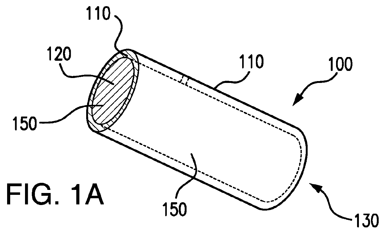
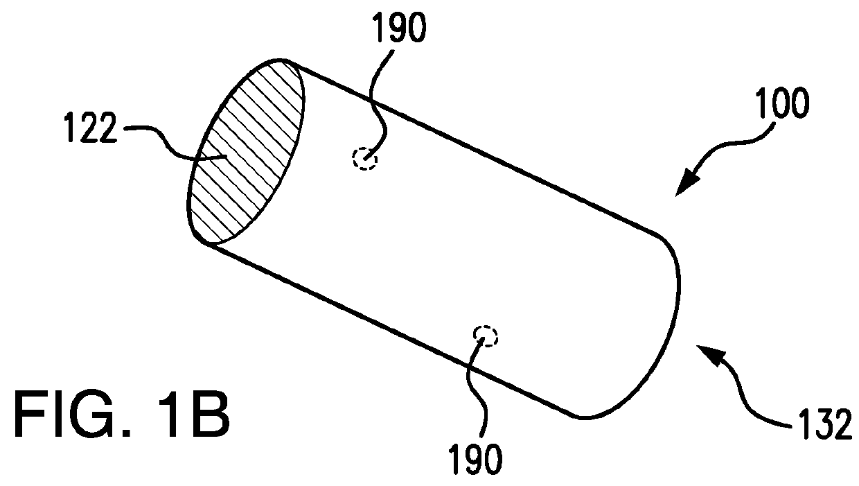
[0149]To model a DDS shell, a 20 AWG miniature polyimide tube was used with an outside diameter of 962 microns, a wall thickness of 76.2 microns, and an inside diameter of 810 microns. The inside cross section area was 0.515 mm2 The polyimide tube was not biodegradable, so in this case, the aim is to test the release capability of core through a fixed area.

[0150]The polymer solution of Example 1 loaded with BSA-FITC was injected into a ˜3″ long segment of the polyimide tube. The tube was incubated at 37° C. for 15 minutes, and the BSA-FITC loaded polymer solution formed a hydrogel. The tube was cut into about 9 mm-long sections. Each 9 mm tube section had an estimated 5.92 mg of the hydrogel is loaded inside the tube. The sections of polyimide tube containing BSA loaded triblock copolymer hydrogel were further incubated at 37° C. for about 15 minutes. Both ends of each tube segment were sealed with wax to prevent any loss of water before the protein release...

example 3 preparation

of BSA-FITC Loaded Hydrogel Core

[0151]The triblock copolymer and BSA-FITC solution in Example 1 was also used a comparative hydrogel. It is difficult to obtain an accurate measurement for the total surface area of the sample because of the curvature in the hydrogel surface, but based on the inside diameter on the vial, the initial release surface area is at least 126.7 mm2, and the release surface area was about constant for the first month of release and shrank gradually. The composition of the hydrogel sol consists of 95.3 mg of triblock copolymer, 4.40 mg of BSA-FITC and water. The total solids concentration was 29.7% by weight. After the sol was placed in a glass vial and incubated at 37° C. for 15 minutes, it formed a gel. To this vial, 2 ml of 1M phosphate buffered solution (PBS) pH=7.4 already incubated at 37° C., was added to the gel and the PBS and the gel clearly remained in two phases at the start and over the course of the in vitro experiment. The time was recorded as ze...


PUM

PropertyMeasurementUnit
lengthaaaaaaaaaa
lengthaaaaaaaaaa
lengthaaaaaaaaaa

Abstract

An ocular implant is provided for an intraocular delivery of a therapeutic biologic agent. The implant may be used intracamerally or intravitreally. The implant may include a sustained-release biodegradable core and a biodegradable shell, wherein the shell has a longer biodegradable half-life than the core. The core may include a biodegradable gel medium, an active therapeutic biologic agent, and a biologic stabilizer. Upon insertion into the anterior chamber or vitreous body of an eye, the therapeutic biologic agent is released over an extended period, that may range from one day to one year. The therapeutic biologic agent may be, for example, tissue-plasminogen activator, an anti-VEGF agent, or another biopharmaceutical. The biodegradable implant may completely dissolve after implantation and need not be removed.

Description

CROSS REFERENCE TO RELATED APPLICATIONS[0001]This application claims priority to U.S. Provisional Patent Application No. 62 / 473,458, filed Mar. 19, 2017, and U.S. Provisional Patent Application No. 62 / 537,931, filed Jul. 27, 2017, and U.S. Provisional Patent Application No. 62 / 581,673, filed Nov. 4, 2017.FIELD OF THE INVENTION[0002]This invention relates to an intraocular sustained release drug delivery implant system and formulation. In particular, this invention relates to an implant that delivers biopharmaceutical agents for treating an ocular condition, by inserting an implant device into an ocular region or site, wherein a therapeutic biologic agent is released over time and the implant is fully resorbable (biodegradable).BACKGROUND[0003]Biologic therapeutics are well known and widely prescribed due to their benefits and efficacy. However, biologic therapeutics (biopharmaceuticals) have several difficulties associated with their use. Biologic therapeutic agents may be associate...

Claims


Application Information

Patent Timeline
Patent Type & Authority Applications(United States)
IPC IPC(8): A61L31/14A61F9/00A61L31/10A61L31/16
CPCA61L31/148A61F9/0017A61L31/10A61L31/16A61L31/145A61F2250/0067A61F2230/0069A61F2210/0004A61L27/34A61L27/52A61L27/54A61L27/58A61L2300/414A61L2430/16C08L67/04
Inventor PAN, DAVIDPAN, BRIAN W.
Owner BIOHEALTHWAYS INC
Why Eureka
Try Eureka