Ocular compositions

a technology of compositions and ocular parts, applied in the field of ocular parts, can solve the problems of low or sub-therapeutic drug levels, difficult access to retina, choroid, etc., and achieve the effects of reducing the risk of choroid toxicity, and improving the ps of the ey

Inactive Publication Date: 2018-11-15
RE VANA THERAPEUTICS LTD
View PDF5 Cites 4 Cited by
  • Summary
  • Abstract
  • Description
  • Claims
  • Application Information

AI Technical Summary

Benefits of technology

[0004]The present invention provides ocular compositions that can be administered to the eye in various forms to achieve controlled release of a therapeutic agent (or drug). The invention allows the flexibility to administer a range of small and large drug molecules including proteins, peptides and gene therapeutics, and maintain their activity for a controlled period of time. The invention also provides methods of treating a number of eye diseases comprising administering the ocular compositions of the invention to a subject in need thereof.

Problems solved by technology

The PS of the eye, which includes the retina, choroid, and vitreous body, is difficult to access due to the recessed location within the orbital cavity.
Therefore, drug delivery to the PS of the eye has remained one of the most challenging tasks for pharmaceutical scientists and retina specialists.
Topical (e.g. eye drops) and systemic (e.g. oral tablets) routes result in low or sub-therapeutic drug levels due to multiple ocular barriers, requiring administration of unnecessarily high concentrations of drug that causes drug-related toxicity and producing low treatment efficacy.
However, this method has shown low intraocular bioavailability due to a delay in drug diffusion through the sclera, systemic clearance and loss of drug before reaching the target tissues (e.g. retina / choroid).
However, this is not a desirable method of drug delivery for several reasons: the need for frequent injections, significant tissue trauma, short half-lives of injected drugs, uncomfortable and painful to patients, requires professional training, causes rise in intraocular pressure (IOP), can result in severe injection-related infections (e.g. endophthalmitis, hemorrhage, and cataract), mechanical injury to the lens and retina, high drug-induced toxicities, and higher costs.
Furthermore, post intravitreal injection patients may require hypotensive treatment to maintain normal IOP, and may even undergo glaucoma surgery.
These risks are dependent upon the needle type, where lower gauge needles causes higher damage to the eye.
To this end, limited controlled release intraocular implants have been engineered that are either injected with hypodermic needles, or surgically sutured to the eye, yet these methods of administration are highly invasive and cause excessive tissue trauma.
Vitrasert®, Retisert®, I-Vation™ and Rh CNTF are non-biodegradable and surgically implanted in the eye, with attendant higher risks for infections, higher cost of administration, increased IOP and low patience compliance.
Iluvien® (non-biodegradable) & Ozurdex® (biodegradable) are injected into the eye by using 25G and 22G needles, respectively, a procedure that takes ˜20 minutes to accomplish, causing considerable pain / discomfort and significant morbidity (subconjunctival haemorrhage, vitreous leak and increased IOP).
However, wear time of these devices (e.g. 5 to 20 min for iontophoresis), degree of discomfort to patients, potential for adequate sustained release, costs and safety of long-term application are yet to be established.
Furthermore, no long-acting or controlled release protein / peptide-based therapies have gained approval in an implant system to date.

Method used


Image

  • Ocular compositions
  • Ocular compositions
  • Ocular compositions

Examples


example 1

, Preparation of ISPcl Gel Formulations

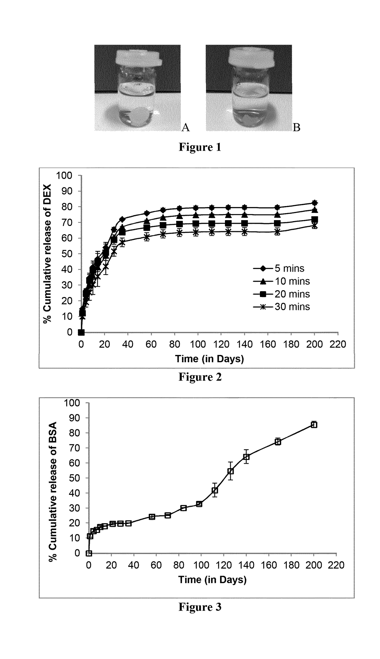
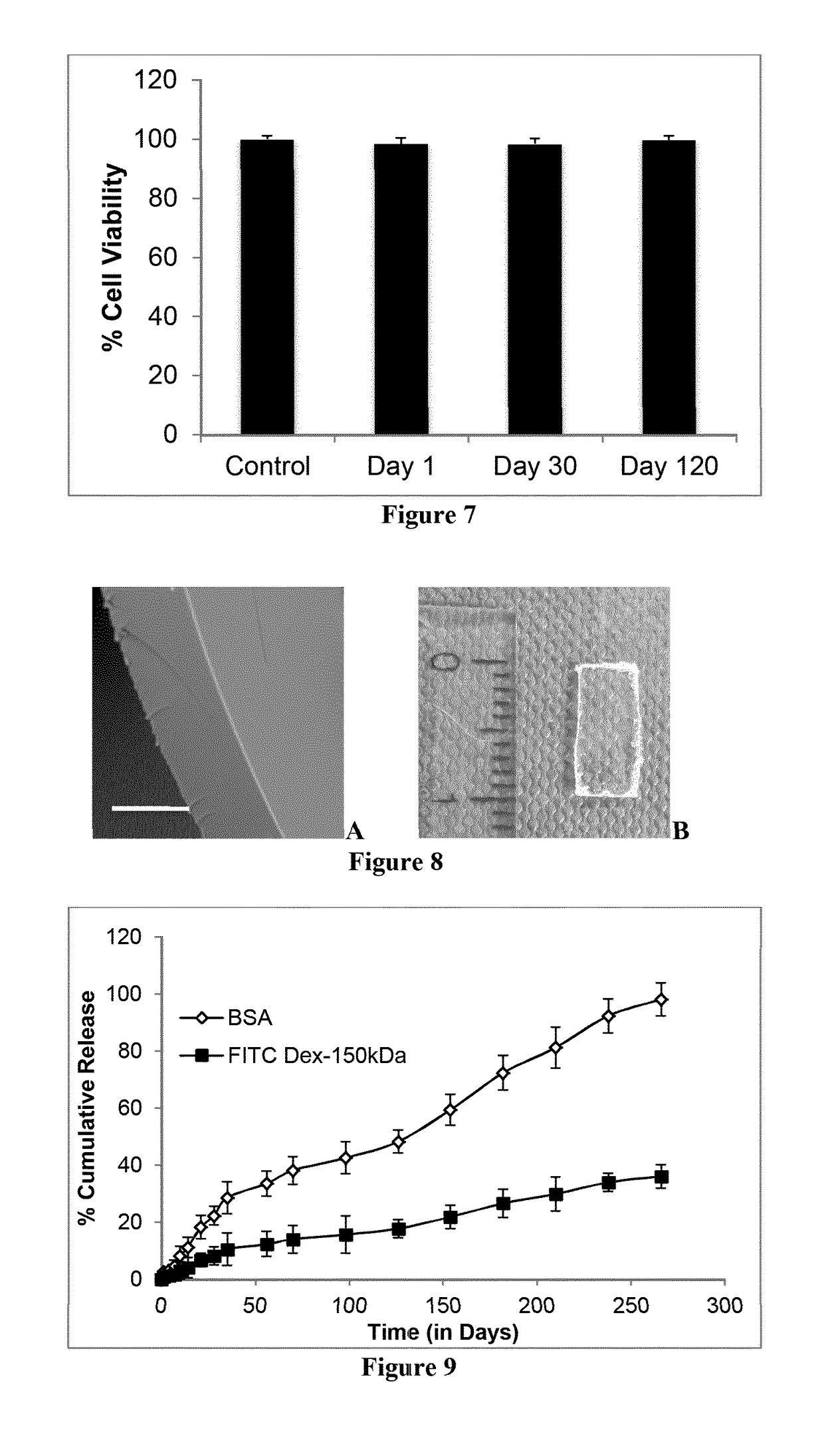
[0215]For preparation of ISPcl, the molecules under investigation (DEX, OVA, BSA, FITC-dextran 150 kDa and BVZ) were firstly added in selected PEGDA (Mw=700 Da) at a concentration of 0.5% w / w. Once the molecules were fully dissolved or suspended, the desired amount of PLGA 75 / 25 was then added to the molecule / PEGDA mixture and left to dissolve at room temperature to produce 30% w / w PLGA formulations. Prior to photocrosslinking predetermined amount of photoinitiator Irgacure® 2959 (2% w / v in 70% ethanol in water as stock solution) was added to the formulation and vortexed for predetermined time to ensure complete mixing.

example 2

, In Vitro Drug Release Study

[0216]For drug release studies selected ISPcl gel formulation were injected (approx. 0.2 g or 0.1 g) into desired amount of PBS (pH 7.3±0.2). Sink conditions were maintained for each drug type. ISPcl were formed by exposing them immediately to 365 nm using bench-top UV light (at 3.1±0.1 mW / cm2, CAMAG, Muttenz, Switzerland) for varying periods of time in PBS. The implants were stored in an incubator (37° C. and 60 rpm) for the duration of the release study. At predetermined time intervals the entire PBS medium was removed and replaced with equal amount of fresh PBS. All the experiments were carried out in triplicates. The concentration of released drug molecule in the PBS samples was analyzed as described below.

[0217]Analysis of DEX and TA samples were carried out using reversed-phase HPLC with UV detection (Agilent 1260 Infinity Quaternary System) using an Agilent Zorbax Eclipse Plus 250 mm C18 column (250 mm length, 4.6 mm internal diameter and 5 μm par...

example 3

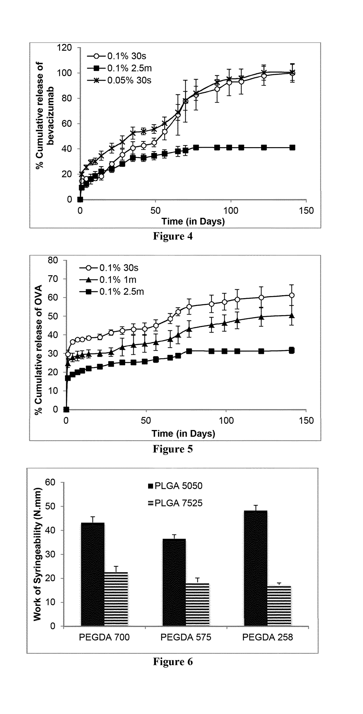
, Syringeability of ISPcl

[0221]Syringeability is a very important parameter in considering whether a formulation is suitable to be delivered via a syringe and needle, especially if the needle in question has a small bore, as would be required for ocular delivery. Therefore, Work of Syringeability (WoS) was investigated to determine the effort that would be required to expel the ISPcl gel formulations through 27G needle that is commonly used in intraocular injections. Briefly, 1 ml disposable medical syringes (Becton, Dickinson and Company, Oxford, UK) were filled with the ISPcl gel formulations to a constant height equivalent to 0.1 ml. Using the Texture Analyser (Stable Micro Systems, Surrey, UK), the content of the syringe was expelled at a rate of 0.5 mm / second. The area under the resultant force-distance plot was used to determine the WoS using the Exponent TA.XT software (Version 4.0). The WoS observed relating to expelling air from a blank syringe was subtracted from the exper...


PUM

PropertyMeasurementUnit
molecular weightaaaaaaaaaa
wavelengthaaaaaaaaaa

Abstract

The invention provides an ocular composition comprising: 99 to 60% (w/w) of a photopolymerizable composition selected from the group of fragments or monomers consisting of polyalkylene glycol diacrylate and polyalkylene glycol dimethacrylate, wherein the photopolymerizable composition has a molecular weight in the range of 100 to 20,000 Dalton; a biodegradable polymer selected from the group consisting of aliphatic polyester-based polyurethanes, polylactides, polycaprolactones, polyorthoesters and mixtures, copolymers, and block copolymers thereof; a photoinitiator; and a therapeutic agent. The composition can be used to form an ocular implant and an in situ ocular implant.

Description

BACKGROUND OF THE INVENTION[0001]Chronic retinal diseases are the leading contributor to visual impairment and blindness worldwide. Loss of sight has a major personal impact on people's daily lives and has a profound economic impact on individuals, families, support agencies, society and the state. The World Health Organization estimates that globally about 285 million people are visually impaired, of which 39 million are blind and 246 million have low vision. Diseases that originate in the posterior segment (PS) or back of the eye lead to permanent loss of vision if left untreated and account for the majority of blindness, such as in age-related macular degeneration (AMD), diabetic retinopathy (DR), diabetic macular edema (DME), cytomegalovirus (CMV) retinitis, retinitis pigmentosa, uveitis and glaucoma. The PS of the eye, which includes the retina, choroid, and vitreous body, is difficult to access due to the recessed location within the orbital cavity. Therefore, drug delivery to...

Claims


Application Information

Patent Timeline
Patent Type & Authority Applications(United States)
IPC IPC(8): A61K9/00A61K47/34A61K47/32A61K47/22A61K47/18A61K9/51C07K16/22A61N5/06
CPCA61K9/0048A61K9/0051A61K9/0024A61K47/34A61K47/32A61K47/22A61K47/183A61K9/5138A61K9/5146A61K9/5153A61K9/5123C07K16/22A61N5/062C07K2317/24C07K2317/76A61K2039/54A61N2005/0661A61K47/08A61K47/14A61K31/573A61K31/58A61K2039/541A61K38/00A61P27/02
Inventor THAKUR, RAGHU RAJ SINGHJONES, DAVIDGUJRAL, CHIRAG
Owner RE VANA THERAPEUTICS LTD
Why Eureka
Try Eureka