Method for diagnosing cardiomyopathies

a cardiomyopathy and cardiomyopathy technology, applied in the field of cardiomyopathy diagnosis, can solve the problems of difficult echo findings, substantial discrepancy in the predictive value of the proportion of treg lymphocytes circulating in blood in patients, and little research on t/sub>h/sub>,

Active Publication Date: 2018-12-13
FUNDACION CENT NACIONAL DE INVESTIGACIONES CARDIOVASCULARES CARLOS III CNIC
View PDF4 Cites 0 Cited by
  • Summary
  • Abstract
  • Description
  • Claims
  • Application Information

AI Technical Summary

Benefits of technology

The present invention provides an in vitro method for diagnosing cardiomyopathy (especially myocarditis) in humans using specific microRNAs (miR-721 and miR155) as indicators. This method can provide a differential diagnosis of cardiomyopathy and acute myocardial infarction, which is crucial for timely treatment and recovery. The method involves comparing the levels of these miRNAs with a reference value or a control indicator of absence of cardiomyopathy. Overall, this invention provides a reliable and accurate tool for the diagnosis and differential diagnosis of cardiomyopathy and myocarditis.

Problems solved by technology

However, little research has been conducted concerning TH17 cells circulating in the blood of patients with myocarditis.
However, there is a substantial discrepancy with respect to the predictive value of the proportion of Treg lymphocytes circulating in blood in patients with acute and chronic heart pathologies (Ammirati et al., 2010; Carvalheiro et al., 2012; Sardella et al., 2007).
The diagnosis of myocarditis made based on clinical, laboratory, ECG, and Echo findings is not always easy.
Moreover, a safe and sensitive non-invasive diagnostic test to confirm the diagnosis of acute myocarditis, compared to other pathologies with myocardial injury like acute coronary disease, is not available nowadays.
However, EMB is not commonly performed due to safety reasons; 6% of wide range complications and 0.4 incidence of death due to perforation.
This often makes these patients undergo coronary angiography and MRI with contrast to rule out ischemia, with consequent damage to the patient, delay in the election of immunosuppressive treatment of choice and high medical expenses associated with coronary angiography and MRI as the only reliable methods for distinguishing these Myocarditis from AMI.
Myocarditis is therefore a diagnostic challenge that does not have a solution in clinical practice.

Method used


Image

  • Method for diagnosing cardiomyopathies
  • Method for diagnosing cardiomyopathies
  • Method for diagnosing cardiomyopathies

Examples


example 1

iRNA-721 and Characterizing its Target Genes

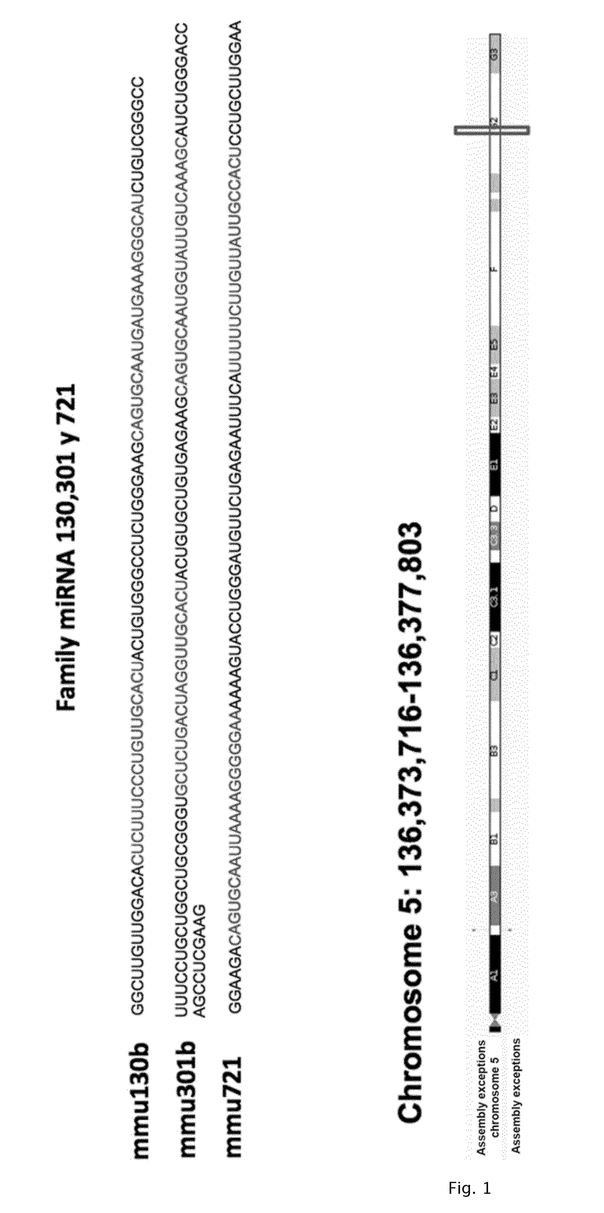
[0105]miRNA-721 was first described in a paper on mouse embryon brain tissue, in which the expression thereof during central nervous system embryogenesis was studied (Wheeler et al., FEBS Letters 2006). In this paper, miRNA was identified as miRY and was subsequently entered in the miR Base as mir-721. This miRNA is located in chromosome 5 within the non-coding region of CUX1 gene. miR-721 belongs to the family of miRNAs miR-130 / 301 / 721 (FIG. 1).

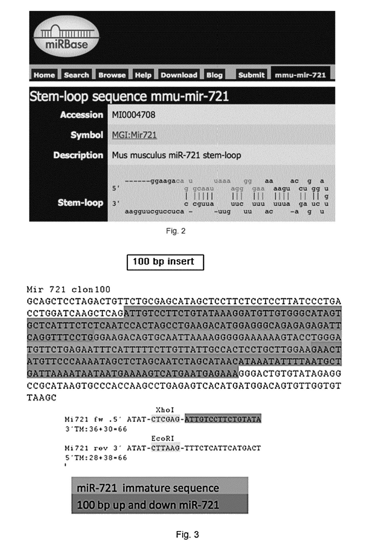
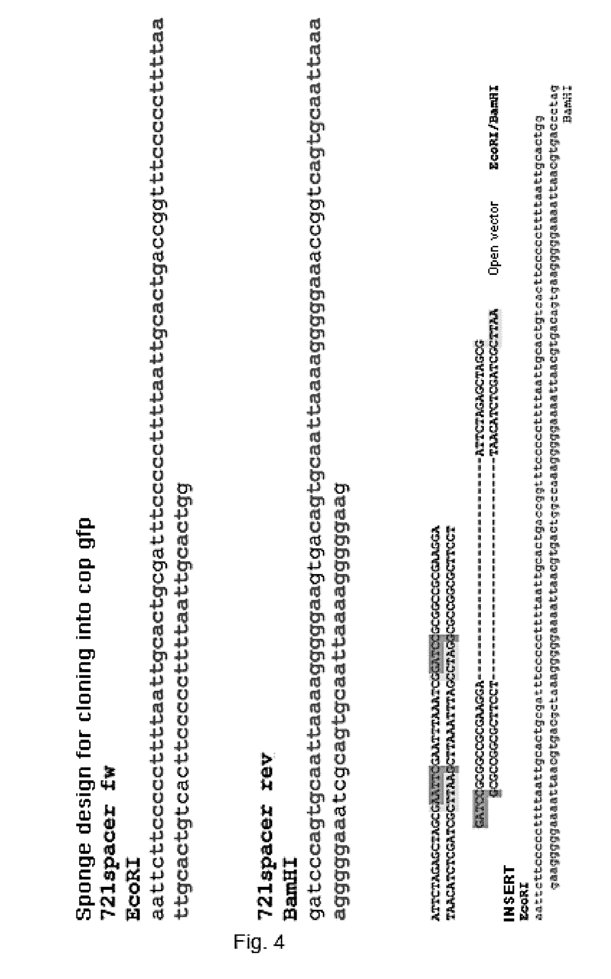
[0106]To study this microRNA in depth and its role in regulating inflammation and therefore in cardiovascular diseases, miRNA was cloned into expression vectors to conduct loss and gain of function assays, as well as for the functional validation of the possible target genes of this miRNA. Since the assays must essentially be conducted in primary cells, retroviral and lentiviral expression vectors have been tested, because they are the most efficient tools for gene expression in mammalian cells giv...

example 2

[0119]2.1. In Vivo Experiments for Validating the Role of miR-721 in the Th17 Cell Effector Function

[0120]Since the study of miR-721 function in the experimental autoimmune myocarditis (EAM) model requires very extensive experiments and working highly inaccessible tissue, the myocardium, a mouse lower limb muscle mouse ischemia model that is widely used in research to study the peripheral arterial disease (PAD) has been developed in the laboratory. To that end, the animals are subjected to a surgery explained next: a 5 mm incision is made in the inguinal area of the left leg allowing exposure of the femoral nerve, vein and artery. The connective tissue is carefully separated, and the vein, artery and nerve are dissected by means of separating the arterial adventitia layer close to the area distal to the femoral artery bifurcation. Once the three components are separated, the femoral artery is ligated by carefully passing thereunder a suture thread and making two surgical knots close...

example 3

MIR721 and MIR155 Expression in the Animal Model of Experimental Autoimmune Myocarditis

[0134]According to the results obtained, CD69-deficient naive T CD4+, T regulator and TH17 lymphocytes have changed the expression pattern of various microRNAs. The CD69 molecule limits the severity of cardiac dysfunction in the model of experimental autoimmune myocarditis (EAM) by means of controlling TH17 responses. To that end, this model is used to investigate the role of the previously mentioned miRNAs in this disease. The murine model of EAM is induced by means of immunization with a peptide derived from the α-myosin heavy chain (MyHCα) specific for heart tissue emulsified with CFA (Complete Freund's Adjuvant), which causes effector TH17 cell activation and differentiation. 21 days after immunization, the mice are in the acute phase of the disease, presenting inflammation in the myocardium and anti-cardiac myosin antibodies in serum. The chronic phase appears after 56 days, characterized by ...


PUM

PropertyMeasurementUnit
diameteraaaaaaaaaa
temperatureaaaaaaaaaa
MRIaaaaaaaaaa

Abstract

The present invention provides an in vitro method for obtaining data useful for diagnosing a cardiomyopathy in a human subject, wherein the method comprises using, as an indicator, the expression levels, obtained from a biological sample isolated from the human subject, preferably a plasma sample, of one or more of the following miRNAs miR-721 and / or miR-155); and obtaining a result of the method by comparing the expression levels of said one or more miRNAs with a reference value or with the expression levels of a control, indicator of the absence of cardiomyopathy.

Description

BACKGROUND OF THE INVENTION[0001]Inflammation of the myocardium or myocarditis is a disease with heterogeneous etiology, oftenly caused by an infectious pathogen (bacteria, virus, fungi or protozoa) (Andreoletti et al., 2009) or by an autoimmune disorder (Frustaci et al., 2009), which can result in dilated cardiomyopathy (Felker et al., 2000). T CD4+ cells are necessary and sufficient for the development of myocarditis (Smith and Allen, 1991; Smith and Allen, 1993). Specifically, TH17 lymphocytes (Sonderegger et al., 2006) and IL-23 cytokine are critical for the development of the pathology, being TH17 cells more immunopathogenic than TH1 cells when they are exogenously transferred (Rangachari et al., 2006). Furthermore, suppression of TH17 cell differentiation through the blocking of the IL-6 signaling pathway prevents the development of EAM (Experimental Autoimmune Myocarditis) (Sonderegger et al., 2008; Yamashita et al., 2011). With respect to IL-17, it has been observed that thi...

Claims


Application Information

Patent Timeline
Patent Type & Authority Applications(United States)
IPC IPC(8): C12Q1/6876
CPCC12Q1/6876C12Q2600/158C12Q2600/178C12Q2525/207C12Q2600/118C12Q1/6883
Inventor MART N FERN NDEZ, MARIA PILARS NCHEZ D AZ, RAQUELMATESANZ MAR N, ADELAJIMENEZ BORREGUERO, LUIS JES SS NCHEZ MADRID, FRANCISCO
Owner FUNDACION CENT NACIONAL DE INVESTIGACIONES CARDIOVASCULARES CARLOS III CNIC
Why Eureka
Try Eureka