Method for separating cell from biological tissue

a biological tissue and cell technology, applied in cell dissociation methods, instruments, artificial cell constructs, etc., can solve the problems of difficult to identify the specific type of collagen and difficult to produce antibodies, and achieve the effect of high biological activity and efficient and stable cell separation

Active Publication Date: 2018-12-20
TOHOKU UNIV +2
View PDF2 Cites 0 Cited by
  • Summary
  • Abstract
  • Description
  • Claims
  • Application Information

AI Technical Summary

Benefits of technology

[0045]Owing to the present invention, a method for efficiently and stably separating cells having a high biological activity from a biological tissue is provided.

Problems solved by technology

However, since many types of collagens are present and collagens are present in a wide variety of multicellular animals, it is difficult to produce antibodies.
Because of these problems, it is difficult to realize identification of the specific type of collagen.

Method used


Image

  • Method for separating cell from biological tissue
  • Method for separating cell from biological tissue
  • Method for separating cell from biological tissue

Examples


reference example 1

ajor Protein-Degrading Enzyme to be Used in Separating Cells or the Like from Biological Tissue

[0070][Identification of Enzyme for Use in Separating Pancreatic Islets from Rat's Pancreas Tissue]

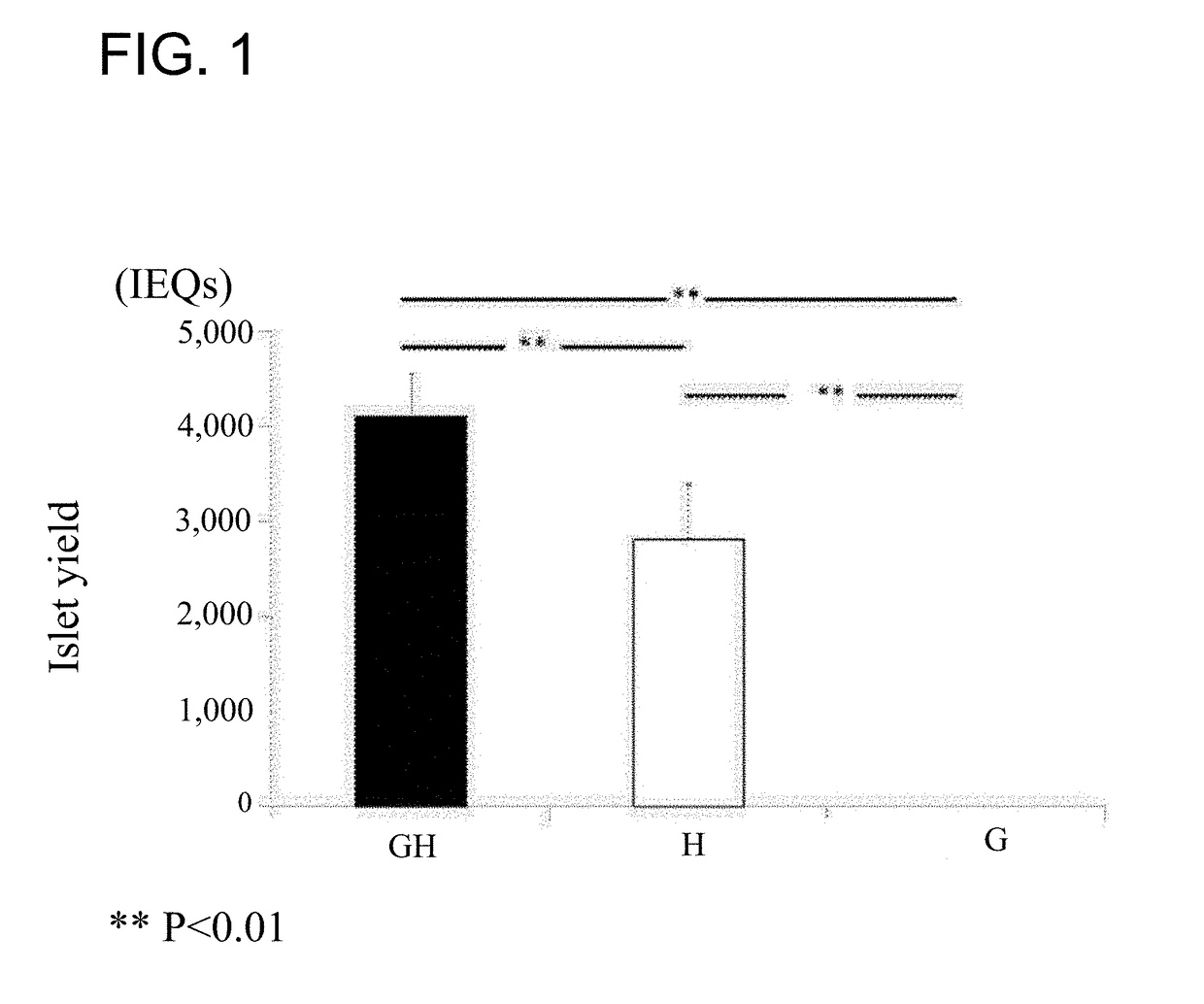
[0071]A male Lewis rat (10 to 13 weeks old) was used. Before the pancreas was excised out, the duodenum was clamped. A degrading-enzyme composition (10 mL, cold Hank's Balanced Salt Solution (HBSS)), which contains at least one of a protein-degrading enzyme: recombinant collagenase G (8.4 mg) and collagenase H (2.9 mg), and a neutral protease: thermolysin (0.3 mg), was injected through the bile duct. After a treatment was performed at 37° C. for 14 minutes, density gradient centrifugation was performed to obtain a fraction of pancreatic islets.

[0072]The case (GH group) where thermolysin, collagenase G and collagenase H were simultaneously added, the case (G group) where thermolysin and collagenase G alone were added, and the case (H group) where thermolysin and collagenase H alone were added,...

reference example 2

ubstrate for Major Protein-Degrading Enzyme

[Identification of Substrate for Collagenase H]

[0076]The pancreas tissue slices (100 mg) of a Lewis rat were treated with 20 mM HEPES (pH8.0) containing a protease inhibitor cocktail (Roche) and 1 mM CaCl2 at 37° C. overnight. Thereafter, the tissue slices were washed with a buffer and subjected to enzyme degradation with a buffer containing collagenase H (0.1 mg / mL) for 10 hours at 37° C.

[0077]After the enzyme degradation, incubation was performed in 50% acetonitrile containing 100 mM TMPP (Sigma-Aldrich) (10 μL) for 30 minutes. Then, acetone precipitation was performed with cold acetone and thereafter centrifugation was performed. The resultant precipitate was dried and digested with trypsin (10 μL / mg) in a 100 mM ammonium carbonate solution, overnight.

[0078]After the digest was treated by ZipTip (Millipore), the resultant peptides were eluted by concentration gradient with 2.5-40% acetonitrile in 10% formic acid and subjected to mass ana...

example 1

n of Cells Depending Upon the Composition of Major Protein of Biological Tissue

[Quantification of Collagen III and Collagen I in Pancreas Tissue and Liver Tissue of Rat]

[0081]Tissue slices of a Lewis rat (10 to 13 weeks old) were prepared. Enzymatic immunoassay using labeled antibodies (Chemicon Merck Millipore) against collagen III and collagen I was performed to compare expression of collagen III and collagen I in the pancreas tissue and liver tissue. As a result, the number of positive staining obtained by immunohistostaining of collagen III and collagen I in the liver tissue was significantly lower than that in the pancreas tissue.

[Separation of Cells from Pancreas Tissue and Liver Tissue of Rat]

[0082]It was predicted that the major protein of a liver tissue may be a protein other than collagen III and collagen I, and thus the amount of collagenase H in the degrading-enzyme composition for liver cell separation may be low compared to for pancreatic islet separation.

[0083]A liver...


PUM

PropertyMeasurementUnit
weight ratioaaaaaaaaaa
timeaaaaaaaaaa
elasticaaaaaaaaaa

Abstract

Provided is a method for efficiently and stably separating cells having a high biological activity from a biological tissue, by using a degrading-enzyme composition, which is prepared by adding an enzyme for degrading a major protein of the biological tissue in an amount determined depending on the composition of the major protein to a predetermined amount of a neutral protease and / or a protease derived from Clostridium sp. According to this method, the type and amount of protein-degrading enzyme to be used for isolating cells can be determined from the composition of a major protein of the biological tissue. Thus, cells having a high biological activity can be efficiently separated while reducing the amount of protein-degrading enzyme to be used.

Description

CROSS-REFERENCE TO RELATED APPLICATIONS[0001]This application is a continuation of U.S. application Ser. No. 14 / 765,020, filed Jul. 31, 2015, which is a national stage application of International Appl. PCT / JP2014 / 000527, filed Jan. 31, 2014, and which claims priority of Japanese Patent Appl. No. 2013-018774, filed Feb. 1, 2013, all of which are hereby incorporated by reference.TECHNICAL FIELD[0002]The present invention relates to a method for separating cells from a biological tissue, and particularly to a method for efficiently and stably separating cells having a high biological activity by controlling the amount of protein-degrading enzyme to be used.BACKGROUND ART[0003]Enzymatic isolation of cells or cell aggregates (cell clusters), which are present in various organs and tissues such as liver, pancreas, kidney, periodontal tissue, skin, cartilage, bone and nervous tissue of multicellular animals such as mammals, birds, reptiles and fish as well as in an ES cell tissue and a fi...

Claims


Application Information

Patent Timeline
Patent Type & Authority Applications(United States)
IPC IPC(8): C12N5/071C12N5/00
CPCC12N2509/00C12N5/0676C12N5/00C12N5/067C12N9/50C12N9/52C12Y304/24007G01N33/56966
Inventor GOTO, MASAFUMIMURAYAMA, KAZUTAKAYAMAGATA, YOUHEIWATANABE, KIMIKO
Owner TOHOKU UNIV
Why Eureka
Try Eureka