
Devices, processes, and systems for determination of nucleic acid sequence, expression, copy number, or methylation changes using combined nuclease, ligase, polymerase, and sequencing reactions

a technology of nucleic acid sequence and process, applied in the field of devices, processes, and systems for determining nucleic acid sequence, expression, copy number, or methylation changes using combined nuclease, ligase, polymerase, sequencing reactions, can solve the problems of not being able to meet current sequencing technology, give the best treatment, and technical challenges, so as to save $300 billion in annual healthcare costs, accurate identification and enumeration of targets

Active Publication Date: 2020-02-06
CORNELL UNIVERSITY
View PDF0 Cites 4 Cited by
  • Summary
  • Abstract
  • Description
  • Claims
  • Application Information

AI Technical Summary

Benefits of technology

This patent describes a method to detect cancer at an early stage, which can save lives and reduce healthcare costs. The method can also monitor the effectiveness of treatments and detect the recurrence of cancer. Infectious disease testing is also described, which can provide accurate results for multiple targets simultaneously. The patent also mentions that the method can be used for non-invasive prenatal testing to identify chromosomal copy anomalies.

Problems solved by technology

Currently, patients with predisposing or early disease are not identified, and those with disease are not given the best treatment—all because of failures at the diagnostic level.
There are several challenges for developing reliable diagnostic and screening tests.
The first challenge is to distinguish those markers emanating from the tumor or fetus that are indicative of disease (i.e. early cancer) vs. presence of the same markers emanating from normal tissue.
This is a challenge that needs to address the biological variation in diseases such as cancer.
Finally, there is the technical challenge to distinguish true signal resulting from detecting the desired disease-specific nucleic acid differences vs. false signal generated from normal nucleic acids present in the sample vs. false signal generated in the absence of the disease-specific nucleic acid differences.
This would require sequencing 300,000 GB of DNA, a task beyond the reach of current sequencing technology, not to mention the enormous data-management issues.
Sequence capture has suffered from dropout, such that maybe 90-95% of the desired sequences are captured, but desired fragments are missing.
Alternatively, PCR amplification provides the risk of introducing a rare error that is indistinguishable from a true mutation.
Further, PCR loses methylation information.
While bisulfite treatment has been traditionally used to determine the presence of promoter methylation, it is also destructive of the DNA sample and lacks the ability to identify multiple methylation changes in cell-free DNA.
A consensus accuracy may be achieved in the presence of high error rates by sequencing the same region of DNA 30 to 100 times. However, a high error rate makes it extremely difficult to identify a sequence variant in low abundance, for example when trying to identify a cancer mutation in the presence of normal DNA.
Therefore, a low error rate is required to detect a mutation in relatively low abundance.
The disadvantage is that this approach is unable to distinguish a true mutation from a PCR error in the early rounds of amplification.
Thus, while the sensitivity of 2% (i.e. detecting one mutant allele in 50 wt alleles) is sufficient for evaluating late-stage cancers prior to making a treatment decision, it is not sensitive enough for early detection.
These methods require extending across the target DNA, and, thus, it would be impossible to distinguish true mutation, from polymerase generated error, especially when copying across a damaged base, such as deaminated cytosine.
Finally, these methods do not provide information on methylation status of CpG sites within the fragment.
Mutations present in only one or a few family members represent sequencing mistakes or PCR-introduced errors occurring late in amplification.
Mutations occurring in many or all members of one family in a pair arise from PCR errors during the first round of amplification such as might occur when copying across sites of mutagenic DNA damage.
The disadvantage of this approach is that many fragments need to be sequenced to obtain at least five members of each strand in a family pair (i.e. minimum of 10 sequence reads per original fragment, but often requiring far more due to fluctuations).
Further, the method has not been tested on cfDNA, which tend to be smaller than fragments generated from intact genomic DNA, and thus would require sequencing more fragments to cover all potential mutations.
Finally, the method does not provide information on methylation status of CpG sites within the fragment.
The disadvantage of this approach is that many fragments need to be sequenced to obtain at least five members of each strand in a family pair (i.e. minimum of 10 sequence reads per original fragment, but often requiring far more due to fluctuations).
Also, the method requires extending across the target DNA, and thus it would be impossible to distinguish true mutation, from polymerase-generated error, especially when copying across a damaged base, such as deaminated cytosine.
Further, the method has not been tested on cfDNA, which tend to be smaller than fragments generated from intact genomic DNA, and thus would require sequencing more fragments to cover all potential mutations.
Finally, the method does not provide information on methylation status of CpG sites within the fragment.
However, the method does not provide the ability to determine copy number changes, nor provide information on methylation status of CpG sites within the fragment.
Ligation reactions can be inefficient, which creates the potential of lower yields when mutational sample input is limited.
Further, this approach does not select for specific targets.
Although the technique is designed to be highly efficient in capturing just the desired fragments, it is also a lengthy, multi-step procedure, with the potential of lower yields when mutational sample input is limited.
Although it may also be adapted to identify low-abundant mutations, the multi-wash procedure creates the potential of lower yields when mutational sample input is limited.
This approach is very rapid and robust; however, it does require the extra step of physically removing initial gene-specific primers, as well as a cleanup / size selection after the second PCR step (presumably to eliminate primer dimers), and it is unclear if this procedure creates the potential of lower yields when mutational sample input is limited.

Method used


Image

  • Devices, processes, and systems for determination of nucleic acid sequence, expression, copy number, or methylation changes using combined nuclease, ligase, polymerase, and sequencing reactions
  • Devices, processes, and systems for determination of nucleic acid sequence, expression, copy number, or methylation changes using combined nuclease, ligase, polymerase, and sequencing reactions
  • Devices, processes, and systems for determination of nucleic acid sequence, expression, copy number, or methylation changes using combined nuclease, ligase, polymerase, and sequencing reactions

Examples


examples

Prophetic Example 1—Use of PCR-PCR-Taqman™ or PCR-PCR Unitaq Detection for Unknown Pathogen Identification and Quantification

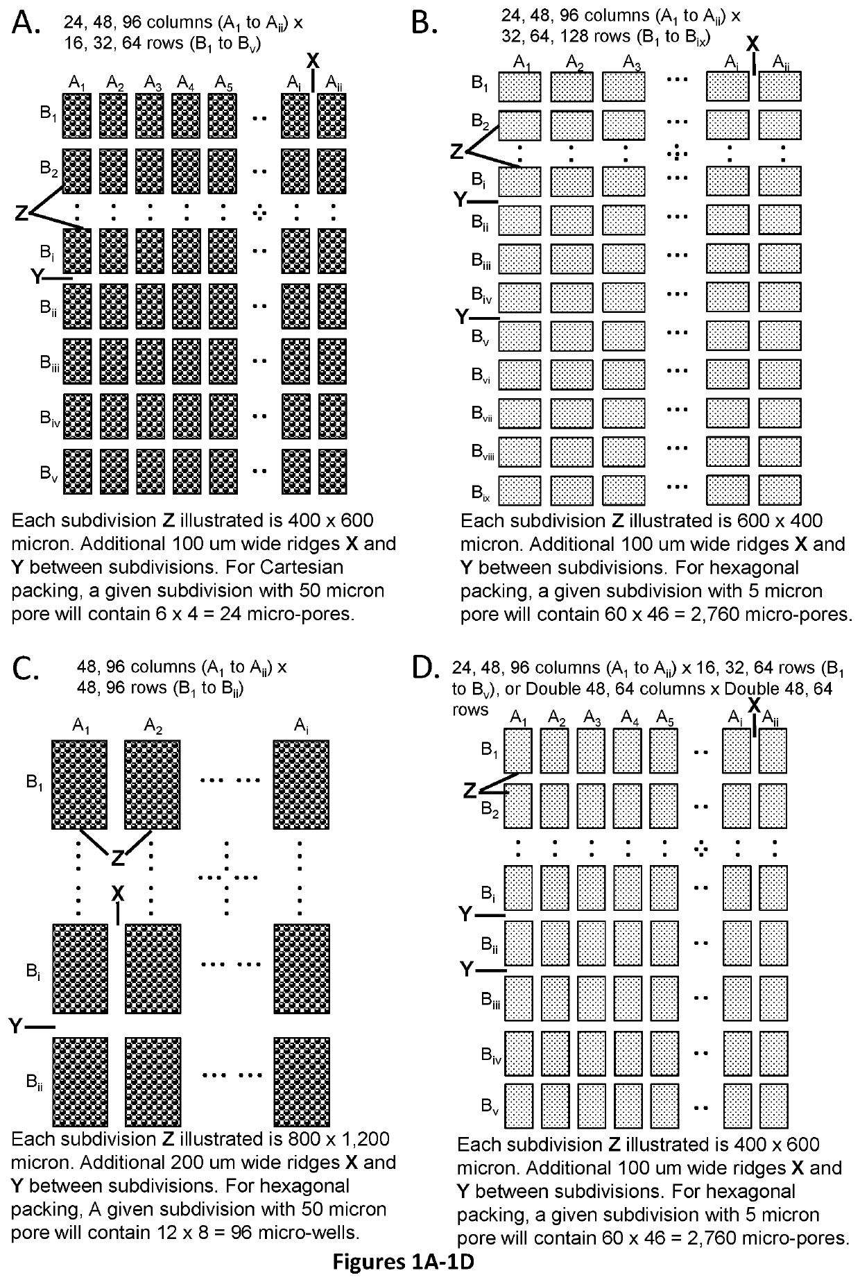
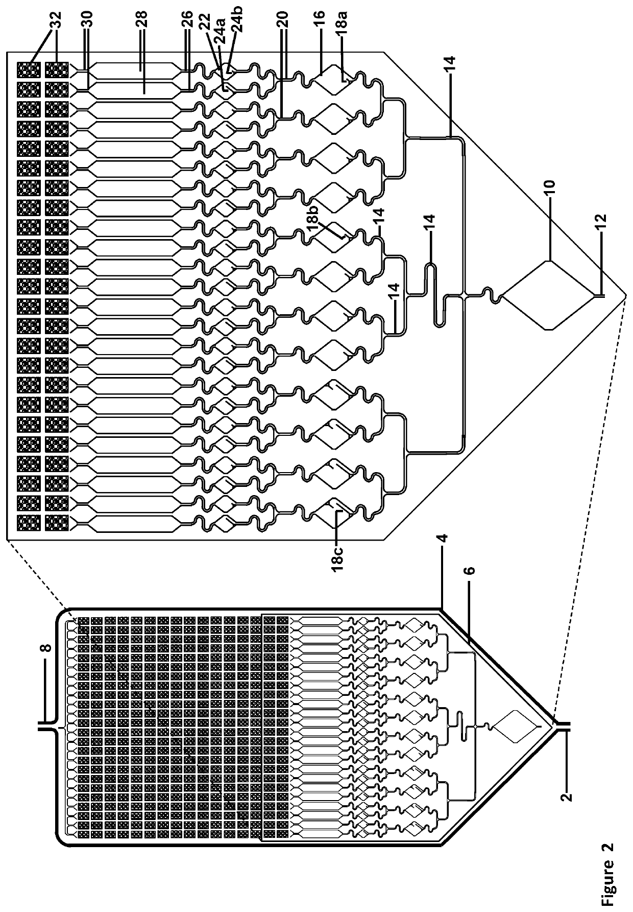
[0307]The assay described below would use a cartridge with 24×16=384 (or optional 768) subdivisions for 9,216 micro-well or micro-pore array format, with 24 micro-wells or micro-pores per subdivision, and 384 micro-wells or micro-pores per column (using pre-spotted array): Please see FIGS. 16, 17, 18, and 24.

[0308]The assay may be designed to detect and quantify 384, 768, or 1,536 potential targets. Preparation of the cartridge would require spotting 24× of either 16, 32, or 64 nested PCR primer pairs on the front side of the array, with adding UniTaq or Universal Tag primer and target-specific probe sets at right angles and drying them down before cartridge assembly.

[0309]1. Initial multiplexed amplification of the sample—384, 768, or 1536 potential targets. Perform 9 cycles of multiplexed PCR in the Initial Reaction chamber, yielding a maximum of 512 copies ...

##ic example 21

Prophetic Example 21—Example of LDR Primer Design with Separate Split UniTaq

Probe (UniSpTq)

[0510]Example of LDR Primer Design with Separate Split UniTaq Probe:

[0511](Tm=65.2) (150 bp total; 60 bp TS DNA)

Upstream LDR primer sequence: (Ai-Bi′-zi-TS) 5′-TCAGTATCGGCGTAGTCACCCGAGTTTCCTTG-A-TCACTTTCGGAC(SEQ ID NO: 46) (Upstream-Target-Sequence; 30 bp)-ribose base-first 4 downstream bases-3′ Block Downstream LDR primer sequence: (TS-zi′-Bj′-Ci′) 5′-(Downstream-Target-Sequence; 30 bp) GTCCGAAAGTGA-A-GCTGACTGTGAGGTGCGGAAACCTATCGTCGA-3′ (SEQ ID NO: 47) 1st UniTaq Primer: (Ai) 5′-TCAGTATCGGCGTAGTCACC-3′ (SEQ ID NO: 48) 2nd UniTaq primer: Ci 5′-TCGACGATAGGTTTCCGCAC-3′ (SEQ ID NO: 49) UniTaq Probe: (F1-Bj,Bi-Q) 5′-F1-CTCACAGTCAGC-A-CAAGGAAACTCG-Q-3′ (SEQ ID NO: 50) Full length PCR product: 5′-TCAGTATCGGCGTAGTCACCCGAGTTTCCTTG-A-TCACTTTCGGAC(SEQ ID NO: 51) (Upstream-Target-Sequence; 30 bp) (Downstream-Target-Sequence; 30 bp) GTCCGAAAGTGA-A-GCTGACTGTGAGGTGCGGAAACCTATCGTCGA-3′ (SEQ ID NO: 52)Notes b...


PUM

PropertyMeasurementUnit
thickaaaaaaaaaa
thickaaaaaaaaaa
diameteraaaaaaaaaa

Abstract

The present invention relates to methods, devices, instruments, processes, and systems for the highly specific, targeted molecular analysis of regions of human genomes and transcriptomes from the blood, i.e. from cell free circulating DNA, exosomes, microRNA, IncRNA, circulating tumor cells, or total blood cells. The technology enables highly sensitive identification and enumeration of mutation, expression, copy number, translocation, alternative splicing, and methylation changes using spatial multiplexing and combined nuclease, ligation, polymerase, and sequencing reactions. Such technology may be used for non-invasive early detection of cancer, non-invasive cancer prognosis, and monitoring both treatment efficacy and disease recurrence of cancer.

Description

[0001]This application claims the priority benefit of U.S. Provisional Patent Application Ser. No. 62 / 478,412, filed Mar. 29, 2017, which is hereby incorporated by reference in its entirety.FIELD OF THE INVENTION[0002]The present invention relates to devices, processes, and systems for determination of nucleic acid sequence, expression, copy number, or methylation changes using combined nuclease, ligation, polymerase, and sequencing reactions.BACKGROUND OF THE INVENTION[0003]Advances in DNA sequencing hold the promise to standardize and develop non-invasive molecular diagnosis to improve prenatal care, transplantation efficacy, cancer and other disease detection and individualized treatment. Currently, patients with predisposing or early disease are not identified, and those with disease are not given the best treatment—all because of failures at the diagnostic level.[0004]In the cancer field, there is a need to develop such technology for early detection, guiding therapy, and monit...

Claims


Application Information

Patent Timeline
Patent Type & Authority Applications(United States)
IPC IPC(8): B01L3/00C12Q1/6869
CPCB01L2300/0864B01L2300/087B01L2400/0622B01L3/502761C12Q1/6869B01L2200/16B01L2200/0668B01L2300/0819B01L2300/165B01L2200/027B01L2300/1805B01L3/50851B01L7/52B01L2300/0681B01L2300/0829B01L2300/0867B01L2300/161B01L3/527B01L2200/0605B01L2200/0642B01L2200/0673B01F33/3017B01F33/813
Inventor BARANY, FRANCIS
Owner CORNELL UNIVERSITY
Learn More