
Bioprinter devices, systems and methods for printing soft gels for the treatment of musculoskeletal and skin disorders

Pending Publication Date: 2020-04-23
NUTECH VENTURES
View PDF0 Cites 10 Cited by
  • Summary
  • Abstract
  • Description
  • Claims
  • Application Information

AI Technical Summary

Benefits of technology

The patent describes a new bioprinter device that can print adhesive scaffolds in real-time at the site of a traumatic injury. This device is handheld and can be used in any setting without the need for an expensive imaging modality. The printed scaffolds adhere to the surrounding tissue, eliminating the risk of slippage. The device is easy to use and can be controlled remotely, making it ideal for treating skin disorders. The invention solves challenges associated with the treatment of VML injuries and offers a solution for eliminating the need for additional surgeries.

Problems solved by technology

Skeletal muscle damage, which can be caused by varied etiologies including crush injuries, burns, and tumors, are a leading cause of loss of independence, disability, and mortality (50).
Regenerative medicine approaches such as growth factor and stem cell delivery have shown some promise, but the majority of these approaches have been limited by low cell engraftment and factor bioavailability (51).
Despite the impressive level of the structural details achievable with 3D bioprinters, the implantation of hydrogel-based constructs has remained a major rate-limiting step.
Moreover, conventional 3D bioprinters are slow in responding to the urgent clinical needs.
For example, in the case of a traumatic injury, it takes several hours to take 3D images from the injury site and reconstruct the SDL file.
The printing process on its own takes several hours.
VML is seen in traumatic injuries or other underlying conditions necessitating surgical intervention and results in an en bloc loss of skeletal muscle with corollary functional impairment (1).
VML is often a result of traumatic extremity injury and results in major social tribulations and economic hardship.
VML injuries, however, are characterized by substantial soft tissue destruction, incomplete tissue regeneration, and extensive fibrosis; all of which limit the recovery of extremity function (3).
Although conventional reconstructive treatments for these injuries can prevent amputation, they are limited in their ability to restore muscle strength and movement.
Muscle tissue transplantation is the current standard clinical VML treatment, but is limited by the availability, functional and wound complications at the donor site, incomplete functional restoration, and high failure rates (1, 2, 4-10).
Additionally, muscle flap transfer is a technically difficult operation and requires a highly skilled surgical team.
Thereby, this approach is principally limited to tertiary care centers (4).
These complications can result in the need for further surgical procedures at the wound site (2).
Although the complications of surgical procedures are avoided, prosthetics must be custom made for each patient, which is expensive (4).
Braces also fail to restore the native structure and function of the muscle and do not work for all types of VML (4, 6).
Due to a lack of definitive treatment, VML often leads to permanent disability and pain, despite multiple interventions (1, 11).
Regenerative medicine approaches such as growth factor and stem cell delivery have shown some promise, but the majority of these approaches have been limited by low cell engraftment or factor bioavailability (1).
Despite the impressive level of structural detail achievable with 3D bioprinters, the implantation of hydrogel-based constructs has remained a major rate-limiting step.
Moreover, conventional 3D bioprinting methods are slow in responding to urgent clinical needs.
Furthermore, the duration of the printing process is highly dependent on the printing method and can take several hours.
Despite the excellent resolution of this bioprinter, it was not feasibly portable for use in remote settings.
Furthermore, a technology that can adequately address the limitations associated with skeletal muscle defects has not been developed.

Method used


Image

  • Bioprinter devices, systems and methods for printing soft gels for the treatment of musculoskeletal and skin disorders
  • Bioprinter devices, systems and methods for printing soft gels for the treatment of musculoskeletal and skin disorders
  • Bioprinter devices, systems and methods for printing soft gels for the treatment of musculoskeletal and skin disorders

Examples


example 1

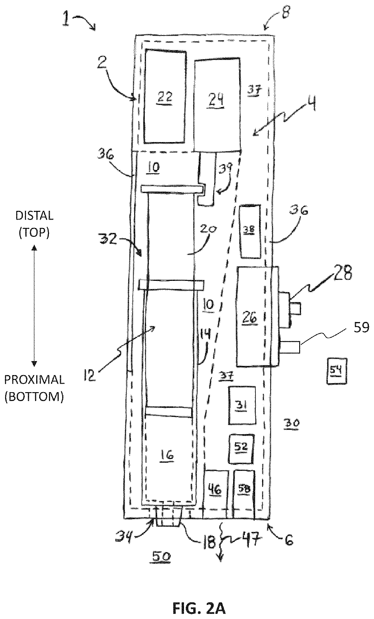
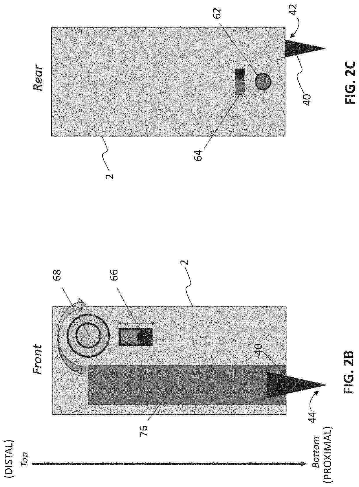
[0070]In an embodiment a pen printer is provided. The pen printer is a device that can be used to print polymer hydrogels for in situ application, and can be easily operated using simple mechanical controls to extrude gels at a consistent rate. This flow can be customized to the desired flow rate. The gels are first loaded into a syringe and placed into the pen printer, where it is then extruded using electrical actuators controlled by a stepper motor. The stepper motor slowly pushes the back of the syringe towards the front, pushing the gels out. Because the gels are loaded into a standard Luer-lock syringe, the nozzle of the syringe can be optimized to the gel being used, using gage syringe tips or tapered tips. While this application can be used across a wide range of hydrogels, current hydrogels may include chitosan / alginate and GelMA-based hydrogels.

[0071]Chitosan / alginate gels are being researched for their structural and antimicrobial properties in skeletal muscle tissue appl...

example 2

[0079]Current strategies for the treatment of VML injuries require acute stabilization of the patient, followed by delayed extremity reconstruction. Reconstructive operations include the implantation of autologous or allogeneic tissues. (13) Tissue engineering strategies, which have not yet been utilized in clinical practice, would also require a second surgical procedure to be implanted. However, an inflammatory reaction and loss of myogenesis factors following VML limit the regenerative capacity of the injured skeletal muscle. Without rapid intervention, the healing response of the body results in fibrosis rather than functional restoration of composite muscle tissue. (3) A robust strategy for VML treatment was created that can be utilized quickly, without the need for access to sophisticated surgical tools. FIGS. 9A-9D illustrate the utilization of a handheld bioprinter for in situ printing of scaffolds. FIG. 9A is a schematic diagram of the in situ bioprinting of cell-laden GelM...

example 3

[0082]The physical and mechanical properties of GelMA can be changed by varying the degree of methacrylation, prepolymer concentration, temperature, photoinitiator (PI) concentration, and the light exposure time and intensity. (37) In the disclosed handheld partially automated bioprinting system, each of the bioink input variables was selected to match the necessary biological and mechanical properties of native skeletal muscle. Following established literature, the use of medium level methacrylate GelMA is mechanically and biologically suitable for muscle tissue engineering applications. (35) The lithium phenyl-2, 4, 6-trimethylbenzoylphosphinate (LAP) was used as the PI, and its concentration was selected to be 0.067% (w / v) following the previous literature. (37) The exposure time selected for printed scaffolds was 20 seconds (the estimated time that printed hydrogels are directly exposed to light). The light wavelength was 365 nm with a flux of 120 lumens at an emitted distance o...


PUM


Abstract

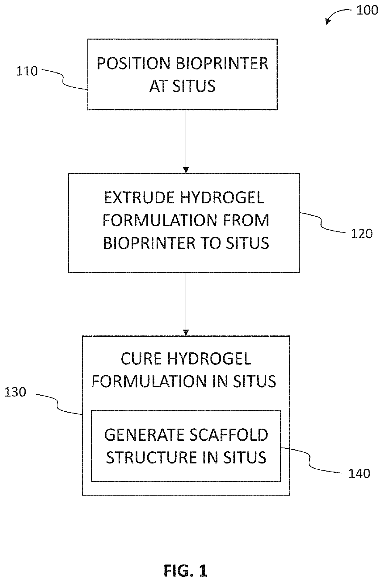
Disclosed are devices, systems and methods that utilize bioprinters to form scaffold structures in situ to facilitate treatment of the musculoskeletal or skin disorders in patients. A method for treating a musculoskeletal disorder of a patient includes positioning a bioprinter at a situs of the musculoskeletal disorder, or a situs of a skin injury, in the patient. The method includes extruding a hydrogel formulation from the bioprinter into the situs. The method includes curing the hydrogel formulation in the situs. The hydrogel formulation has a composition effective to generate, upon the curing, a scaffold structure in the situs to facilitate muscle hypertrophy, or skin wound healing, in the situs.

Description

CROSS-REFERENCE TO RELATED APPLICATIONS[0001]This application claims the benefit of U.S. Provisional Application No. 62 / 748,124, filed Oct. 19, 2018, which is incorporated herein by reference in its entirety.STATEMENT AS TO FEDERALLY SPONSORED RESEARCH[0002]This invention was made with government support under GM126831 and AR073822 awarded by the National Institutes of Health. The government has certain rights in the invention.TECHNICAL FIELD[0003]The present disclosure relates to medical devices for treatment of musculoskeletal and skin disorders, and more particularly to devices, systems and methods that utilize bioprinters to form scaffold structures in situ to facilitate treatment of the musculoskeletal and skin disorders in patients.BACKGROUND[0004]Skeletal muscle damage, which can be caused by varied etiologies including crush injuries, burns, and tumors, are a leading cause of loss of independence, disability, and mortality (50). Volumetric muscle loss (VML), specifically, is...

Claims


Application Information

Patent Timeline
IPC IPC(8): C12M3/00C12N5/00C12M1/26
CPCB33Y30/00C12M33/00B33Y80/00C12N5/0062B33Y70/00C12M21/08B33Y10/00B33Y50/02A61M5/178A61F2/0063
Inventor TAMAYOL, ALIMOSTAFAVI, AZADEHRUSSELL, CARINAWILLIAMS, TYRELLSINHA, INDRANILQUINT, JACOB
Owner NUTECH VENTURES
Learn More