Improvements in or relating to cell analysis

a cell analysis and cell technology, applied in the field of cell analysis, can solve the problems of limiting the number of cells that are analysed, low method sensitivity, labour intensive, etc., and achieve the effect of high throughput and high sensitivity

Pending Publication Date: 2020-07-23
UNIV OF WESTERN AUSTRALIA
View PDF0 Cites 3 Cited by
  • Summary
  • Abstract
  • Description
  • Claims
  • Application Information

AI Technical Summary

Benefits of technology

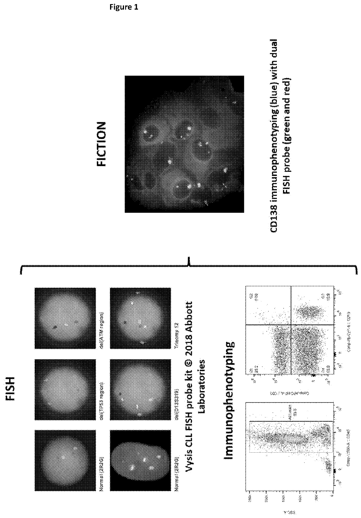
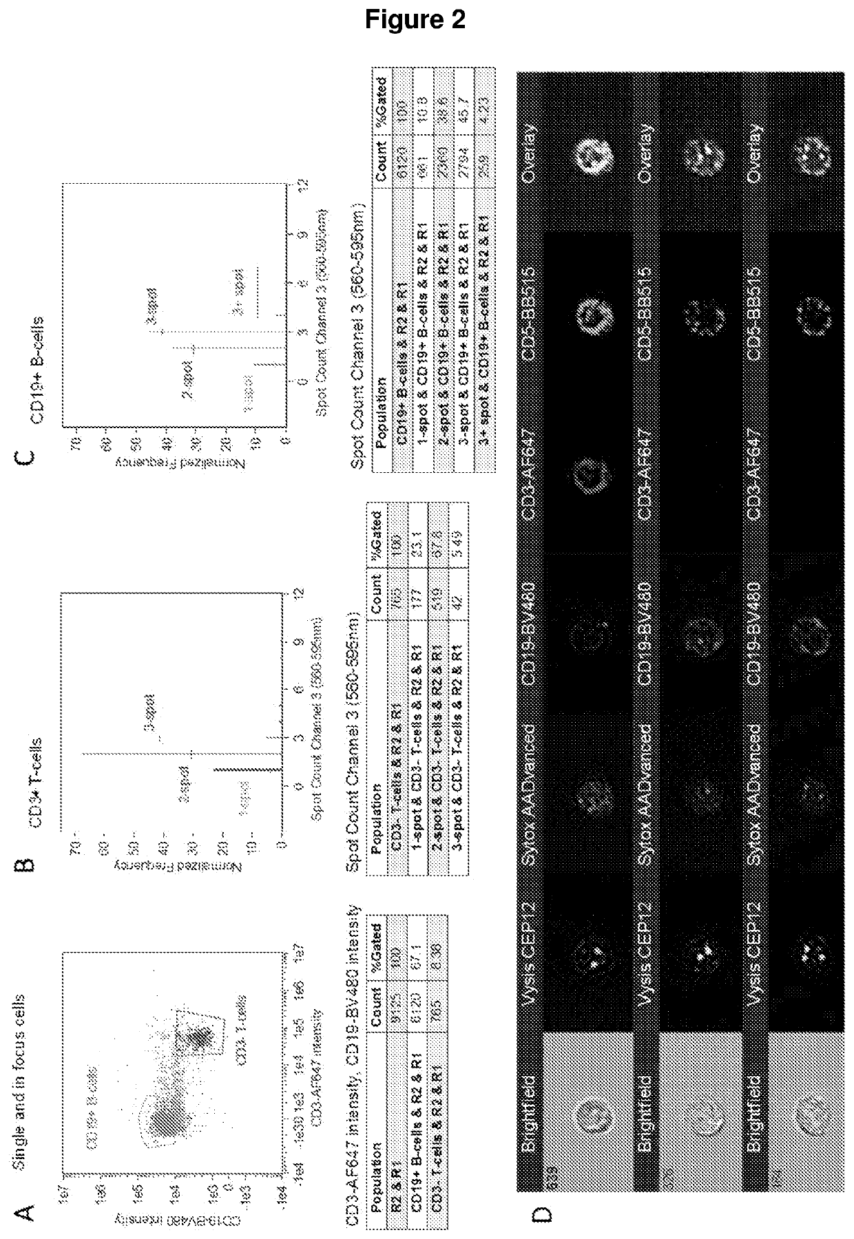
[0021]Since imaging flow cytometers have multispectral image capabilities, they offer the capacity for “immuno-flowFISH”, i.e. immunoFlSH of cells in suspension. Immuno-flowFISH has the capability to provide an integrated automated high-throughput single platform test for the identification of genomic aberrations in cells identified by phenotype, resulting in higher sensitivity.
[0022]Combining FISH on cells in suspension (FISH-IS) with cell phenotyping enables locus-specific probes (i.e. pr...

Problems solved by technology

Hence the sensitivity of the method is low.
Further, while the FISH method for the visualisation of chromosomal abnormalities in cells is important for the diagnostic assessment of malignant diseases (especially haematological malignancies) and non-malignant diseases (e.g. antenatal diagnosis of genetic disease), it is labour intensive and does not specifically identify the cell of interest—only nuclei are visualised and these could be derived from any cell type.
Although the combination of immunophenotyping and genetic analysis (i.e. FICTION) increases the specificity of FISH detection of chromosomal a...

Method used


Image

  • Improvements in or relating to cell analysis
  • Improvements in or relating to cell analysis
  • Improvements in or relating to cell analysis

Examples


example results

[0355]The first fixative tested in the protocol was a methanol based buffer of 70% methanol / 4% formaldehyde / 5% acetic acid, which was a combination of the Carnoy's fixative used for traditional FISH and FISH-IS (as, for example, described in Minderman H, Humphrey K, Arcadi J K, et al. Image Cytometry-Based Detection of Aneuploidy by Fluorescence In Situ Hybridization in Suspension. Cytometry Part A 2012; 81A:776-784) and the 4% formaldehyde fixative used in the immuno-S-FISH protocol (as, for example, described in K. Fuller, S. Bennett, H. Hui, A. Chakera and W. Erber. Development of a robust immuno-S-FISH protocol using imaging flow cytometry. Cytometry Part A. 2016; 89A:720-730).

[0356]Experiments were performed with CD3-BB515 immunophenotyping (FIG. 6) and chromosome 1 or 12 enumeration probe (CEP1 or CEP12), which hybridise to highly repetitive human satellite DNA sequences located near the centromere of the chromosome, on healthy donor nucleated cells. FISH was expected to resul...

example 2

owFISH Compared to Earlier Prior Art Technology

[0366]The following example presented in Table 9 compares Immuno-flowFISH technology against Immuno-S-FISH Technology. While Table 10, provides the broad working ranges for analysis of a peripheral blood mononuclear cell (PBMC) preparation and the highly preferred optimum conditions for that methodology.

immuno-S-FISHComparative variations in theProtocol(Fuller et al., CPA 2016)Immuno-flowFISH 2018protocolsPeripheral blood 1.Ficoll-paque density 1.RBC lyse with BDThe RBC lyse step is preferable tomononuclear cellcentrifugation (this limitsPharmLyse a buffereddensity gradient purification for(PBMC) preparationusefulness of theammonium chloride-baseddiagnostic applications as there isprotocol for minimallysing reagent at pHless cell loss which is essential forresidual disease7.1-7.4minimal residual disease detection.detection) on fresh 2.Wash cells in PBS (noRBC is commonly used in flowperipheral blood onlyFBS or BSA)cytometry.Bone marrow ...


PUM

PropertyMeasurementUnit
Temperatureaaaaaaaaaa

Abstract

The present invention provides a method for cell analysis, comprising: preparing a blood sample comprising nucleated cells having surface, cytoplasmic or nuclear antigens (markers); antibody staining the cell markers; fixing and permeabilising the cells; FISH probe hybridising to chromosomes in the cells; performing imaging flow cytometry on the cells; analysing data obtained from performing imaging flow cytometry; and diagnosing, prognosing or monitoring a medical condition based on the data analysis.

Description

TECHNICAL FIELD[0001]The present invention relates to cell analysis. In a particular non-limiting aspect, the invention relates to diagnostic and prognostic methods for tissue sample and or cell analysis. For example, the invention provides methods of analysis for the diagnosis, prognosis or monitoring of medical conditions or therapeutic conditions.BACKGROUND ART[0002]The following discussion of the background art is intended to facilitate an understanding of the present invention only. The discussion is not an acknowledgement or admission that any of the material referred to is or was part of the common general knowledge as at the priority date of the application.[0003]Cytogenetic analysis is an integral component in the assessment of many disorders such as malignancies, syndromes with abnormal numbers of chromosomes such as Trisomy 21 (Down syndrome), and chimerism in transplant recipients.[0004]Diagnostic laboratories currently use fluorescence in situ hybridisation (FISH) analy...

Claims


Application Information

Patent Timeline
IPC IPC(8): C12Q1/6841C12Q1/6827G01N21/64G01N33/53
CPCC12Q1/6827C12Q1/6841G01N21/64G01N33/53G01N33/5091G01N33/582G01N1/30G01N21/6486C12Q2565/626C12Q2563/107C12Q2565/102G01N33/5094G01N33/57426G01N2015/1006G01N15/147G01N15/1475
Inventor ERBER, WENDY NAOMIFULLER, KATHRYN AMANDAHUI, HENRY YEUK LAM
Owner UNIV OF WESTERN AUSTRALIA
Why Eureka
Try Eureka