
Micropatterned hydrogel for cell cultures

a cell culture and hydrogel technology, applied in the field of micropatterned hydrogels, can solve the problems of limited treatment options, no known medical treatment effectively addressing the underlying molecular, and the failure of cartilage repair to regenerate hyaline articular cartilage, etc., to facilitate cell adhesion and promote adhesion.

Pending Publication Date: 2020-10-08
SOUTH DAKOTA BOARD OF REGENTS
View PDF1 Cites 4 Cited by
  • Summary
  • Abstract
  • Description
  • Claims
  • Application Information

AI Technical Summary

Benefits of technology

The patent describes a way to make a surface that can attract specific types of cells and keep them attached to it for a long time. This is done by using a special surface coating made of a chemical called polydopamine. This technique can help scientists study the behavior of specific cells and potentially develop new treatments for diseases.

Problems solved by technology

To date, surgical restoration techniques used for cartilage repair do not regenerate hyaline articular cartilage.
There are currently no known medical treatments that effectively address the underlying molecular causes of OA.
Current pharmaceutical treatment options are limited to the use of analgesics like non-steroidal anti-inflammatory drugs (NSAID) and intra-articular corticosteroid injections to reduce the pain associated with inflammation, which only provides temporary relief and can have negative consequences with long-term use.
However, they are also associated with concerns of ethical issues regarding the treatment of animals, cost and management issues, anatomical differences of cartilage in animals compared to humans, and age variations of animal species at the time of testing.
Two-dimensional (2D) cell culture techniques are particularly unsuitable for articular chondrocytes.
Under standard 2D culture conditions, however, chondrocytes tend to develop an artificially-induced fibroblastic phenotype after expansion or more than approximately 10 days in culture, which is known to alter their behavior.
Three-dimensional (3D) scaffolds have shown promise for promotion of phenotype maintenance of articular chondrocytes and for chondrogenesis of mesenchymal stem cells (MSCs), however, although the past decade has realized significant progress in the development of many types of three-dimensional (3D) cell culture systems, these techniques are all inherently limited in their utility by restricted oxygen diffusion, restricted and non-uniform penetration of both small molecule and macromolecule treatment agents, and limited optical penetration depth.
There remains a paucity of such systems that are well-suited for high-resolution optical imaging of large numbers of adherent cells (e.g., using high-throughput techniques).

Method used


Image

  • Micropatterned hydrogel for cell cultures
  • Micropatterned hydrogel for cell cultures
  • Micropatterned hydrogel for cell cultures

Examples


example 1

te Morphology Influences Internal Architecture

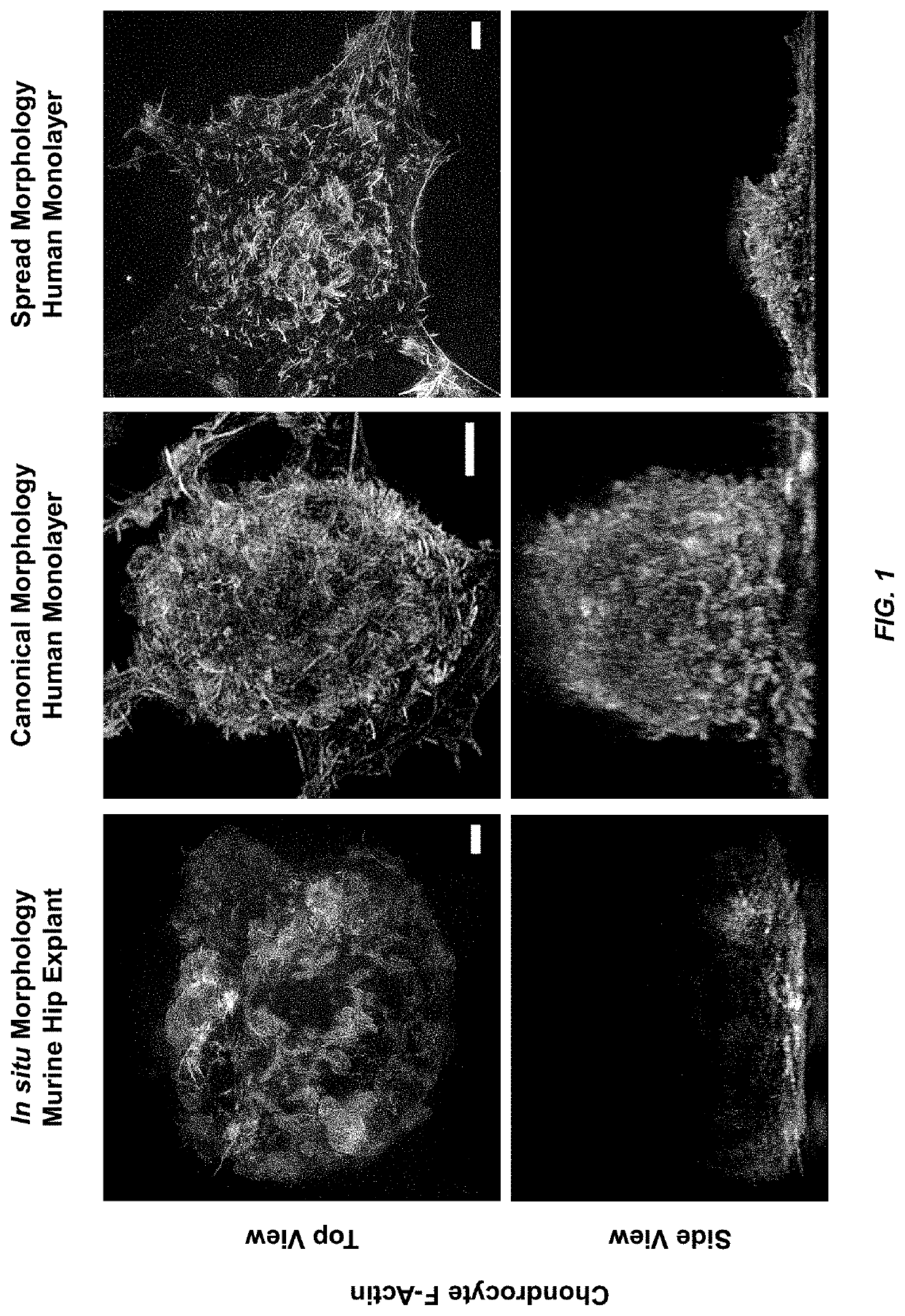
[0059]When plated on standard 2D platforms, chondrocytes tend to rapidly lose their canonical spheroidal morphology due to the adherent chondrocyte cells being on a hard and flat substrate, and adopt a fibroblastic phenotype within 10-14 days of culture. These morphological changes can result in substantial changes to chondrocyte architecture, including the length, density, and distribution of cortical actin fibers. The in situ chondrocyte shown in FIG. 1 (left) can be seen to have an actin network with length and density more similar to the chondrocyte displaying the canonical phenotype in FIG. 1 (middle), than to that of the chondrocyte in FIG. 1 (right) which has a spread morphology that is more typical of chondrocytes in standard monolayer culture. Note that the in situ chondrocyte in FIG. 1 (lower left) is not flat, but that the turbidity of the tissue prevented imaging its full thickness, thereby illustrating the difficulties inher...

example 2

te Diameter

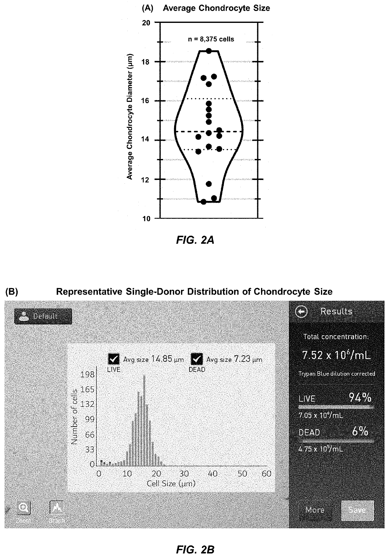
[0060]The diameter of 8,375 chondrocytes from 18 independent donors was measured using a Countless II FL cell counter. As shown in FIG. 2A, human chondrocytes were found to have a mean diameter of 14.6 μm±2.1 μm (S.D.), and this data represented the diameter measurements from 8,375 cells over n=18 individual donors. For each donor, the cell counter provided direct measurements of cell density (i.e., number), viability (based on trypan blue exclusion, circularity, and diameter), and average diameter, as well as a pictographic histogram of the distribution of diameters within the sample. The chondrocyte diameter distribution of average donors depicted a Full Width at Half Maxima (FWHM) of 12-18 μm, as shown in FIG. 2B. These measurements served as the basis for the selection of CellWell diameters of 12, 15, and 18 μm.

example 3

Design and Manufacturing

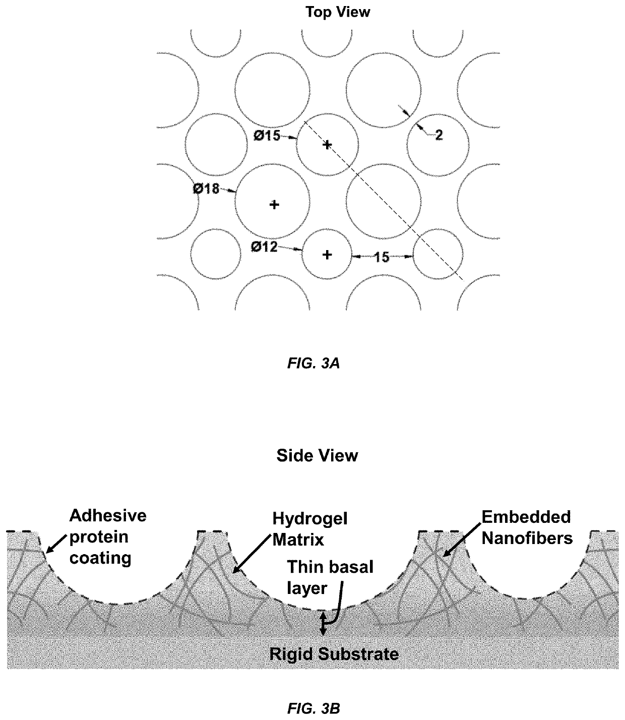
[0061]Computer-aided design (CAD) files were generated in SolidWorks consisting of an array of circles of 12, 15, and 18 μm diameters and used to generate the photomask pattern, as shown in FIG. 3. The design was chosen such that the distance between any two consecutive wells varied from 2 μm to 15 μm.

[0062]Micropatterned silicon wafers were obtained from the Utah Nanofab core lab at the University of Utah, USA, and standard contact lithography techniques were utilized to generate PDMS CellWell stamps. PDMS stamps were sterilized in an autoclave at 121° C. for 23 minutes. “Containment chambers” were microfabricated with 15 μm-tall walls, in which the CellWell casting process took place. These walls were thus constructed to be ˜8 μm taller than the hemispheroids in the stamps to provide room for several microns of material to separate the basal surface of the cells from the underlying cover glass without adding excessive bulk that can confound imaging experime...


PUM

PropertyMeasurementUnit
diameteraaaaaaaaaa
distanceaaaaaaaaaa
diameteraaaaaaaaaa

Abstract

The present disclosure describes hydrogels which are micropatterned with a network of wells for cell culture. In a preferred embodiment, the micropatterned hydrogels are embedded with a nanomaterial. Further described are methods of forming the micropatterned hydrogels and methods of culturing cells in the micropatterned hydrogels. The hydrogels can be natural or synthetic.

Description

CROSS-REFERENCE[0001]This application is related to and claims priority under 35 U.S.C. § 119 to U.S. Provisional Application Ser. No. 62 / 828,857 filed on Apr. 3, 2019 and entitled “MICROPATTERNED HYDROGEL FOR CELL CULTURES”; the entire contents of this patent application are hereby expressly incorporated herein by reference.TECHNICAL FIELD[0002]The present disclosure relates to micropatterned hydrogels. Preferably, the hydrogels are micropatterned with a plurality of wells for cell cultures.BACKGROUND[0003]Osteoarthritis (OA) is a painful disease of the articular joints that is primarily characterized by the degradation of the extracellular matrix (ECM) in the articular cartilage. To date, surgical restoration techniques used for cartilage repair do not regenerate hyaline articular cartilage. Although symptoms can improve temporarily after surgical repair, 85% of patients progress to failure within 7.5 years or less. There are currently no known medical treatments that effectively ...

Claims


Application Information

Patent Timeline
Patent Type & Authority Applications(United States)
IPC IPC(8): C12M1/12C12N5/077
CPCC12N2533/30C12N5/0655C12M25/14C12M25/02C12N2535/10C12N2533/76C12N2533/00C12M25/06C12M23/20
Inventor WOOD, SCOTTSARASWAT, RAM
Owner SOUTH DAKOTA BOARD OF REGENTS
Learn More