

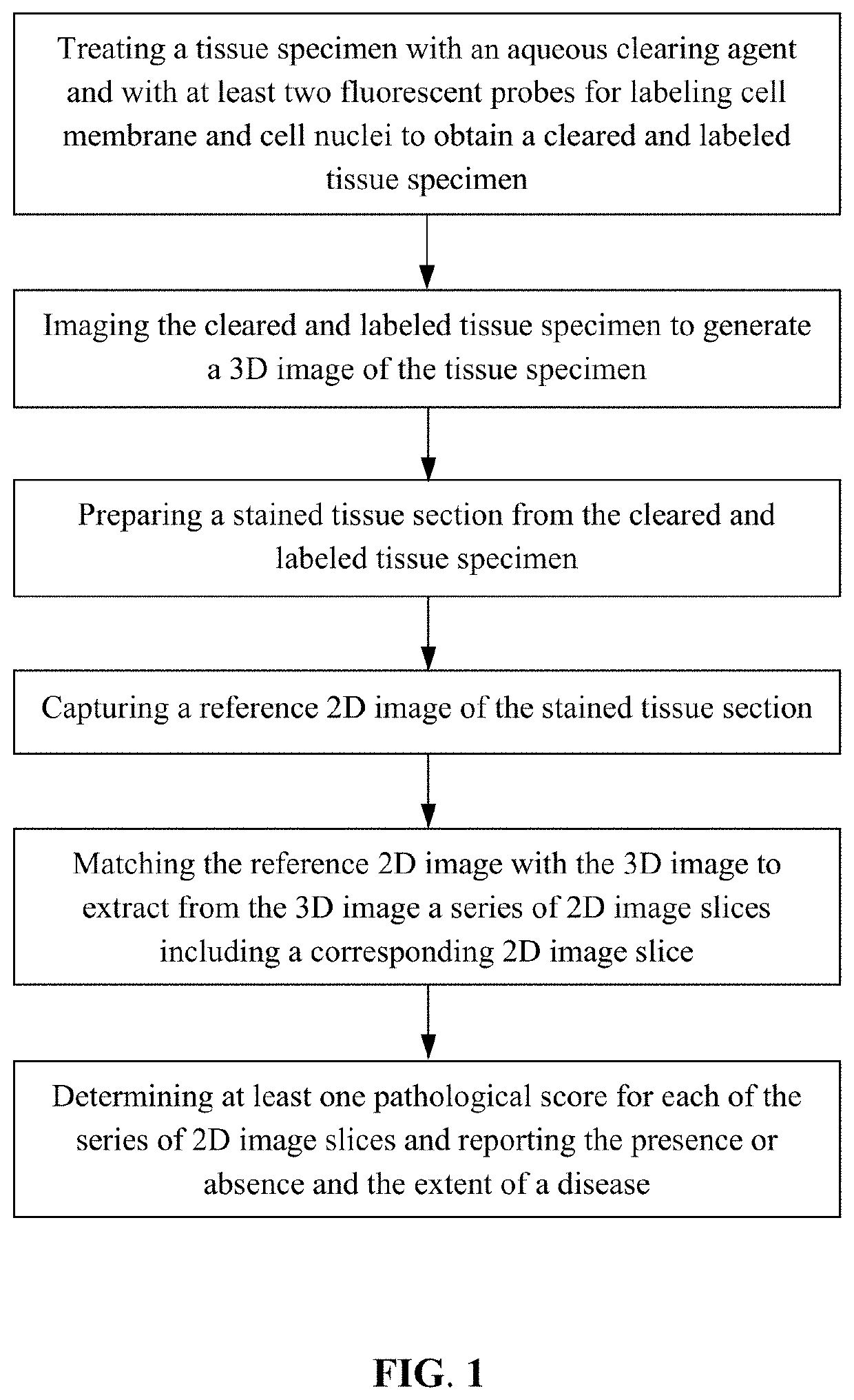
Method for analyzing tissue specimens
a tissue specimen and tissue technology, applied in the field of histopathologic diagnosis, can solve the problems of tissue distortion from sample preparation, reducing the accuracy of diagnostic decisions, and the representation of samples (i.e., stained sections), so as to facilitate operation performance, improve diagnostic accuracy, and facilitate the effect of operation
- Summary
- Abstract
- Description
- Claims
- Application Information
AI Technical Summary
Benefits of technology
Problems solved by technology
Method used
Image

Examples
example 1
Diagnostic Report Based on a Breast Tissue Specimen
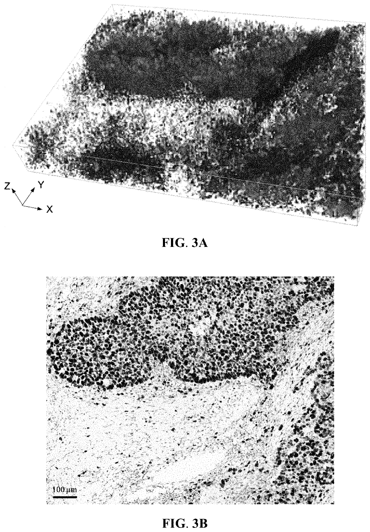
[0067]A fresh breast tissue specimen was collected from a female patient suffering from breast cancer. The tissue specimen, first fixed with 4% formaldehyde, was then permeated with 0.1-1% Triton X-100. Subsequently, the tissue specimen was stained with SYTO 16 and DiD to label cell nucleus and cell membrane, respectively. Each labeling was carried out at room temperature for 8 hours, followed by labeling of Ki-67 by treating the specimen with rabbit anti-Ki-67 primary antibody (VENTANA) at room temperature for about 10 hours and with Alexa Fluor 405-conjugated goat anti-rabbit IgG secondary antibody (Thermo Fisher Scientific) at 4° C. for about 16 hours. The labeled specimen was then immersed in an aqueous clearing agent with a refractive index of about 1.45 at room temperature for about 3 hours to obtain a cleared and labeled specimen. The aqueous clearing agent was prepared by mixing 30-50% meglumine diatrizoate and 10-30% trieth...
example 2
Diagnostic Report Based on a Lung Tissue Specimen
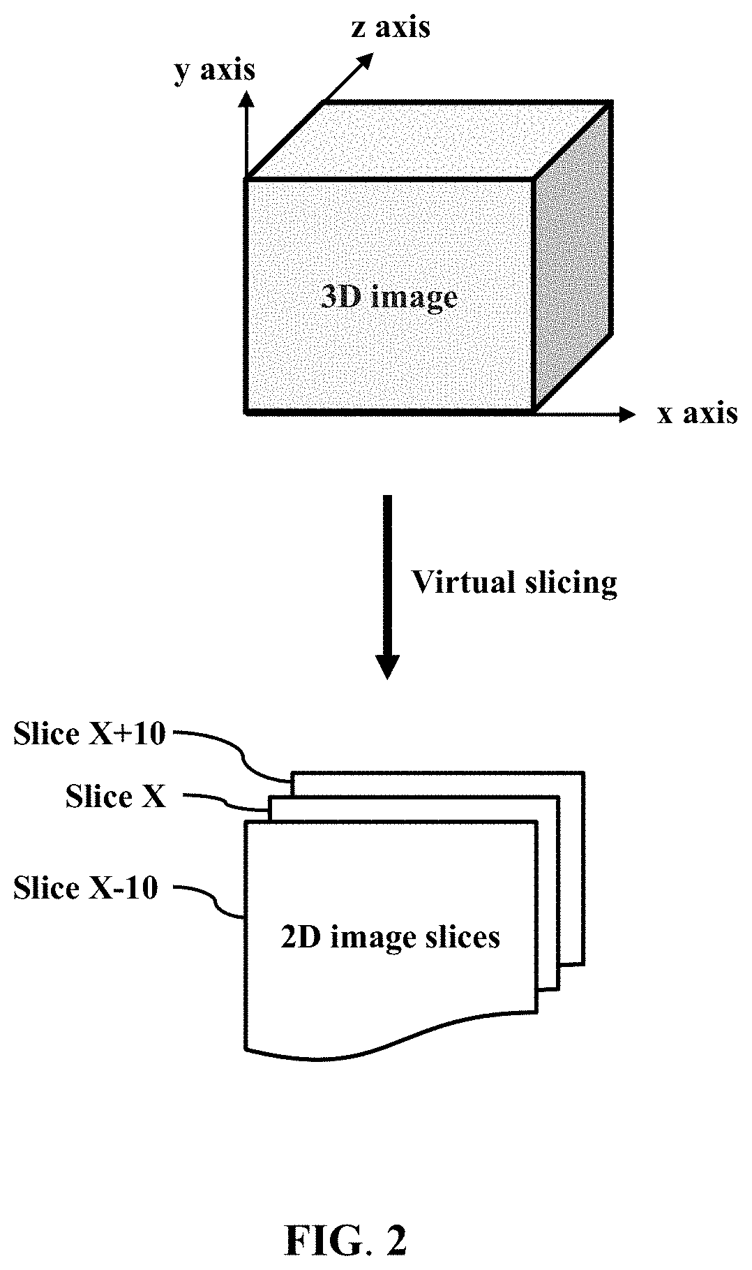
[0072]A fresh lung tissue specimen was collected from a patient suffering from lung cancer. The tissue specimen, first fixed with 4% formaldehyde, was then permeated with 0.1-1% Triton X-100. Subsequently, the tissue specimen was stained with SYTO 16 and DiD to label cell nucleus and cell membrane, respectively. Each labeling was carried out at room temperature for 6-8 hours. The labeled specimen was then immersed in an aqueous clearing agent with a refractive index of about 1.45 at room temperature for about 3 hours to obtain a cleared and labeled specimen. The aqueous clearing agent was prepared by mixing 30-50% meglumine diatrizoate and 10-30% triethanolamine in distilled water. The cleared and labeled tissue specimen, with a thickness of about 200 μm, was imaged from the top surface to the bottom surface with a LSCM system (LSM780; Zeiss) to obtain about a hundred successive 2D images of the specimen that were then used to generat...
PUM
 Abstract
Description
Claims
Application Information