Cell encapsulation compositions and methods for immunocytochemistry

a cell encapsulation and composition technology, applied in the field of cell encapsulation compositions and immunocytochemistry, can solve the problems of time-consuming, difficult pelleting, and easy loss of small cell mass during supernatant removal, and achieve the effect of easy attribution

Pending Publication Date: 2021-08-05
THE UNIV OF BRITISH COLUMBIA
View PDF0 Cites 1 Cited by
  • Summary
  • Abstract
  • Description
  • Claims
  • Application Information

AI Technical Summary

Benefits of technology

The patent describes a method to repeatedly label and evaluate cells using a process called immunocytochemistry. The cells are first encapsulated in a special material and then labeled with fluorescent antibodies. The locations of the cells are recorded. Later, the cells are treated to make the fluorescent labels inactive and labeled again with different antibodies. This process makes it easy to attribute signals from multiple markers to a specific cell. The method can be repeated to evaluate multiple markers simultaneously. Overall, this technique provides a reliable and repeatable way to study cells and the molecules they express.

Problems solved by technology

When working with non-adherent cells, the additional step of centrifuging the cells into a pellet to remove the supernatant by pipetting or pouring1,2 is also required and can be time consuming.
When there are fewer cells, pelleting becomes more challenging and the smaller mass of cells is more easily lost during supernatant removal.
This approach works well for adherent cells grown in culture, but the adhesives are typically ineffective for primary cells or suspension cells grown in culture.
While both primary cells and cultured cells can be effectively adhered to a glass slide, but this process may still result in significant losses.
Furthermore, Cytospin™ is a serial process performed one sample at a time, which has limited capacity for high-throughput screening studies involving large numbers of samples14.
Consequently, analyzing these cells requires imaging over many microscopy fields in order to detect a sufficient number of cells, which is particularly challenging when searching for rare cells, such as CTCs.

Method used


Image

  • Cell encapsulation compositions and methods for immunocytochemistry
  • Cell encapsulation compositions and methods for immunocytochemistry
  • Cell encapsulation compositions and methods for immunocytochemistry

Examples


example 1

ion of Hydrogel Cell Encapsulation Compositions

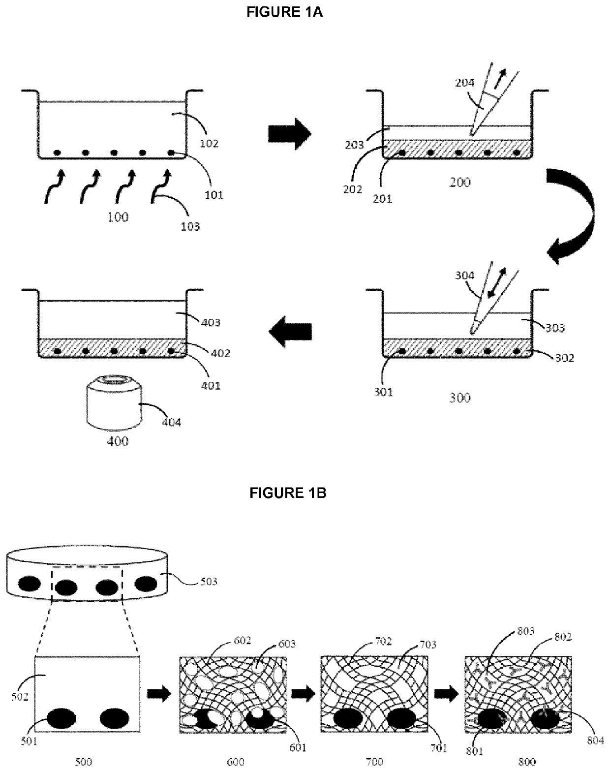
[0090]To prevent damage to the cells and their DNA, a photo-initiator, Irgacure™ 2959, was selected based on its transparency and ability to absorb long wave UV light (>350 nm). To reduce cytotoxicity, the concentration of Irgacure™ 2959 was limited to 0.1% (w / v). However, an alternative cross-linking agent may be used provided and depending on the crosslinking agent chosen may be used at a greater concentration. The thickness, porosity, and mechanical stability of the PEGDA hydrogel can be optimized either by varying their molecular weight or by mixing with poly (ethylene glycol) (PEG) and PBS. The hydrogel porosity can be optimized to encapsulate and affix cells to the surface of an imaging well plate, while allowing antibodies to diffuse through the pores and reach the cells. The mechanical stability of the photo-polymerized hydrogel is important to withstand pipette manipulation during the staining process while the thickness of the...

example 2

and Image Acquisition Using ICC Composition

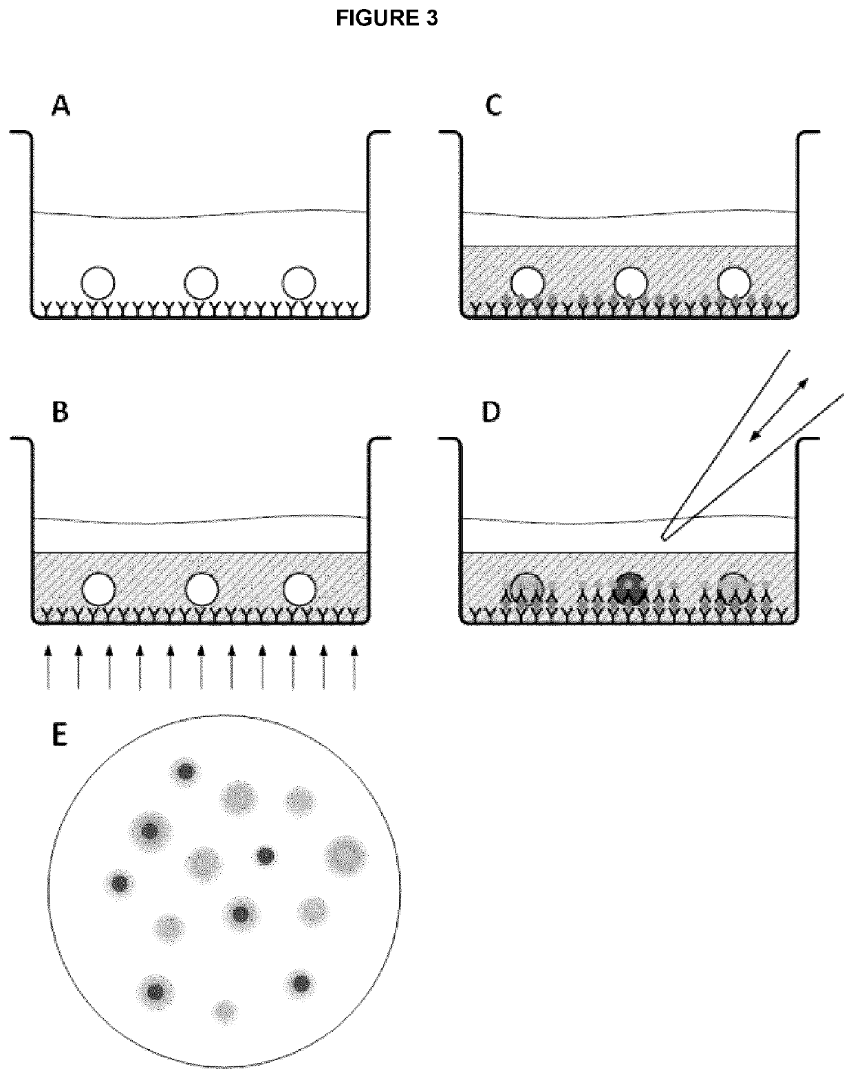
[0099]To investigate the efficiency of ICC stain as well as image quality of encapsulated cells, we used a standard ICC protocol, according to the manufacturer's guideline40, for staining cells and compared the staining of encapsulated cells to non-encapsulated cells. However, instead of using centrifugation to remove the excess antibody stains, supernatant from each washing step may simply be removed by pipetting. Image acquisition in macroporous hydrogels, after polymerization, has traditionally proven to be difficult due to the large pore sizes29. To determine if the PEG porogen influences image quality, we imaged encapsulated cells before and after photo-polymerization. Prior to polymerization, the hydrogel was transparent, but became lightly opaque after photo-polymerization. However, this color change had no effect on the visualization of unstained or stained cells by bright field microscopy (data not shown). The comparison of PEGDA h...

example 3

ation of Cell Loss in Immunocytochemistry

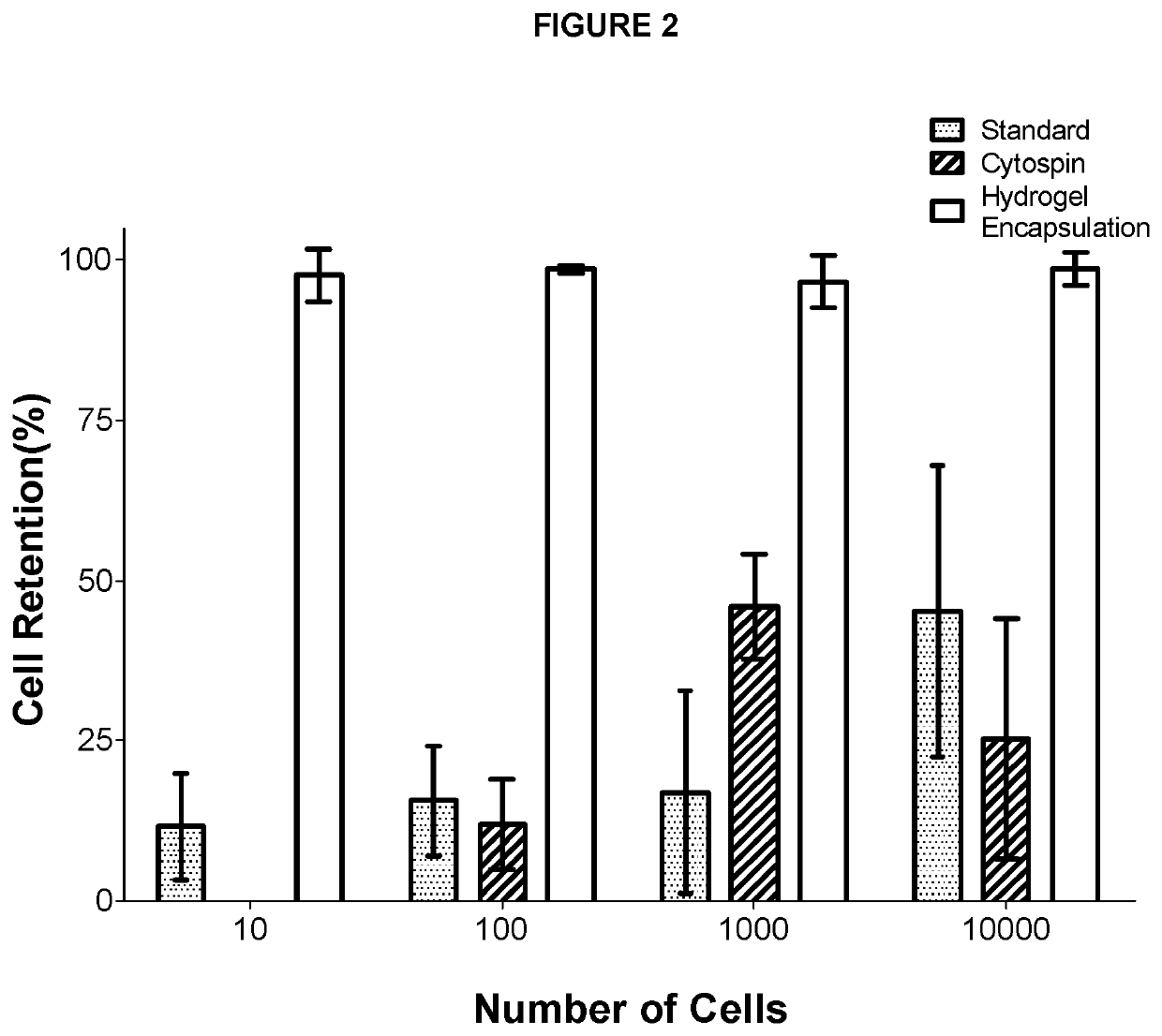
[0101]To quantify cell loss during ICC, cells were counted before and after ICC for sample sizes of 10, 100, 1,000, and 10,000 cells using three different protocols: 1) traditional ICC performed on 384-well imaging plates, 2) ICC performed on cells adhered to microscope slides using cytospin, and 3) ICC performed on PEGDA hydrogel encapsulated cells. Two individuals counted encapsulated cells in each image and the results were averaged to limit any error resulting from manual counting. Traditional ICC and CytoSpin™ showed a staggering amount of cell loss for cell samples ranging from 10 cells to 10,000 cells (FIG. 2). On the other hand, the current cell encapsulating hydrogel ICC compositions and methods limited cell loss to 1-3% showing improved cell retention during staining, washing, and centrifugation for all sample sizes.


PUM

PropertyMeasurementUnit
Temperatureaaaaaaaaaa
Lengthaaaaaaaaaa
Timeaaaaaaaaaa

Abstract

Provided herein are compositions comprising: a scaffold polymer having one or more acryloyl groups or one or more methacryloyl groups; optionally a porogen and a crosslinking agent, compositions that upon crosslinking form a hydrogel for use in cell encapsulation and methods for immunocytochemistry of encapsulated cells. Scaffold polymers used are selected from: Poly(ethylene glycol) diacrylate (PEGDA); Poly(ethylene glycol) dimethylacrylate (PEGDMA); Poly(ethylene glycol) methyl ether acrylate (PEGMEA); Poly(ethylene glycol) methacrylate (PEGMA); and Poly(ethylene glycol) methyl ether methacrylate (PEGMEMA), and porogens selected from: Poly(ethylene glycol) (PEG); Chitosan; Agarose; Dextran; Hyaluronic acid; Poly(methyl methacrylate) (PMMA); Cellulose and derivatives thereof; Gelatin and derivatives thereof; and Acrylamide and derivatives thereof. The invention also provides, at least in part, compositions for forming a porous hydrogel around a cell suitable for immunostaining of cells within the hydrogel.

Description

CROSS REFERENCE TO RELATED APPLICATION[0001]This application claims the benefit of U.S. Provisional Patent Application Ser. No. 62 / 666,371 filed on 3 May 2018, entitled “REAGENT AND PROCESS FOR LOSSLESS IMMUNOCYTOCHEMISTRY”.FIELD OF THE INVENTION[0002]The invention relates to cell encapsulation compositions and methods for immunocytochemistry. The invention also provides compositions for forming a porous hydrogel around a cell suitable for immunostaining of cells within the hydrogel.BACKGROUND OF THE INVENTION[0003]Immunocytochemistry (ICC), or immunofluorescence, are a variety of assays for phenotyping cells based on protein expression and localization established by labeling using antibodies having a detectable tag. An ICC assay will often involve the steps of fixation, permeabilization, blocking, and immunostaining. Each of these steps is followed by at least one washing step, where reagent solutions are exchanged. When working with non-adherent cells, the additional step of cent...

Claims


Application Information

Patent Timeline
IPC IPC(8): G01N33/531G01N1/30C08L71/02C08L51/08C08J3/075
CPCG01N33/531G01N1/30C08L71/02C08L51/08C08J2451/08C08J2371/02C08J2351/08C08J2471/02C08J3/075C08L1/02C08L89/06A61L27/52G01N33/54353A61L27/56A61L27/18A61L2300/64A61L2300/62C08L1/08C08L5/08C08L5/12C08L5/02C08L33/14C08J9/26C08J2333/14C08J2205/022C08J2207/10C08J2201/026C08J3/28C08F299/00G01N33/554C08G65/3322C08J2333/06C08J2433/06C08L33/06C08F20/18C08F18/08
Inventor MA, HONGSHENLEE, JEONG-HYUN
Owner THE UNIV OF BRITISH COLUMBIA
Why Eureka
Try Eureka