Fluorescent probe for use in detection of brain tumor

a fluorescent probe and brain tumor technology, applied in the field of brain tumor fluorescence probes, can solve the problems of inability to extend the excision to the surrounding brain, glioma often becomes more malignant, and rarely achieve complete cure, etc., to achieve the effect of improving the removal ratio, high sensitivity, and increasing the possibility

Pending Publication Date: 2022-10-27
THE UNIV OF TOKYO
View PDF0 Cites 0 Cited by
  • Summary
  • Abstract
  • Description
  • Claims
  • Application Information

AI Technical Summary

Benefits of technology

[0023]According to the fluorescent probe of the present invention, a brain tumor can be specifically and sensitively detected and labeled semi-instantaneously. The fluorescent probe of the present invention can be directly and topically applied or sprayed to a brain tumor, and therefore has an exceptional effect of acknowledging the presence of a brain tumor or increases the possibility to be identified and imaged accurately, quickly and with high sensitivity by a simple method regardless of the dosage form.
[0024]Thus, improvement of the removal ratio and improvement of the overall survival rate by labeling an affected part such as a brain tumor during surgery can be expected. In addition, it is also possible to solve the problem that currently, there is not a fluorescent probe that can be labeled for low-grade glioma.
[0025]In addition, a detection method using the fluorescent probe of the present invention is superb in usefulness in that the detection procedure is simple, the detection can be performed by visible light which is safe for living bodies, and the amount of the fluorescent probe to be used is very small.

Problems solved by technology

Even in low-grade glioma, complete cure is rarely achieved, and glioma often becomes more malignant in several years and fatal in 5 to 10 years.
In glioma surgery, complete removal is a good prognostic factor, but excision cannot be extensively extended to the surrounding brain because of functional localization.
It is often difficult to achieve both improvement of the removal ratio and preservation of the brain function, which are in a trade-off relationship.
However, methods using 5-ALA have issues such as sensitivity-specificity limitations, necessity of preoperative internal administration, a problem about fluorescence duration associated with metabolism, difficulty of re-administration, and inability to perform fluorescent labeling in the case of low-grade tumors (e.g. Non-Patent Literatures 1 and 2).
In addition, in conventional methods of tumor visualization, it is difficult to achieve both improvement of the removal ratio and preservation of the cranial nerve function, which are in a trade-off relationship.

Method used


Image

  • Fluorescent probe for use in detection of brain tumor
  • Fluorescent probe for use in detection of brain tumor
  • Fluorescent probe for use in detection of brain tumor

Examples


example 1

[0077]1. Synthesis of Fluorescent Probe

[0078]Hydroxymethyl rhodamine green (HRMG) having various dipeptide sites was synthesized in accordance with the reaction scheme described in Example 1 of WO 2016 / 006678. Compounds having different dipeptide sites were obtained by variously changing the Fmoc-amino acid used in the synthesis of compound A7 in Example 1.

[0079]The synthesized compound is one in which in the aforementioned formula (I), all of R1, R2, R3, R4, R5, R6, R7, R8 and R9 are hydrogen atoms, X is O, Y is a methylene group, and a combination of P1 and P2 is as follows.

TABLE 1CompoundP1P2YK190ArginineProlineYK213HistidineGlycineYK19TyrosineGlycineYK281Methionine sulfoxideAcetylleucine

example 2

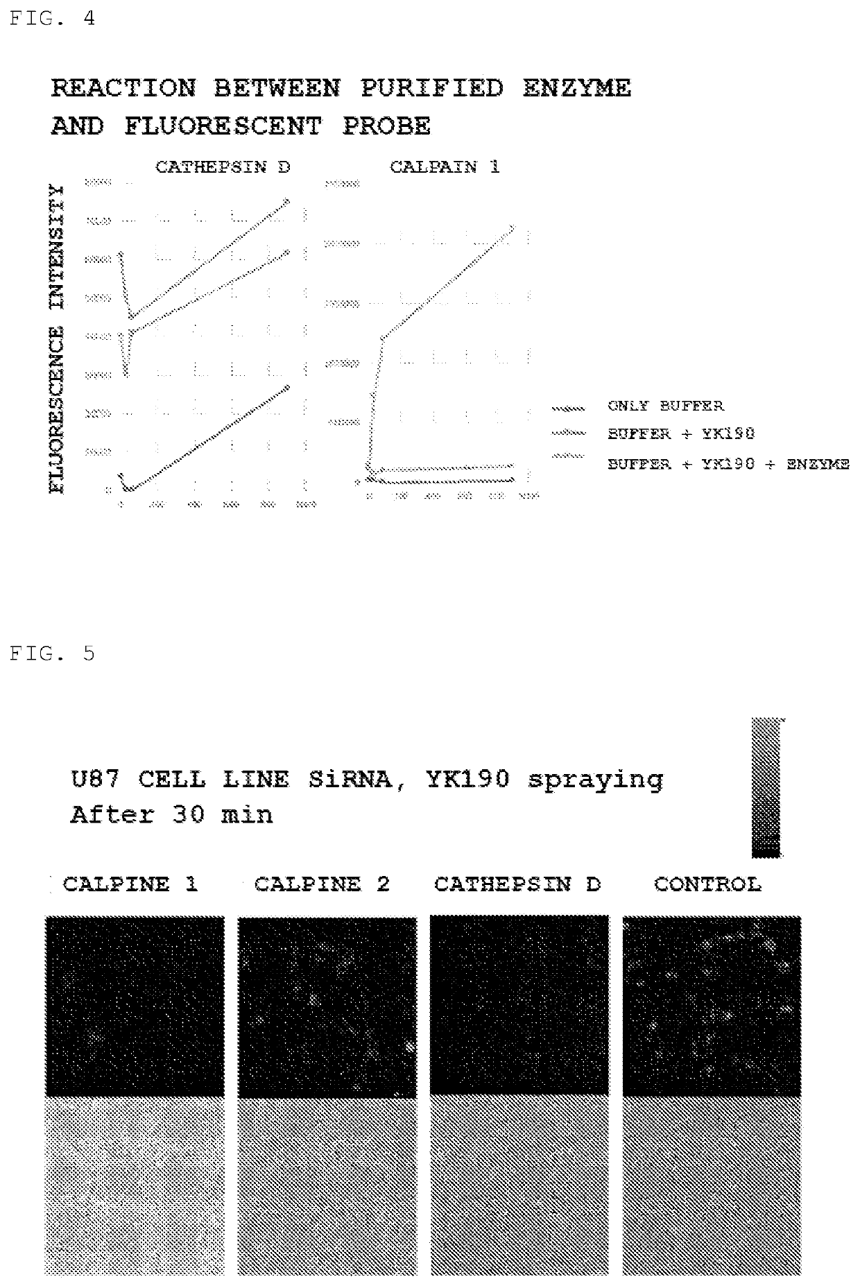
[0080]2. Screening by Fluorescent Probe

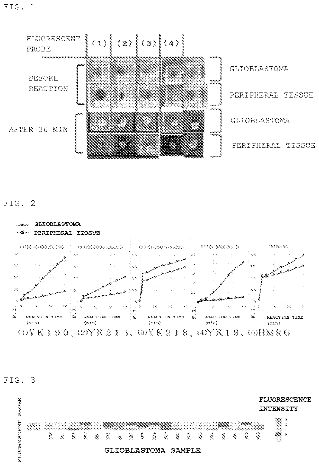
[0081]The fluorescent intensity of fresh glioblastoma and peritumoral tissue were compared using the fluorescent probes YK190, YK213, YK19, and YK281 synthesized in Example 1. As a comparative example, hydroxymethyl rhodamine green (HRMG) having no dipeptide site was used.

[0082]50 μM probe solutions (200 μL) in PBS, containing 0.5% v / v DMSO as a co-solvent was added dropwise to each specimen, and the fluorescence intensity was measured over time using Maestro In Vivo Imaging System Ex.[0083]Excitation wavelength: 465 nm[0084]Emission wavelength: 515 nm

[0085]As a result, as shown in FIGS. 1 and 2, fluorescent labeling was performed in a tumor-specific manner as compared with the surrounding tissue for (1) YK190 and (2) YK213, whereas there was no significant difference as compared with the surrounding tissue for (3) YK218. (5) HRMG is a comparative example having no dipeptide site on the side chain. As shown in FIG. 2, both YK190 and YK213 exhib...

example 3

[0087]3. Identification of Protease

[0088]Next, the protease involved in the fluorescent response obtained in Example 2 was identified.

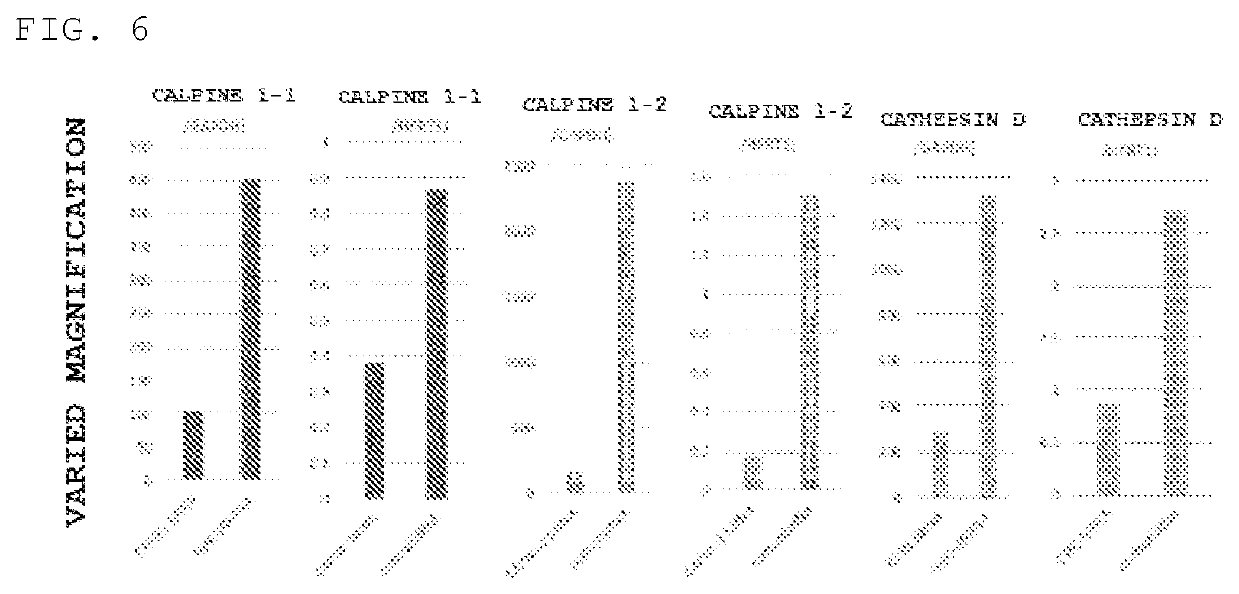
[0089]Protease analysis was performed by a DEG (diced electrophoresis gel) assay and peptide mass fingerprinting using LC MS / MS (liquid chromatograph-mass spectrometry) and as candidate enzymes, cathepsin D, cytosolic nonspecific dipeptidase, calpain 1 and cytosolic aminopeptidase were identified.

[0090]For these candidate enzymes, a change in fluorescence intensity was detected using inhibitors (SNJ-1945 and Amastatin), and the results showed that fluorescence suppression by SNJ-1945 occurred for calpain 1 and cathepsin D.

[0091]For calpain 1 and cathepsin D purified enzymes, a change in fluorescence intensity by the reaction with the fluorescent probe YK190 was measured, and the results showed that, as shown in FIG. 4, a significant increase in fluorescence intensity was observed. Therefore, it was suggested that the fluorescent response to glioblasto...


PUM

PropertyMeasurementUnit
wavelengthaaaaaaaaaa
fluorescence wavelengthaaaaaaaaaa

Abstract

Provided is a novel fluorescent probe which can be used in a spray manner, has an outstandingly high sensitivity-specificity with instantaneousness, and enables detection of a brain tumor.
A fluorescent probe for detecting a brain tumor, including a compound of the following formula (I) or a salt thereof:
wherein P1 represents an arginine residue, a histidine residue or a tyrosine residue, P2 represents a proline residue or a glycine residue, where P1 is linked to an adjacent N atom by forming an amide bond, and P2 is linked to P1 by forming an amide bond; R1 represents a hydrogen atom, or 1 to 4 identical or different substituents each independently selected from the group consisting of an optionally substituted alkyl group, a carboxyl group, an ester group, an alkoxy group, an amide group and an azide group; R2, R3, R4, R5, R6 and R7 each independently represent a hydrogen atom, a hydroxyl group, an optionally substituted alkyl group or a halogen atom; R8 and R9 each independently represent a hydrogen atom or an alkyl group;
X represents O, Si(Ra)(Rb), Ge(Ra)(Rb), Sn(Ra)(Rb), C(Ra) (Rb) or P(═O)(Ra); Ra and Rb each independently represent a hydrogen atom, an alkyl group or an aryl group; and Y represents a C1-C3 alkylene group.

Description

TECHNICAL FIELD[0001]The present invention relates to a fluorescent probe for detecting a brain tumor. More specifically, the present invention relates to a fluorescent probe capable of specifically detecting and labeling a brain tumor by applying the fluorescent probe to a tissue specimen, and a detection method using the fluorescent probe.BACKGROUND ART[0002]Glioma account for about 30% of primary brain tumors, and the median survival duration in glioblastoma, the most malignant type of glioma, is about 1.5 years. Even in low-grade glioma, complete cure is rarely achieved, and glioma often becomes more malignant in several years and fatal in 5 to 10 years. In glioma surgery, complete removal is a good prognostic factor, but excision cannot be extensively extended to the surrounding brain because of functional localization. It is often difficult to achieve both improvement of the removal ratio and preservation of the brain function, which are in a trade-off relationship.[0003]Visua...

Claims


Application Information

Patent Timeline
Patent Type & Authority Applications(United States)
IPC IPC(8): G01N33/533C07K5/06C09B11/24G01N33/574
CPCG01N33/533C07K5/06C09B11/24G01N33/57484G01N2333/96466G01N2333/96472C07K5/06026C07K5/06165C12Q1/37A61K49/0041G01N2800/7028A61K49/0056A61K49/006
Inventor KITAGAWA, YOSUKETANAKA, SHOTASAITO, NOBUHITOKURIKI, YUGOKAMIYA, MAKOURANO, YASUTERU
Owner THE UNIV OF TOKYO
Why Eureka
Try Eureka