Method and apparatus for ultrasound guided intravenous cannulation

a technology of intravenous cannulation and ultrasound guidance, which is applied in the field of use of ultrasound guidance for locating and cannulating peripheral blood vessels, can solve the problems of limited number of medications that can be administered, jeopardizing the patient's life, and the conventional ultrasound system is very complex and complicated, and a lot of delay and sum circuitry for dynamic focusing

Inactive Publication Date: 2000-10-17
PATACSIL ESTELITO G +1
View PDF8 Cites 209 Cited by
  • Summary
  • Abstract
  • Description
  • Claims
  • Application Information

AI Technical Summary

Problems solved by technology

Alternative routes for administration of emergency medications during cardiopulmonary resuscitation other than intravenous route have been demonstrated in practice but only a limited number of medications can be administered.
Alternate routes of administration such as central venous catheter insertion carries big risks or complications that may even jeopardize the patient's life.
Conventional ultrasound systems are very complex systems involving a lot of delay and sum circuitry for dynamic focusing.
Only these sonographers can technically operate these complex machines.
The large console's use for routine intravenous insertion guidance is high impractical.
Rack mounted ultrasound systems needs a lot filters for clutter cancellation and requires millions of operations per second to be implemented and therefore requires hundreds of chips and dissipates hundreds of watts of power.
Although it is smaller in size compared to the rack mounted console, its main purpose and function is for medical diagnostics and was designed to allow use of multiple transducer systems with

Method used


Image

  • Method and apparatus for ultrasound guided intravenous cannulation
  • Method and apparatus for ultrasound guided intravenous cannulation
  • Method and apparatus for ultrasound guided intravenous cannulation

Examples


Embodiment Construction

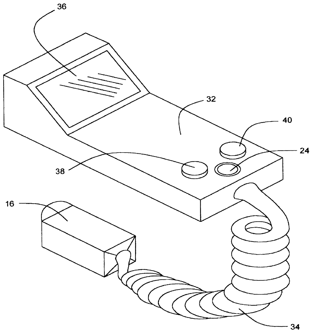
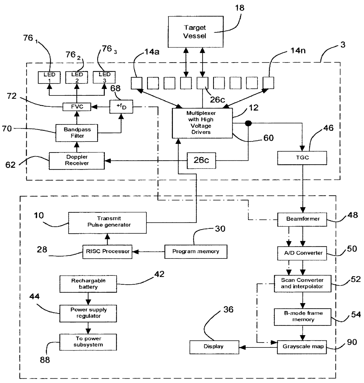
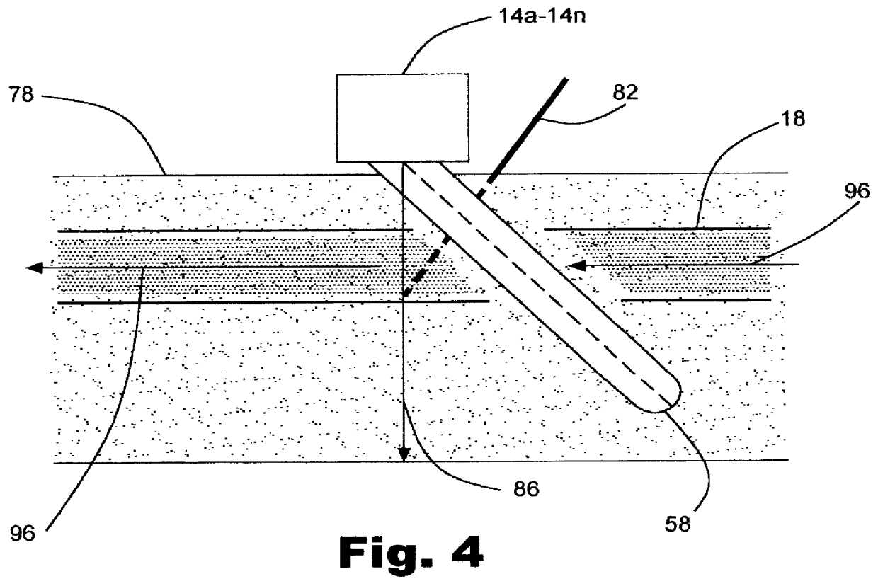
FIG. 1 is a schematic pictorial view of a preferred embodiment of the invention where it is shown as a dual mode intravenous cannulation guidance system wherein the whole architecture is packaged in a handheld housing coupled to a small transcutaneous scanhead by a coiled cable. Packaging such elaborate circuitry, such as in bulky architecture in prior art designs, to a small housing enclosure is possible and has been demonstrated in the U.S. Pat. Nos. 5,722,412 and 5,590,658. It involves judicious implementation of integrated circuitry wherein main image processing functions are integrated in small chips such as the implementation of application specific IC's or high speed, low power CMOS microchip technology. The preferred embodiment of the invention employs funtional integration on microchips which involves the pulse generator 10, multiplexers 12 integrated with high voltage drivers to drive the transducer elements of the array 14a-14n, where "n" represents the total number of el...


PUM


Abstract

A dual mode handheld ultrasonic device is provided for guiding a venous access catheter into a patient's peripheral vein. It provides B-mode imaging with a predetermined aperture and operating frequency which locates and displays a gray scale cross-sectional image of the target blood vessel. A single doppler beam in a separate mode detects the same blood vessel and creates a single scanline image superimposed to such B-mode cross-sectional image. Simultaneously, the intensity of the positive doppler shift detected by the single doppler beam as it hits the target blood vessel activates a plurality of light emitting diode (LED) indicator lights with varying voltage requirements mounted in the scanhead. Activated LED indicator lights forms an arrow pointing inferiorly perpendicular to the target vessel which guides a physician or a paramedical professional to the precise catheter insertion spot on a patient's extremity while simultaneously viewing the target blood vessel's cross-section on the display screen.

Description

BACKGROUND--FIELD OF INVENTIONThe present invention relates to the use of ultrasound guidance for locating and cannulating peripheral blood vessels which can be performed by both medical and paramedical professionals such as registered nurses, licensed vocational nurses, paramedics, laboratory phlebotomists, researchers, portamedics and emergency room and critical care staff etc.Intravenous cannulation is an invasive procedure wherein an intravenous catheter is blindly inserted into the lumen of a peripheral blood vessel through the patient's skin. The procedure is performed by the above-mentioned health care providers primarily for drawing blood from patient's arm for diagnostic purposes or for administration of medications and intravenous fluids to treat or save a patient's life. Successful cannulation is highly dependent on the dilatation of the target blood vessel caused by tourniquet application above the level of the insertion site. The dilatation displaces the overlying skin ...

Claims


Application Information

Patent Timeline
IPC IPC(8): A61B8/08
CPCA61B8/06A61B8/13A61B8/0841A61B8/0833A61B5/489
Inventor PATACSIL, ESTELITO G.PATACSIL, AMELIA V.
Owner PATACSIL ESTELITO G
Why Eureka
Try Eureka