
Apparatus and method for separating and purifying polynucleotides

a polynucleotide and apparatus technology, applied in the field of apparatus and method for separating and purifying polynucleotides, can solve the problems of inability to achieve many methods are slow, laborious and inefficient, and many methods are incapable of achieving the separation of fragments based on the length of the pair, and yield only minute quantities of separated materials. , to achieve the effect of rapid and efficient base-p

Inactive Publication Date: 2001-07-24
ADS BIOTEC INC
View PDF31 Cites 70 Cited by
  • Summary
  • Abstract
  • Description
  • Claims
  • Application Information

AI Technical Summary

Benefits of technology

It is an object of this invention to provide a method and apparatus for rapid and efficient base-pair length size separation of double-stranded DNA (dsDNA) and for effecting size-based separation of single-stranded oligonucleotides and single-stranded DNA fragments, RNA, plasmids and the like.
Liquid chromatography (LC) is a powerful technique for the separation of nucleic acids due to its high resolving capability, short analysis time, and ease of recovery of DNA fragments for subsequent studies. The WAVE.RTM. System combines the precision of ion-pair reversed-phase LC with automated sampling, data acquisition and reporting functions. Fragments can easily be collected and used in downstream experiments such as subcloning, amplification and sequencing.

Problems solved by technology

Traditional methods such as ion exchange chromatography, high pressure reverse phase chromatography, gel electrophoresis, capillary electrophoresis and the like are slow, laborious and inefficient, and they require the services of a highly skilled chromatographic expert.
Furthermore, many methods are incapable of effecting a base-pair length size based separation of these fragments and are capable of yielding only minute quantities of separated materials.
The ability to detect mutations in single-stranded polynucleotides, and especially in DNA fragments which have been amplified by PCR, presents a somewhat different problem since DNA fragments containing mutations are generally the same length as their corresponding wild type (defined herein below) but differ in base sequence.
Sometimes, exact replication fails and an incorrect base pairing occurs, which after further replication of the new strand, results in single-stranded DNA offspring containing a heritable difference in the base sequence from that of the parent.
Such an alteration in the amino acid sequence may be completely benign or may inactivate a protein or alter its function to be life threatening or fatal.
However, these methods cannot distinguish DNA fragments which differ in base sequence, but have the same base pair length.
Therefore, gel electrophoresis cannot be used directly for mutation detection.
This is a serious limitation of GEP.
However, these gel-based techniques are operationally difficult to implement and require highly skilled personnel.
In addition, the analyses are lengthy and require a great deal of set up time.
Therefore, the analysis time of longer DNA fragments can be often be untenable.
However, in certain cases, separations of homoduplexes and heteroduplexes are poorly resolved.
Artifacts and impurities can interfere with the interpretation of DHPLC separation chromatograms in the sense that it may be difficult to distinguish between an artifact or impurity and a putative mutation (Underhill, et al., Genome Res. 7:996 (1997)).
The accuracy, reproducibility, convenience and speed of DNA fragment separations and mutation detection assays based on HPLC have been compromised in the past because of HPLC system related problems.
However, the separation of mixture components can be compromised.
However, only small improvements have been achieved with these efforts.
While these procedures lead to overall improvements in nucleic acid purity relative to original unpurified materials, they suffer from negative characteristics.
Gel electrophoresis suffers from a lack of automation, incomplete separation of distinct fragments, as well as incomplete recovery of fragments.
While solid-phase extraction procedures lend themselves to automation, they also can suffer from incomplete separation of distinct fragments and from incomplete recovery of fragments.
P-217 filed May 13, 1999, this problem is exacerbated by the fact that such samples may contain a very small population of cells containing mutant DNA in the presence of a very large predominantly normal cell population containing, for example, wild type DNA.
That is to say, the concentration of mutant DNA may be too low to detect in absolute terms.
Increasing the amount of mutant DNA by PCR amplification of the sample would not solve the problem described above.
However, these visual methods are largely qualitative, time-consuming, and costly.
However, these methods cannot distinguish point mutations from homoduplexes having the same base pair length.
In addition to the deficiencies of denaturing gel methods mentioned above, these techniques are not always reproducible or accurate since the preparation of a gel slab and running an analysis can be highly variable from one operator to another.

Method used


Image

  • Apparatus and method for separating and purifying polynucleotides
  • Apparatus and method for separating and purifying polynucleotides
  • Apparatus and method for separating and purifying polynucleotides

Examples


example 1

Size Fractionization of DNA Restriction Fragments with MIPC DNA Fragment Analysis System

One application of the WAVE.RTM. System is accurate and rapid sizing of DNA fragments. This feature can be exploited for the isolation of DNA fragments. Here we describe the method and buffer gradients used for size fractionation and isolation of restriction fragments generated by digestion of pUC 18 with HaeIII. The nine fragments isolated range in size from 80 to 587 base pairs.

Liquid chromatography (LC) is a powerful technique for the separation of nucleic acids due to its high resolving capability, short analysis time, and ease of recovery of DNA fragments for subsequent studies. The WAVE.RTM. System combines the precision of ion-pair reversed-phase LC with automated sampling, data acquisition and reporting functions. Fragments can easily be collected and used in downstream experiments such as subcloning, amplification and sequencing.

Table D shows fragment sizing of pUC 18 plasmid cut with Ha...

example 2

Preparation of Spin Column

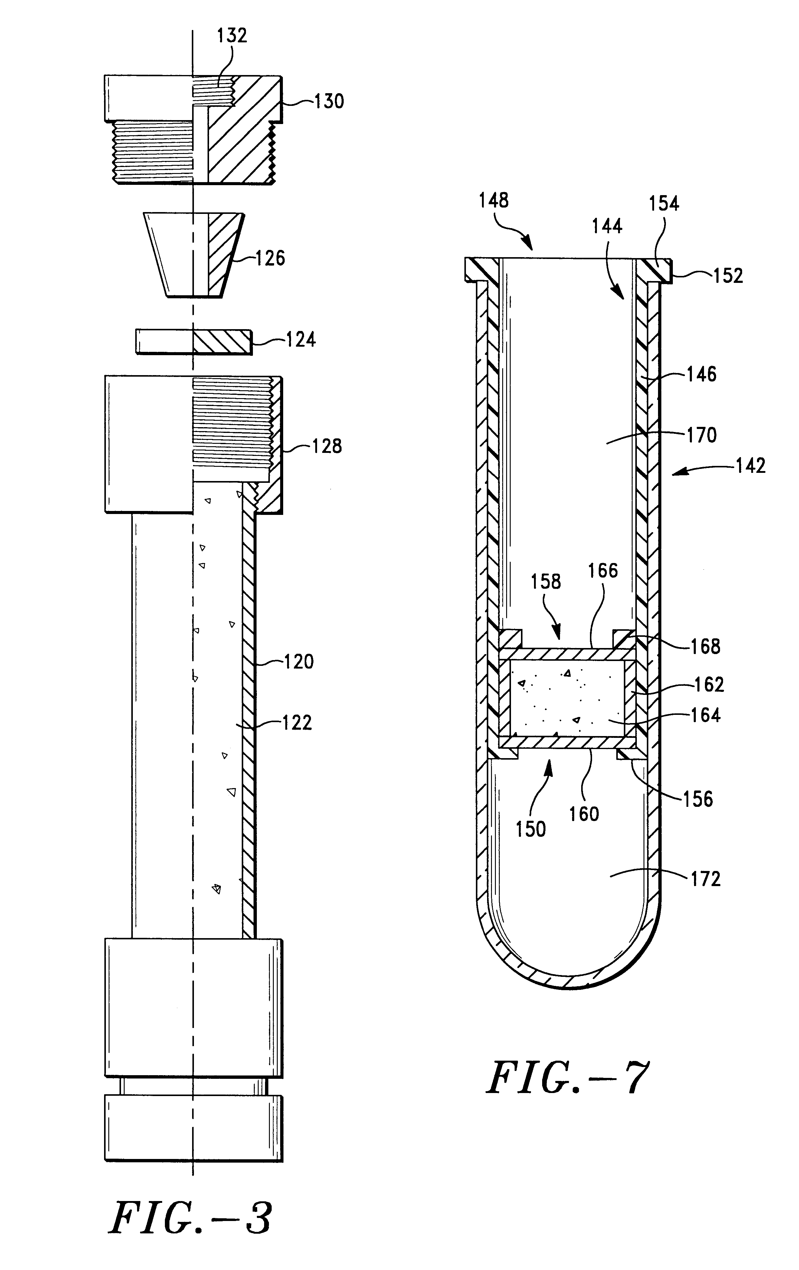
A 50 mg portion of resin is added to each spin column (See schematic representation of column in FIG. 7) while pulling a vacuum on the columns. The sides of the columns were tapped to remove resin from the walls. A polyethylene filter was placed on the top of the resin in each vial, followed by a retaining ring, with gentle tapping with a hammer to position the retaining ring securely against the filter. The spin columns were washed with an aqueous solution containing 50% acetonitrile (ACN) and 0.1 M triethylammonium acetate (TEAA). The vials were then washed with an aqueous solution of 25% ACN and 0.1 M TEAA and then with an aqueous solution of 0.1 M TEAA.

example 3

Separation of PUC 18 Msp I with Spin Column

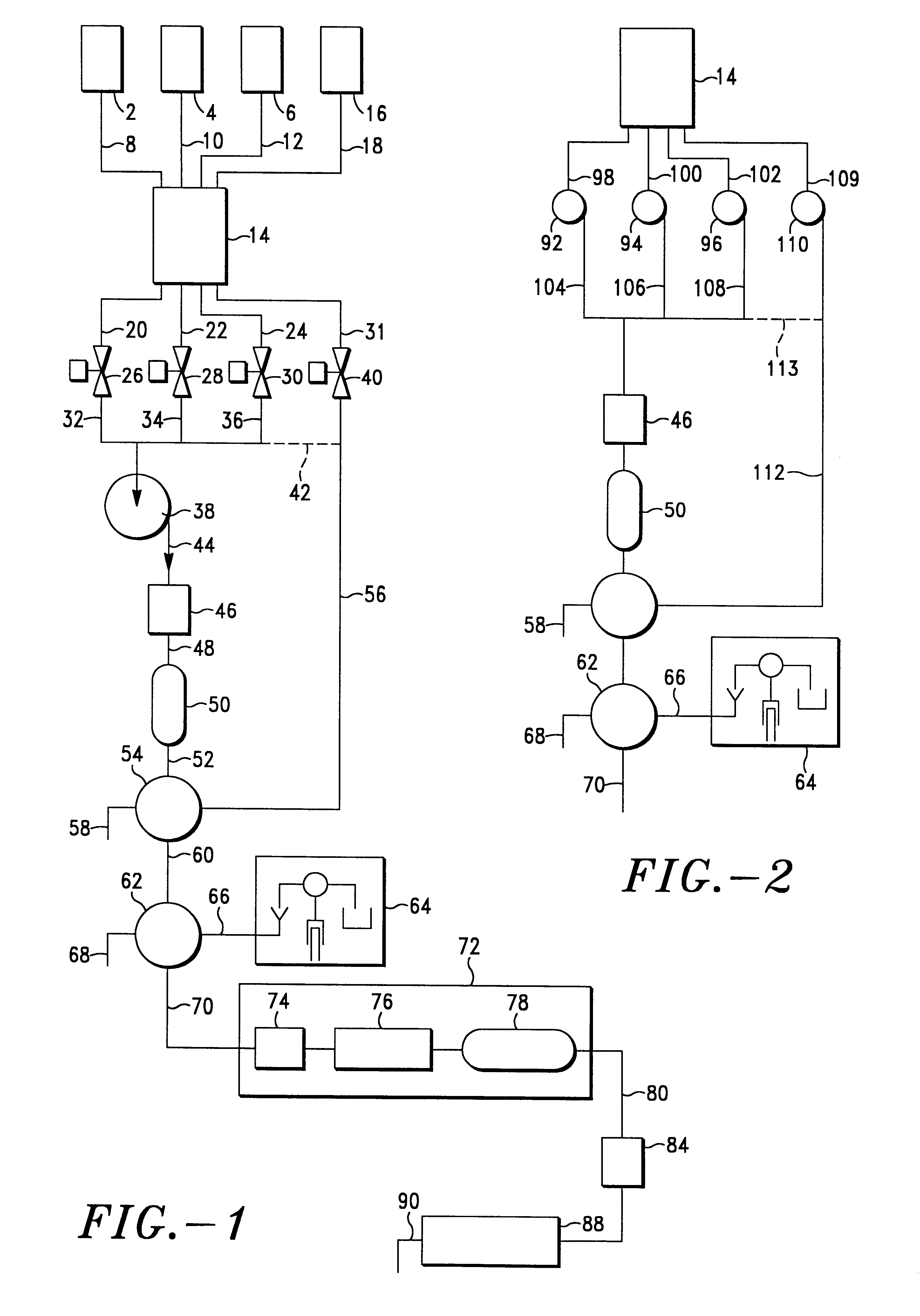
A sample solution was prepared by diluting 35 ml stock pUC 18 Msp I to 1 ml with 0.1 M TEAA (1 ml total volume). A 400 ml aliquot (corresponding to 6.6 mg loaded on the column) was selected. Base pair length separation of the solution was performed using the WAVE separation system (Transgenomic, Inc., Omaha, Nebr.) described in FIGS. 1-3, and the chromatograms obtained for two of the columns are shown in FIG. 22 for the pUC 18 Msp I standard.

An aliquot of the sample solution was pipetted into separate spin columns and left standing for 5 min. Each vial was centrifuged at 5000 rpm for 5 min. Then 400 .mu.l of freshly prepared aqueous solution containing 9.5% ACN and 0.1 M TEAA was pipetted into each spin column, each vial was left standing for 5 min, each vial was centrifuged at 5000 rpm for 5 min, and the filtrate was analyzed using the WAVE separation system. The chromatogram obtained for the eluant is shown in FIG. 23 for the pUC 18 Msp I...


PUM

PropertyMeasurementUnit
Fractionaaaaaaaaaa
Fractionaaaaaaaaaa
Timeaaaaaaaaaa

Abstract

A method for removing a target DNA fragment having a predetermined base-pair length from a mixture of DNA fragments comprises the following steps. A mixture of DNA fragments which may contain the target DNA fragments is applied to a separation column containing media having a nonpolar, nonporous surface, the mixture of DNA fragments being in a first solvent mixture containing a counterion and a DNA binding concentration of driving solvent in a cosolvent. The target DNA fragments are separated from the media by contacting it with a second solvent solution containing a counterion and a concentration of driving solvent in cosolvent which has been predetermined to remove DNA fragments having the target DNA fragment base pair length from the media. The target DNA fragments can be collected and optionally amplified. When the method is being applied to collect a putative fragment, if present, no DNA fragments having the base pair length of the target DNA could be present in the mixture. Alternatively, DNA fragments having the base pair length of the target DNA are present in the mixture. The disclosure also describes an ambient or low pressure device for separating polynucleotide fragments from a mixture of polynucleotide fragments comprises a tube having an upper solution input chamber, a lower eluant receiving chamber, and a fixed unit of separation media supported therein. The separation media has nonpolar separation surfaces which are free from multivalent cations which would react with counterion to form an insoluble polar coating on the surface of the separation media.

Description

This invention relates to an apparatus and method for separating, isolating, and purifying polynucleotides. In particular, this invention relates to methods and devices for separating targeted polynucleotide fragments having a predetermined base-pair length or range of base pair lengths, and for separating and purifying polynucleotides with both high pressure and low pressure devices.A need exists for rapid and efficient procedures for isolating, separating and purifying single-stranded oligonucleotides and single-stranded DNA fragments, RNA single-stranded DNA fragments, plasmids and the like. Traditional methods such as ion exchange chromatography, high pressure reverse phase chromatography, gel electrophoresis, capillary electrophoresis and the like are slow, laborious and inefficient, and they require the services of a highly skilled chromatographic expert. Furthermore, many methods are incapable of effecting a base-pair length size based separation of these fragments and are ca...

Claims


Application Information

Patent Timeline
IPC IPC(8): B01D15/08C07H21/04C08B5/02C08B5/00C07H21/00C12P19/00C12Q1/68C12P19/34
CPCB01J20/287B01D15/366
Inventor GJERDE, DOUGLAS T.HANNA, CHRISTOPHER P.TAYLOR, PAUL D.LEGENDRE, JR., BENJAMIN L.HAEFELE, ROBERT M.
Owner ADS BIOTEC INC
Learn More