Microfabrication of membranes containing projections and grooves for growing cells

a technology of growing cells and microfabrication, which is applied in the field of cell growth and culture, can solve the problems of insufficiently addressed in vitro approaches, cell culture systems producing significant detachment, and inability to provide good indication of cell function in vivo, so as to facilitate cell orientation, facilitate cell adhesion, and enhance cell differentiation

Inactive Publication Date: 2005-09-13
THE BOARD OF TRUSTEES OF THE UNIV OF ILLINOIS
View PDF14 Cites 44 Cited by
  • Summary
  • Abstract
  • Description
  • Claims
  • Application Information

AI Technical Summary

Benefits of technology

[0016]Also provided is a cell culture model for the growth and development of muscle cells comprising a membrane for the growth of cells comprising a microtextured polymer membrane having projections of between about 1 μm to about 100 μm in size and longitudinal grooves; wherein the polymer membrane comprises a surface modification to

Problems solved by technology

Although a much focused research topic in cell physiology, there are some fundamental issues in experimental set-up of muscle cell cultures which have not been adequately addressed.
However, such in vitro approaches are ineffective at providing a good indication of cell function in vivo for a number of reasons.
Firstly, these cell culture systems produce significant detachment between the membrane that is being stretched and the overlying substrata.
Secondly, unlike the complex three-dimensional force effects seen in vivo, the traditional in vitro culture systems forces are transmitted in only one direction.
Furthermore, the complex three-dimensional arrangement of myocytes, and in particular, cardiac myocytes as found in vivo, is usually

Method used


Image

  • Microfabrication of membranes containing projections and grooves for growing cells
  • Microfabrication of membranes containing projections and grooves for growing cells
  • Microfabrication of membranes containing projections and grooves for growing cells

Examples


example 1

Materials and Methods

[0112]The present example provides details of materials and methods employed throughout the application and in the Examples presented herein below.

Polymeric Microtextured Membrane Preparation

[0113]Control silicone membranes are prepared using silicone membranes (Specialty Manufacture, MI) that are pre-treated with ION HCl for 2 hours before coating with laminin to allow the cells to adhere. The microtextured polymeric membranes will be fabricated by a technique where photolithographically defined silicon wafers are used as templates or molds for reproducing complimentary images on desired polymers. This enables the reproduction of precise surface architectures and geometries.

[0114]Silicone microtextured surfaces are produced using a method developed in the Desai laboratory as described herein throughout. Starting with a clean silicon wafer, approximately 1 ml of UV light sensitive negatice photoresist is spun on the wafer for 30 seconds at 1500 rpm. This results...

example 2

Use of Surface Microtopography to Determine Cell Attachment and Shape

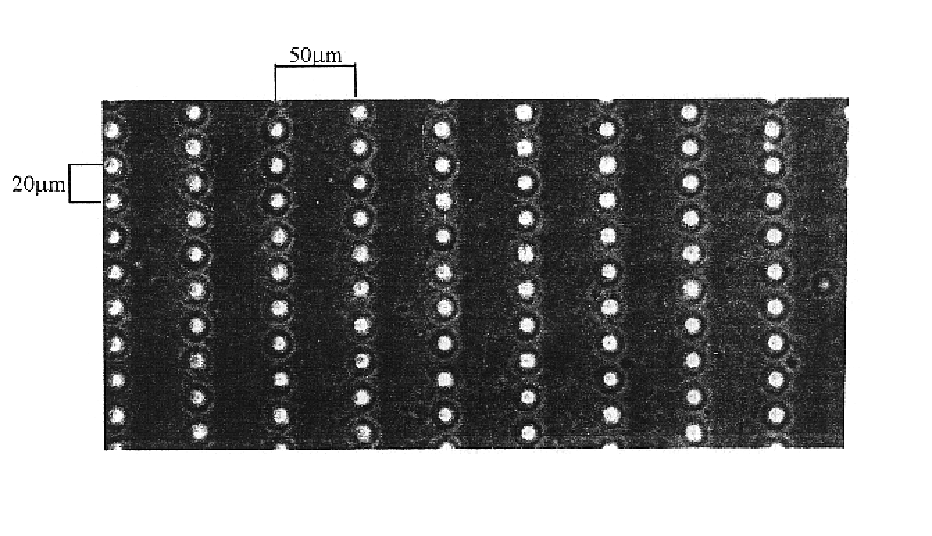
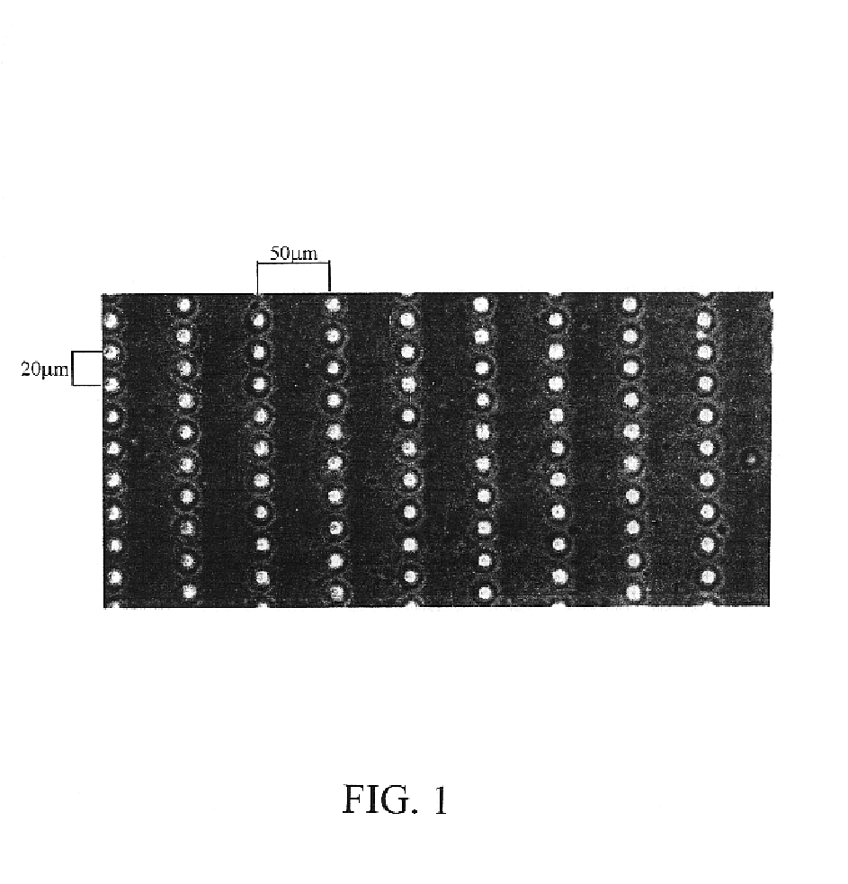
[0126]Microtextured membranes are created using photolithography and microfabrication techniques described herein above. FIG. 1 shows micro-pegged silicone membrane of the present invention viewed with phase microscopy. This membrane has rows of micro-pegs, each 10 μm high spaced 30 μm center to center along the row with 100 μm between the rows (center to center).

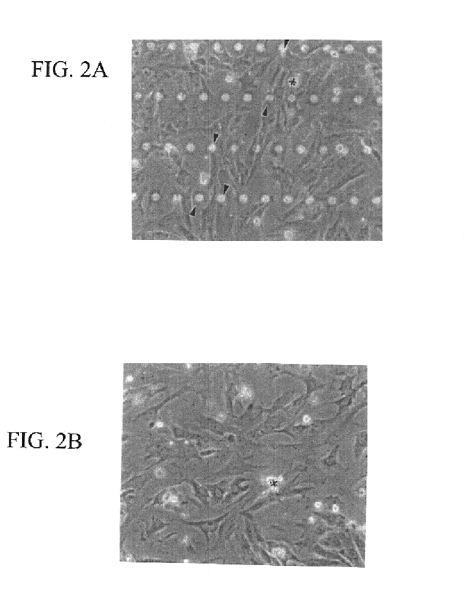
[0127]Primary rat neonatal cardiac myocytes were plated on laminin coated flat silicone membranes or those with micro-pegs 10 μm high to allow for perpendicular attachment. The changes in morphology were assessed by comparing the frequency of peg attachment and cell height and this reveals excellent myocyte shape and peg adhesion. FIG. 2A shows cardiac myocyte cultures growing on a “pegged” silicone membrane coated with laminin. This membrane has rows of micro-pegs, each 10 μm high (seen as rows of bright circles) spaced 30 μm center to center along the ro...

example 3

Altering the Surface Chemistry of the Microtextured Membranes

[0130]This Example deals with chemical bonding protocols which alter the surface chemistry of microtextured silicone and other substrata to promote attachment, adhesion-dependent cell signaling and growth of cardiomyocytes in culture.

[0131]Covalent attachment of peptides to the silicone surfaces are used, as described above and depicted schematically in FIG. 7A. The GRGDSP (SEQ ID NO:1) peptide sequence is known to activate the integrin binding mechanism of various cell lines (Xiao et al., Langmuir 14: 5507-5516, 1998). The peptide functionalized silicone membranes were characterized by radiolabelling and x-ray photoelectron spectroscopy. 125I radiolabelling of the peptide was performed, then the peptide was bound to the silicone surface. FIG. 7B shows the I) C(1s) and II) N(1s) core level x-ray photoelectron spectra for the silicone surfaces at various stages of preparation (letters on spectra correspond to those in FIG. ...


PUM


Abstract

The present invention provides a novel substrate for use in growing cells and for the study of mechanobiology. The membrane of the present invention comprises appropriate microtopography and surface chemical modifications to facilitate the production of adherent and oriented cells that phenotypically resemble cells in vivo.

Description

[0001]The present application claims the benefit of priority of U.S. Provisional application No. 60 / 235,094 filed Sep. 25, 2000. The entire text of that specification is incorporated herein by reference.FIELD OF THE INVENTION[0002]The present invention relates generally to the field of cell growth and culture. More particularly, the present invention provides novel methods and compositions for the growth of cells in an anatomically correct adult phenotype in vitro.BACKGROUND[0003]Cells in the body respond to extracellular stimuli, that are both biochemical and mechanical in nature (Vandenburgh, Am. J. Physiol 262:R350-355, 1992; Buckley, Bone Miner. 4:225-236, 1988; Brunette, Cell Science, 69:35-45, 1984; Harris, 3. Biomech Engineering, 106:19-24, 1984). Both endothelium and muscle respond dynamically to mechanical stimuli and serve as signal transduction interfaces. Although a much focused research topic in cell physiology, there are some fundamental issues in experimental set-up o...

Claims


Application Information

Patent Timeline
IPC IPC(8): C12N5/06C12N5/00A61K35/12C12N5/077
CPCC12N5/0068C12N5/0657A61K35/12C12N2503/00C12N2533/30C12N2533/52C12N2535/10
Inventor RUSSELL, BRENDADESAI, TEJAL A.HANLEY, LUKE
Owner THE BOARD OF TRUSTEES OF THE UNIV OF ILLINOIS
Why Eureka
Try Eureka