Methods for the isolation and analysis of cellular protein content

a cell protein and isolation technology, applied in the field of cell protein isolation and analysis methods and devices, can solve the problems of complex proteomic research, ineffective quantitative data, and inability to provide immunohistochemistry a solution to this problem, and achieve the effect of rapid and reliable identification of source tissu

Inactive Publication Date: 2005-11-29
UNITED STATES OF AMERICA
View PDF14 Cites 38 Cited by
  • Summary
  • Abstract
  • Description
  • Claims
  • Application Information

AI Technical Summary

Benefits of technology

[0010]The present invention describes devices and methods for extracting proteins from samples of microdissected cells, and applying various analytic processes to the extracted proteins, such as immunoassays, 1D and 2D gel electrophoresis characterization, Western blotting, Matrix Assisted Laser Desorption Ionization / Time of Flight (MALDI / TOF), liquid chromatography quadrapole ion trap electrospray (LCQ-MS), and Surface Enhanced Laser Desorption Ionization Spectroscopy (SELDI). These methods allow for direct comparison of qualitative and quantitative protein content of tumor cells and normal cells from the same tissue sample. The methods also allow for investigation of protein characteristics of tumor cells, such as binding ability and amino acid sequence. The present methods also provide, through the use of protein fingerprinting, a rapid and reliable way to identify the source tissue of a tumor metastasis.

Problems solved by technology

Proteomic research has been complicated by the fact that cells undergoing the progression to a diseased state may constitute less than 5% of the volume of a tissue biopsy sample.
Immunohistochemistry cannot provide a solution to this problem, because it can provide only the location of the targeted antigen.
This technique is ineffective at providing quantitative data because it is not possible to calibrate the intensity of immunohistochemical staining with the actual number of antigen molecules in the stained tissue cells.
Additionally, many antibodies cannot differentiate between pro-enzyme and active enzyme species.
Immunologically based studies cannot easily provide a complete picture of a cell's protein contents.

Method used


Image

  • Methods for the isolation and analysis of cellular protein content
  • Methods for the isolation and analysis of cellular protein content
  • Methods for the isolation and analysis of cellular protein content

Examples


example 1

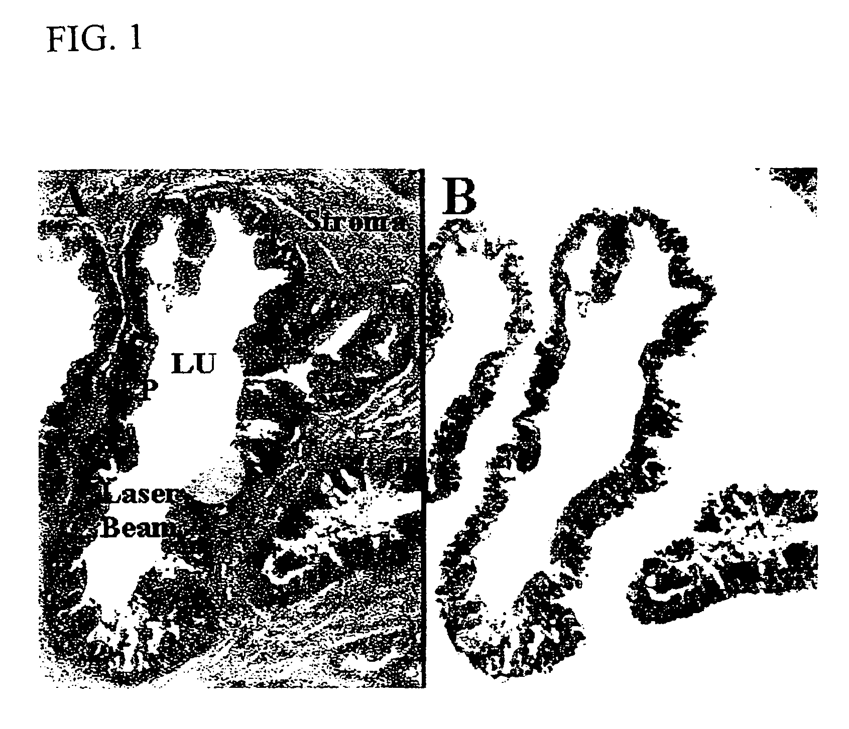
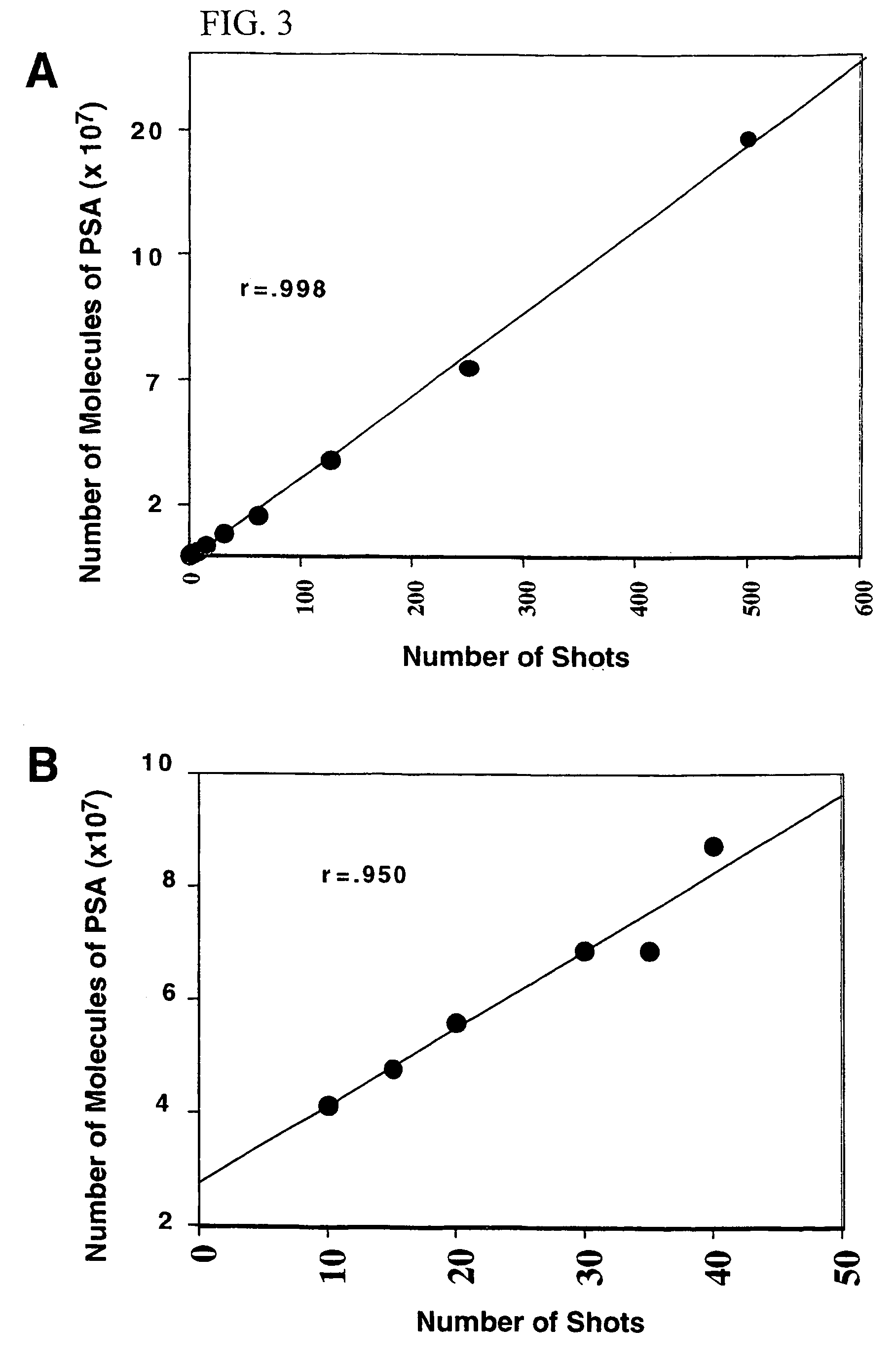
Quantification of Intracellular PSA from Benign and Malignant Prostate Epithelium by Immunoassay

Case Materials

[0048]Tissue was obtained following an IRB approved protocol from both the Urologic Oncology Branch in the National Cancer Institute, Bethesda, Md. and the Mayo Clinic in Rochester Minn. After surgery, the tissue samples were snap frozen in liquid nitrogen. The tissue was then embedded in Optical Coherence Tomography (O.C.T.) compound (Tissue Tek, Miles, Elkhart, Ind.) and stored at −80° C. Cases were selected based on the histology present in the tissue sections so that normal glands, Prostate Intraepithelial Neoplasia (PIN), and adjacent carcinoma, could be compared within the same patient. Prostate tissue cases were selected to include ample stroma to serve as a negative control. Lung tissue was used as a second negative control.

Sectioning and Staining

[0049]The O.C.T embedded tissue blocks were cut into 8 μm sections with a cryostat. After cutting, the sections were immed...

example 2

Comparative Protein Analysis of Normal and Cancerous Esophageal Epithelium Patients and Tissue Samples

[0064]The two specimens studied were from patients who presented to the Shanxi cancer Hospital in Taiyuan, Shanxi Province, People's Republic of China and diagnosed with esophageal cancer. Both patients were considered candidates for curative surgical resection. Both cases were stage two squamous cell carcinomas of the esophagus.

Microdissection

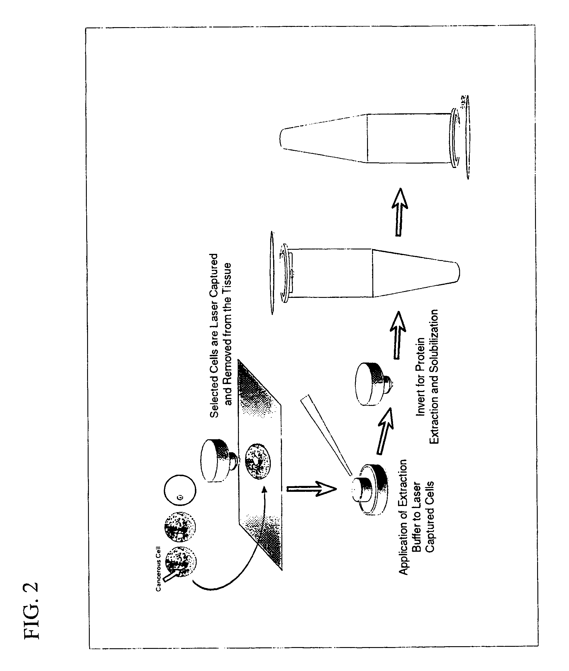
[0065]Frozen section slides were prepared from each case and microdissected by LCM (Pixcell 100, Arcturus Engineering, Mountain View, Calif.) by selectively aiming for and capturing normal epithelium or tumor cells. LCM was performed as previously described except AEBSF (Boeringer Manheim) was added to the staining baths at a final concentration of 2 mM to inhibit proteases. In each case 50,000 cells were procured. Based on careful review of the histologic sections each microdissection is estimated to contain >98% of desired cells (normal vers...

example 3

Comparative Protein Analysis of Normal and Cancerous Prostate Epithelium and In Vitro Prostate Cell Lines

Patients and Tissue Samples

[0090]Tissue from prostate cancer patients undergoing radical prostatectomy at the Clinical Center of the National Cancer Institute (Bethesda, Md.) and at the Mayo Clinic (Rochester, Minn.) was used. Tissue from the peripheral zone of the prostate was procured, immediately snap frozen, and stored at −80° C. Matching normal and tumor cell lines were prepared from the prostatectomy specimens obtained at the NCI and immortalized as described by Bright et al., Cancer Res. 57: 995–1002 (1997). LnCaP and PC3 cells were purchased from the American Type Culture Collection (Manassas, Va.). Microdissection was performed and the cells were prepared for 2D-PAGE analysis as described above in Example 2. 2D-PAGE gels were done using alpha-tubulin to normalize the sample load, as described in Example 2. The anti-alpha tubulin antibodies, used at 1:1000, and HRP-couple...


PUM

PropertyMeasurementUnit
thicknessaaaaaaaaaa
volumesaaaaaaaaaa
diameteraaaaaaaaaa

Abstract

The present invention describes devices and methods for performing protein analysis on laser capture microdissected cells, which permits proteomic analysis on cells of different populations. Particular disclosed examples are analysis of normal versus malignant cells, or a comparison of differential protein expression in cells that are progressing from normal to malignant. The protein content of the microdissected cells may be analyzed using techniques such as immunoassays, 1D and 2D gel electrophoresis characterization, Western blotting, liquid chromatography quadrapole ion trap electrospray (LCQ-MS), Matrix Assisted Laser Desorption Ionization / Time of Flight (MALDI / TOF), and Surface Enhanced Laser Desorption Ionization Spectroscopy (SELDI). In addition to permitting direct comparison of qualitative and quantitative protein content of tumor cells and normal cells from the same tissue sample, the methods also allow for investigation of protein characteristics of tumor cells, such as binding ability and amino acid sequence, and differential expression of proteins in particular cell populations in response to drug treatment. The present methods also provide, through the use of protein fingerprinting, a rapid and reliable way to identify the source tissue of a tumor metastasis.

Description

CROSS REFERENCE TO RELATED APPLICATIONS[0001]This is the National Stage of International Application No. PCT / US00 / 04023, filed Feb. 16, 2000, and claims the benefit of U.S. Provisional Application No. 60 / 120,288, filed Feb. 16, 1999. The provisional application is incorporated herein in its entirety.[0002]By this amendment the specification has been changed to reflect prior related applications. No new matter is added by this amendment.BACKGROUND OF THE INVENTION[0003]1. Field of the Invention[0004]The present invention relates to methods and devices for the analysis of cell samples where the samples are pure populations or subpopulations of desired types. In particular, the present invention allows for direct comparison of protein content and protein characteristics between proteins isolated from tumor and normal cells from the same tissue sample.[0005]2. Discussion of the Background[0006]Proteomics is the study of variations in cellular protein levels between normal and disease st...

Claims


Application Information

Patent Timeline
Patent Type & Authority Patents(United States)
IPC IPC(8): G01N1/18G01N1/28G01N1/30G01N21/00G01N21/75G01N33/53G01N33/68
CPCG01N33/6803Y10T436/255Y10T436/25375G01N2001/284
Inventor LIOTTA, LANCE A.SIMONE, NICOLEEMMERT-BUCK, MICHAELPETRICOIN III, EMMANUEL F.
Owner UNITED STATES OF AMERICA
Why Eureka
Try Eureka