Bone xenografts

a bone xenograft and bone technology, applied in bone implants, prostheses, medical science, etc., can solve the problems of less than optimal bone defect filling, low bone density, and inability to transplant autologous bone tissue for large defects,

Inactive Publication Date: 2005-12-06
APERION BIOLOGICS
View PDF43 Cites 26 Cited by
  • Summary
  • Abstract
  • Description
  • Claims
  • Application Information

AI Technical Summary

Benefits of technology

[0020]As used herein, the term “portion”, as in, for example, a portion of bone or second surface carbohydr

Problems solved by technology

Transplantable autologous bone tissue for large defects, particularly in children, is often unavailable, however.
In addition, autologous bone transplantation may result in postoperative morbidity such as pain, hemorrhage, wound problems, cosmetic disability, infection or nerve damage at the donor site.

Method used


Image

  • Bone xenografts
  • Bone xenografts
  • Bone xenografts

Examples


example 1

Assessment of Response in Mice to Implanted Bone Treated with α-Galactosidase and Demineralized Bone Matrix Gel Containing Osteoinductive Factor

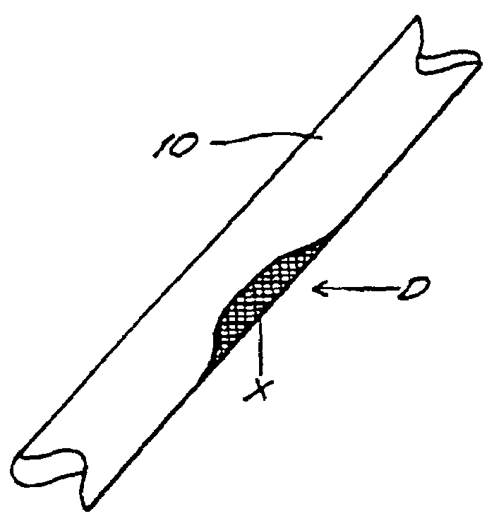
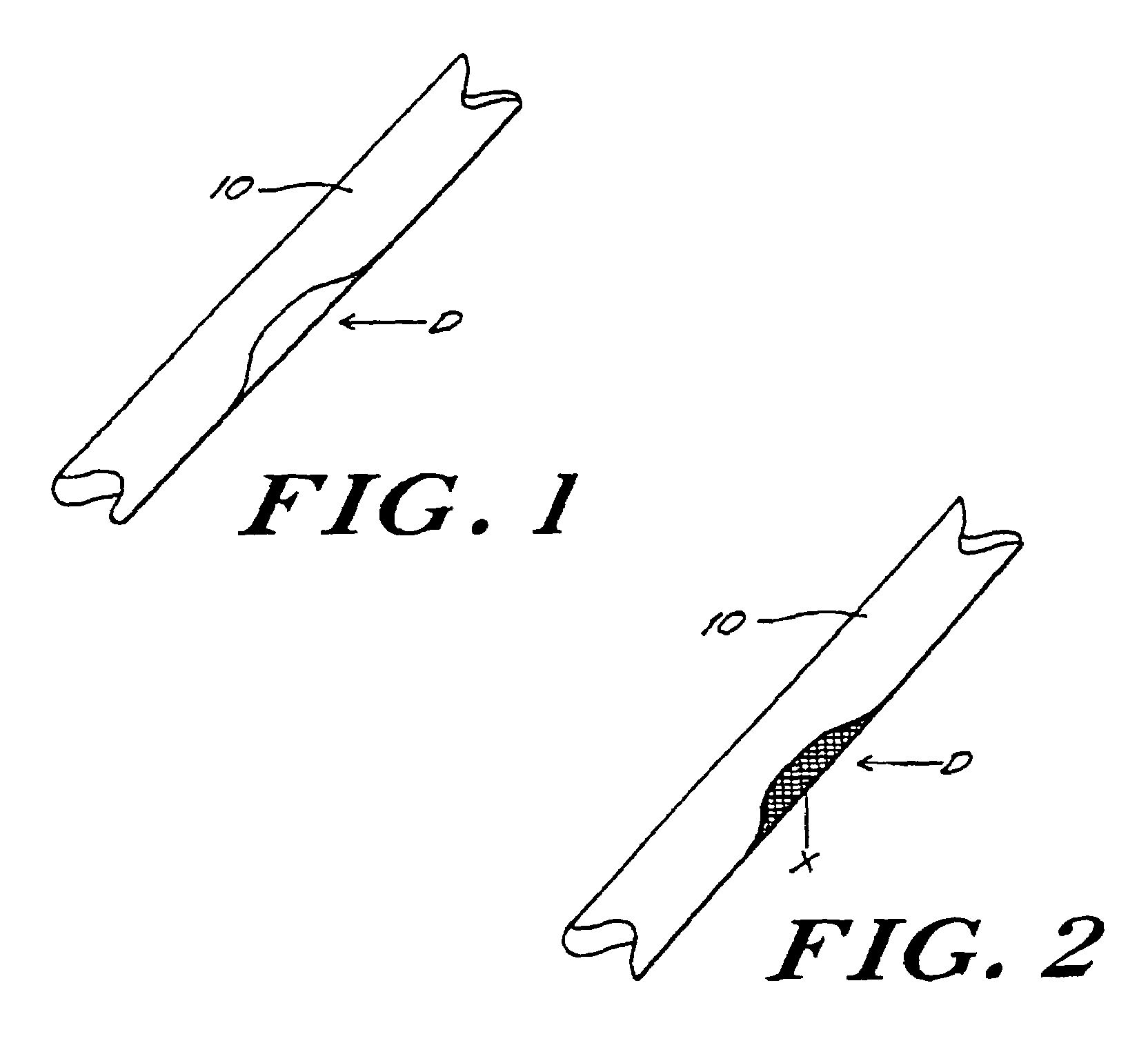
[0062]In this example, porcine bone implants are treated with α-galactosidase to eliminate α-galactosyl epitopes and impregnated with demineralized bone matrix gel containing osteoinductive factor to stimulate the conversion of soft tissue formers in the target defect to osseous tissue formers. The implants are transplanted into mice and the response to the implants is assessed. An exemplary bone portion 10 with a defect D is shown in FIG. 1.

[0063]Porcine bone implants are sterilely prepared and surrounding attached soft tissues surgically removed. The bone specimens are washed for at least five minutes with an alcohol, such as ethanol or isopropanol, to remove synovial fluid and lipid soluble contaminants.

[0064]The bone specimens are frozen at a temperature ranging from about −35° C. to about −90° C., and preferably at a temperature up to a...

example 2

Assessment of Primate Response to Implanted Bone Treated with α-Galactosidase and Demineralized Bone Matrix Gel Containing Osteoinductive Factor

[0074]In this example, porcine bone implants are treated with α-galactosidase and demineralized bone matrix gel containing osteoinductive factor, the implants are transplanted into cynomolgus monkeys, and the primate response to the bone implants is assessed, as described in Example 1. After the bone xenografts are explanted and tissue is harvested for possible immunologic testing, the animals are allowed to recover and are monitored closely until the incisions have healed and the gait of the animals is normal.

example 3

Assessment of Response in Mice to Implanted Bone Treated with α-Galactosidase, Demineralized Bone Matrix Gel Containing Osteoinductive Factor, Fucosyl and Fucosyltransferase

[0075]In this example, porcine bone implants are treated with α-galactosidase to eliminate α-gal epitopes, as described in Example 1. The implants are further treated with fucosyl and fucosyl transferase to cap carbohydrate chains with fucosyl. Fucosyltransferase facilitates the transfer of fucosyl to the xenograft. The fucosyl links to and thus caps the carbohydrate chains. Capping with fucosyl interferes with the ability of the subject's immune system to recognize the xenograft as foreign. The implants are transplanted into mice, and the response to the bone implants is assessed.

[0076]Porcine bone implants are prepared as described in Example 1 including the α-galactosidase treatment. Prior to implantation into the mice, however, the implants are further treated with a predetermined amount of fucosyl and fucosy...


PUM


Abstract

The invention provides an article of manufacture comprising a substantially non-immunogenic bone xenograft for implantation into humans. The invention further provides a method for preparing a bone xenograft by removing at least a portion of a bone from a non-human animal to provide a xenograft (X); washing the xenograft in saline and alcohol; subjecting the xenograft to a cellular disruption treatment; and treating the xenograft with a glycosidase to remove surface carbohydrate moieties. The invention also provides an article of manufacture produced by the above identified method of invention. The invention further provides a bone xenograft for implantation into a human including a portion (10) of a bone from a nonhuman animal, wherein the portion has substantially no surface carbohydrate moieties which are susceptible to glycosidase digestion. Each xenograft of the invention has substantially the same mechanical properties as a corresponding native bone.

Description

[0001]This application is a 371 of PCT / US99 / 05661 filed Mar. 15, 1999, which claims benefit of Ser. No. 60 / 078,136 filed Mar. 16, 1998, and claims benefit of Ser. No. 60 / 100,755 filed Sep. 17, 1998.FIELD OF THE INVENTION[0002]The present invention relates to the field of bone transplantation, and in particular, to replacement and repair of damaged or defective human bone using a substantially immunologically compatible bone from a non-human animal.BACKGROUND OF THE INVENTION[0003]Human bone, a hard connective tissue consisting of cells embedded in an extracellular matrix of mineralized ground substance and collagen fibers, (Stedman's Medical Dictionary, Williams & Wilkins, Baltimore, Md. (1995)), is the most frequently transplanted tissue in humans. J. M. Lane et al. Current Approaches to Experimental Bone Grafting, 18 Orthopedic Clinics of North America (2) 213 (1987). In the United States alone more than 100,000 bone graft or implant procedures are performed every year to repair o...

Claims


Application Information

Patent Timeline
IPC IPC(8): A61F2/28A61F2/30A61L27/36
CPCA61F2/28A61L27/3608A61L27/365A61L27/3683A61L27/3687A61F2/3094A61F2002/2835
Inventor STONE, KEVIN R.
Owner APERION BIOLOGICS
Why Eureka
Try Eureka