Method of creating biological and biosynthetic material for implantation

a biosynthetic material and biosynthetic technology, applied in the field of biosynthetic material creation, can solve the problems of thrombosis and occlusion, lumenal narrowing, and inability to eliminate the degree of calcification

Inactive Publication Date: 2006-04-04
KRYOCOR PTY LTD
View PDF14 Cites 25 Cited by
  • Summary
  • Abstract
  • Description
  • Claims
  • Application Information

AI Technical Summary

Benefits of technology

[0019]Thus, microarchitectural alignment of the tissue is maintained without damaging the tissue.
[0024]Thus, the treating of the tissue in this manner has been found to provide a material with an intact microarchitecture and cellular lining which inhibits in vivo calcification, tissue ingrowth and tranemural angiogenesis, and provides a microenvironment for the adhesion of transplanted living cells. Additionally, haemocompatibility, compliance and flexibility can be varied according to the desired end use by varying the process within the functional range.

Problems solved by technology

Storage in 50% alcohol prior to implantation reduced but did not eliminate the degree of calcification.
An unwanted aspect is the uncontrolled nature of tissue ingrowth particularly in vascular conduits of 6 mm internal diameter and less which causes lumenal narrowing, thrombosis and occlusion with subsequent graft failure.

Method used


Image

  • Method of creating biological and biosynthetic material for implantation
  • Method of creating biological and biosynthetic material for implantation
  • Method of creating biological and biosynthetic material for implantation

Examples


example 1

[0041]The method of treating tissue according to this embodiment was applied to bovine ureters for use as vascular prostheses as follows.

[0042]Bovine ureters were collected under hygienic conditions and transported to the laboratory at 4° C. Physiological saline was pumped via the pelvic end of the ureter. The distal or bladder end was temporarily occluded and the pressure of the saline within the lumen was increased to 115 mm Hg. The diameter of the ureter at the mid point was recorded. A compr ssible silicon mandrel slightly narrower in diameter than the ureter and mounted on a blunt nosed steel introducer was inserted a traumatically into the pressure filled ureter. The metal introducer was removed. Excess tissue was removed from the exterior of the ureter. The ureter was then tethered to the silicone at one end, allowing some movement along the silicone. The mounted ureter was placed into a rotating drum at 12 rpm at a temperature of 21° C. The concentration of glutaraldehyde wa...

example 2

[0045]In vivo inhibition of calcification was assessed in greyhound dogs as follows.

[0046]Grafts of 6 mm internal diameter and not less than 10 cm in length prepared from bovine ureters according to this embodiment were implanted into the aorto-iliac position with the distal aorta to the proximal anastomosis ligated. Grafts of 4 mm internal diameter and not less than 6 cm in length were implanted as femoral interposition grafts.

[0047]No anti-coagulation or antiplatelet therapy was used.

[0048]Periodic angiograms were taken at predetermined intervals to verify patency and detect aneurysms, dilatation or stenoses. The animals were sacrificed at predetermined intervals, or at graft closure or animal distress.

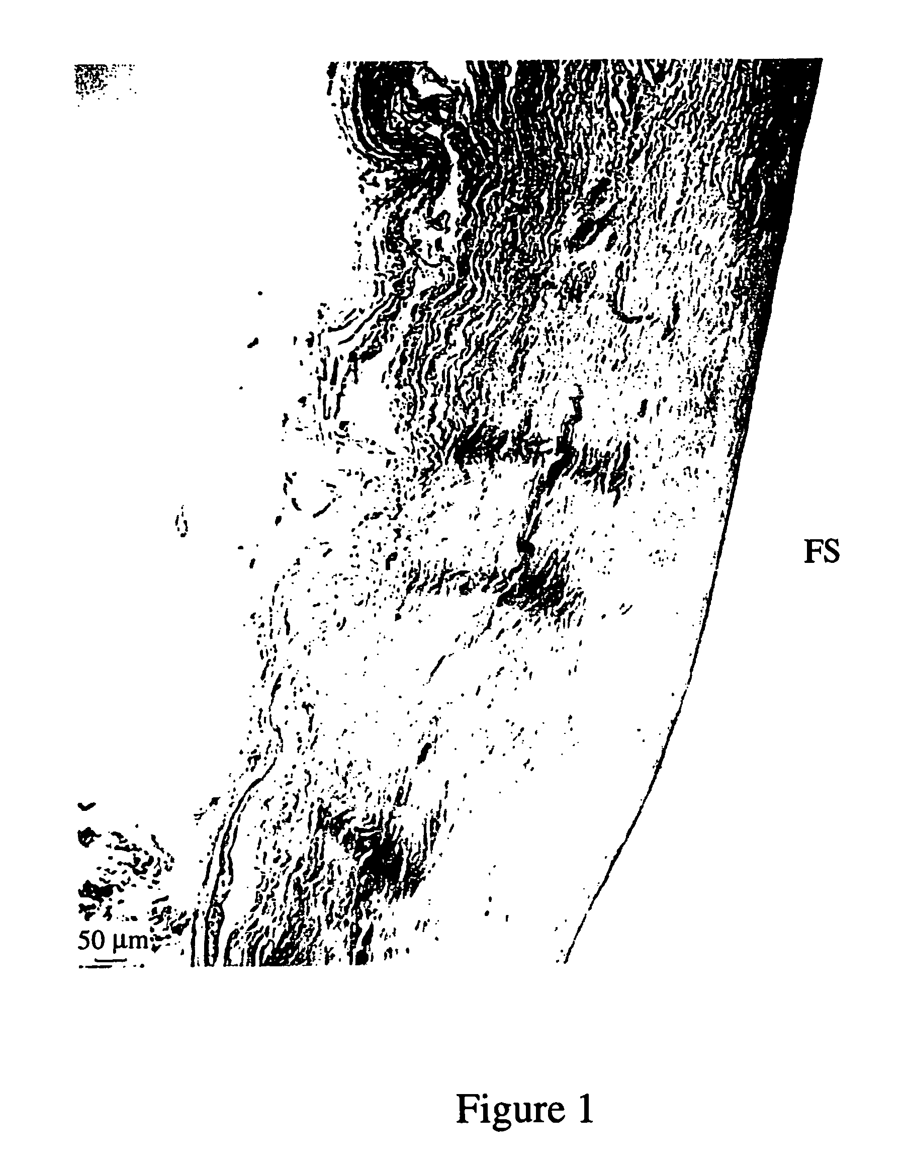
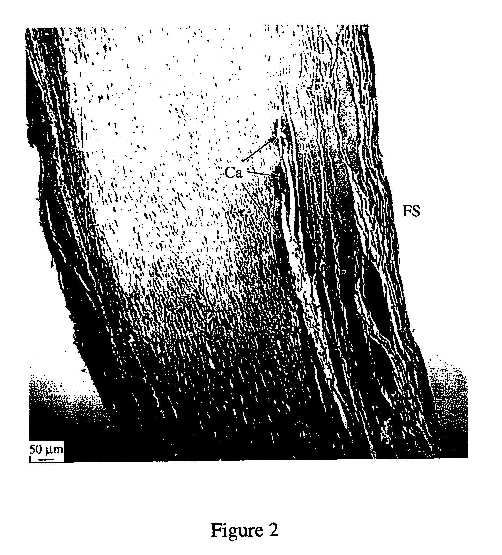
[0049]Finally, macro- and microscopic examination of the explants were performed.

[0050]When patency, mural integrity and luminal integrity of implanted material processed by the current invention were compared with that of U.S. Pat. No. 4,466,139 neither was compromised. However, wh...

example 3

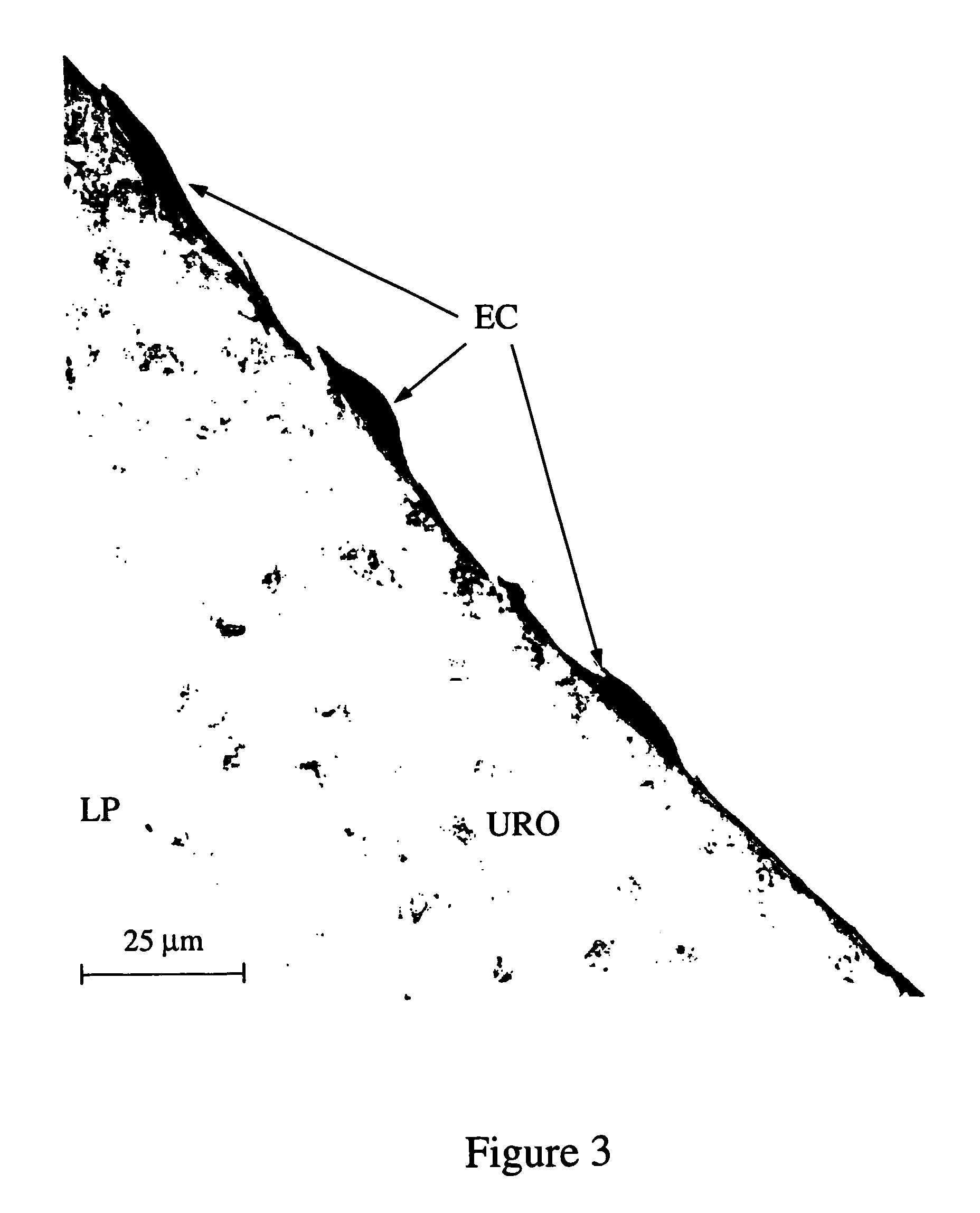
[0052]The attachment and adherence of human saphenous vein endothelial cells transplanted onto the surface of the processed bovine ureter were evaluated as follows.

[0053]Human saphenous vein endothelial cells were isolated, harvested and seeded using published or modified techniques. Human saphenous vein endothelial cells were isolated from inverted varicosed human saphenous vein segments obtained from the operating room and transported to the laboratory in chilled Hanks Balanced Salt Solution (HBSS, Sigma Australia) containing ampicillin (100 μg / mL), gentamycin (50 μg / mL), amphotericin B (2.5 μg / mL), L-glutamine (2.5 mM) and sodium bicarbonate (0.23%) and stored at 4° C. The ends of the veins were ligated with surgical silk. A modified enzymatic harvesting technique was utilized whereby the veins were immersed in 10 mL of 2 mg / mL collagenase (Worthington type IV, Sigma) for 10 minutes at 37° C. then vortexed on an automatic speed for 1 minute at 10 second intervals. The collected c...


PUM

PropertyMeasurementUnit
time periodaaaaaaaaaa
temperatureaaaaaaaaaa

Abstract

A method of creating an implantation material from biological and tissue engineered biosynthetic and biological tissue of autogenic, allogenic and xenogenic origin suitable for implantation into humans or animals as surgical and vascular prostheses. The resultant material inhibits in vivo calcification and provides a non-porous biomatrix which is impervious to angiogenesis and tissue ingrowth and suitable for the adhesion and retention of transplanted living cells such as endothelial cells, without the need for additional extracellular matrix protein coating. The method, which incorporates a gradual increase in glutaraldehyde concentration from 0% to no more than 5% weight / volume in a pH which is gradually changed from acid to alkaline at room temperature, maintains the micro-architecture and the cellular lining of the material. Additionally flexibility, compliance and haemo-compatibility along with strength and durability can be varied according to the end use. The glutaraldehyde penetrates the material gradually and completely, cross linking the collagenous components of the tissue.

Description

BACKGROUND TO THE INVENTION[0001]1. Field of the Invention[0002]The present invention relates to a method of creating biological and biosynthetic material suitable for implantation to replace or augment damaged, diseased or absent tissues, structures or organs, of particular but by no means exclusive application in the treatment of autogenic, allogenic or xenogenic material of mammalian origin, to maintain a microarchitectural organization and inhibit in vivo calcification, tissue ingrowth and transmural angiogenesis. In one embodiment, as may be determined by the end use, transplanted living cells can be attached and retained under in vivo conditions.[0003]2. Description of Related Art[0004]Tissue stabilization and fixation using, for example, glutaraldehyde to impart strength, neutralize antigenic sites and sterilize biosynthetic and biological tissue for human implantation has been described previously. U.S. Pat. Nos. 4,319,363, 4,466,139 and 4,681,588 (Ketharanathan) disclose th...

Claims


Application Information

Patent Timeline
Patent Type & Authority Patents(United States)
IPC IPC(8): A61K35/44A61L2/00A61L27/00A61L27/36
CPCA61L2/0035A61L27/3691A61L27/3683A61L2/0088A61L27/3687
Inventor KETHARANATHAN, VETTIVETPILLAI
Owner KRYOCOR PTY LTD
Why Eureka
Try Eureka