
Cartilage composites and methods of use

a technology of cartilage and composites, applied in the field of new neocartilage compositions, can solve the problems of chondrocyte dedifferentiation and formation of fibrocartilage, inability to grow human articular cartilage using traditional cell culture methods such as tissue-culture plastic surfaces and serum-containing growth media, and failure to achieve in vitro neocartilage production

Inactive Publication Date: 2006-08-08
ZIMMER INC
View PDF20 Cites 28 Cited by
  • Summary
  • Abstract
  • Description
  • Claims
  • Application Information

AI Technical Summary

Problems solved by technology

Attempts to grow human articular cartilage using traditional cell culture methods such as growing chondrocytes on tissue-culture plastic surfaces using serum-containing growth media have proved unsuccessful.
Without a readily available replacement tissue, recent methods of articular cartilage repair have focused on biological resurfacing of cartilage defects with either a prosthetic device or with live chondrocytes.
These methods, like in vitro neocartilage production, have been less than completely successful.
It has recently been shown, however, that neither the autogenous nor the allogenic transplant method results in consistent growth of articular cartilage in vivo, but rather results in chondrocyte dedifferentiation and formation of fibrocartilage.
Because of the reduced aggrecan content of fibrocartilage, it cannot withstand the same biomechanical stresses as articular cartilage.
Fibrocartilage degenerates with use, and its formation following joint repair may promote joint dysfunction and permanent disability.
Despite intensive research efforts to ascertain the biological basis of osteoarthritis, its development and progression remain poorly understood.
Thus, animal models are inadequate for evaluating the efficacy of potential disease modifying agents in human osteoarthritis.
A shortage of normal articular cartilage for studying articular cartilage disease and articular cartilage response to natural and synthetic compounds exists because the only source of healthy articular cartilage currently available is from deceased adult donors which may show degenerative changes.

Method used


Image

  • Cartilage composites and methods of use
  • Cartilage composites and methods of use
  • Cartilage composites and methods of use

Examples


example 1

Preparation and Culture of Immature and Adult Human Chondrocytes

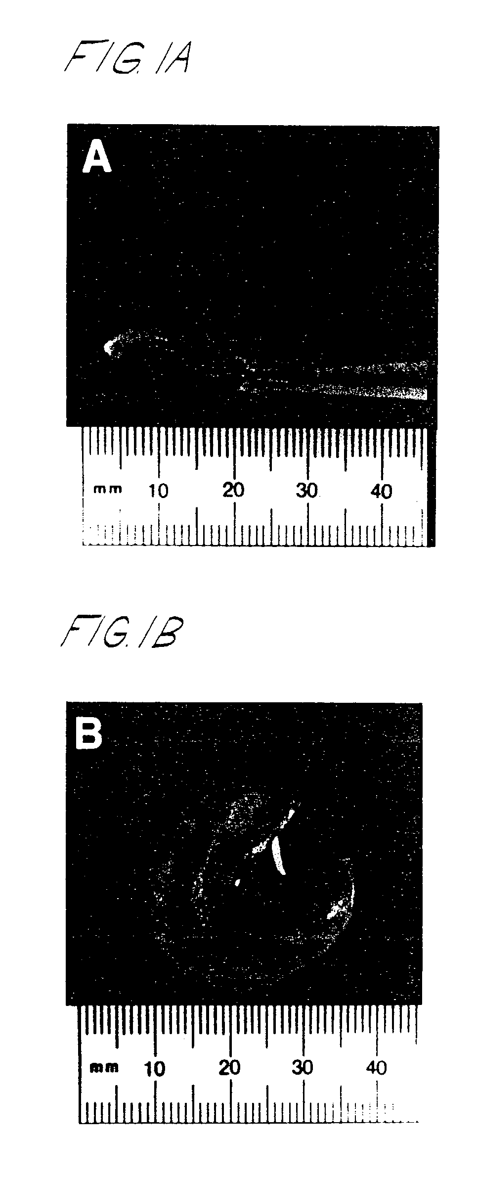
[0128]Chondrocytes were isolated within 8–24 hours of death from the upper region of the tibial plateau and femoral condyle of intact fetal bones (18–21 weeks gestation, Advanced Bioscience Resources, Inc., Alameda, Calif.). Hyaline cartilage from infant knees (≦8 months) was removed from the metaphysis of both the proximal tibia and distal femur using rib cutters and transferred to sterile serum-free DMEM, containing antibiotics (2×) and 1% bovine serum albumin, for chondrocyte isolation as described below. Additional samples of more mature cartilage (12–58 years) were processed similarly. All of these cartilage tissues were provided by Mid-America Transplant Association (St. Louis, Mo.). All tissues were transported on wet ice prior to use.

[0129]Skin from fetal limbs was removed and stored at −20° C. for preparation of collagen standards (i.e., types I and III) via limited pepsinization. Skeletal muscle and other conn...

example 2

Biochemical Assessment of Neocartilage Formation

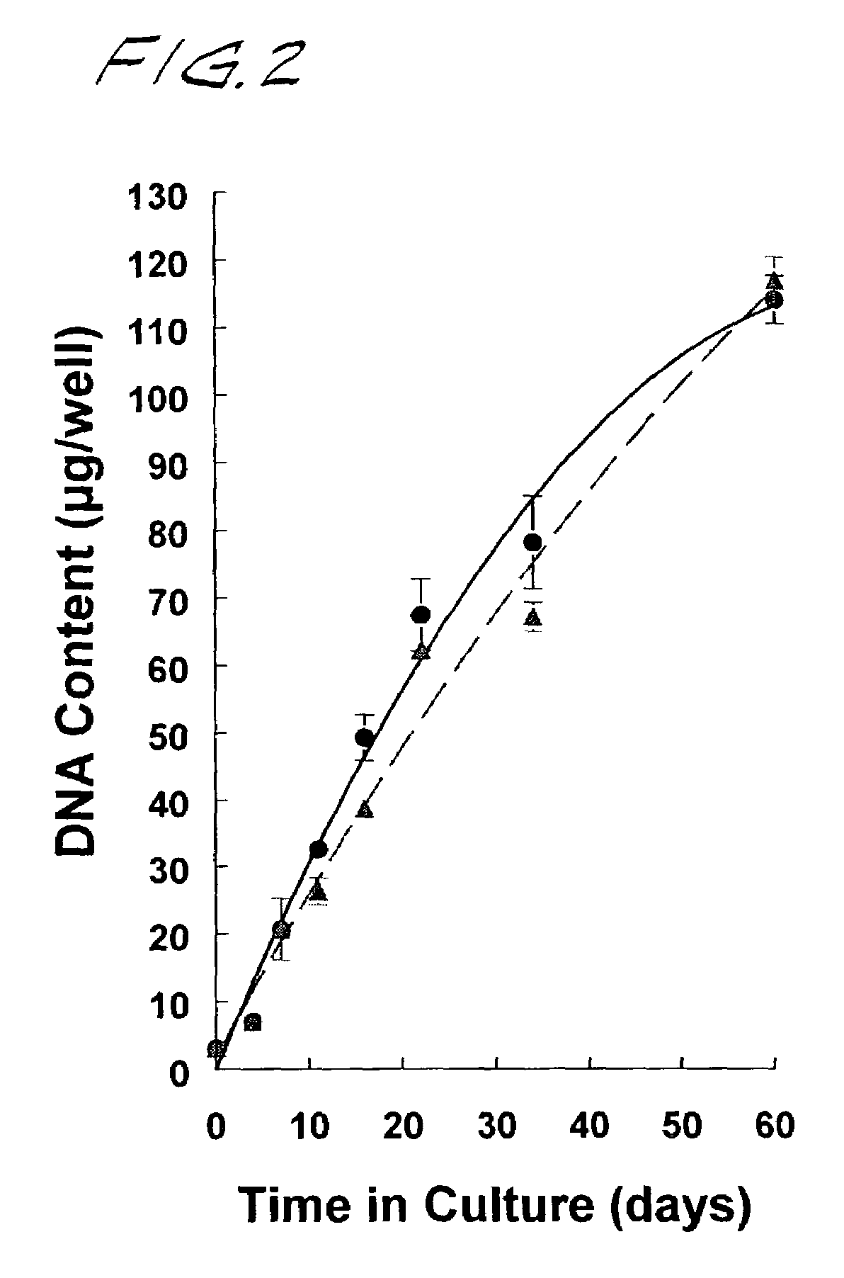
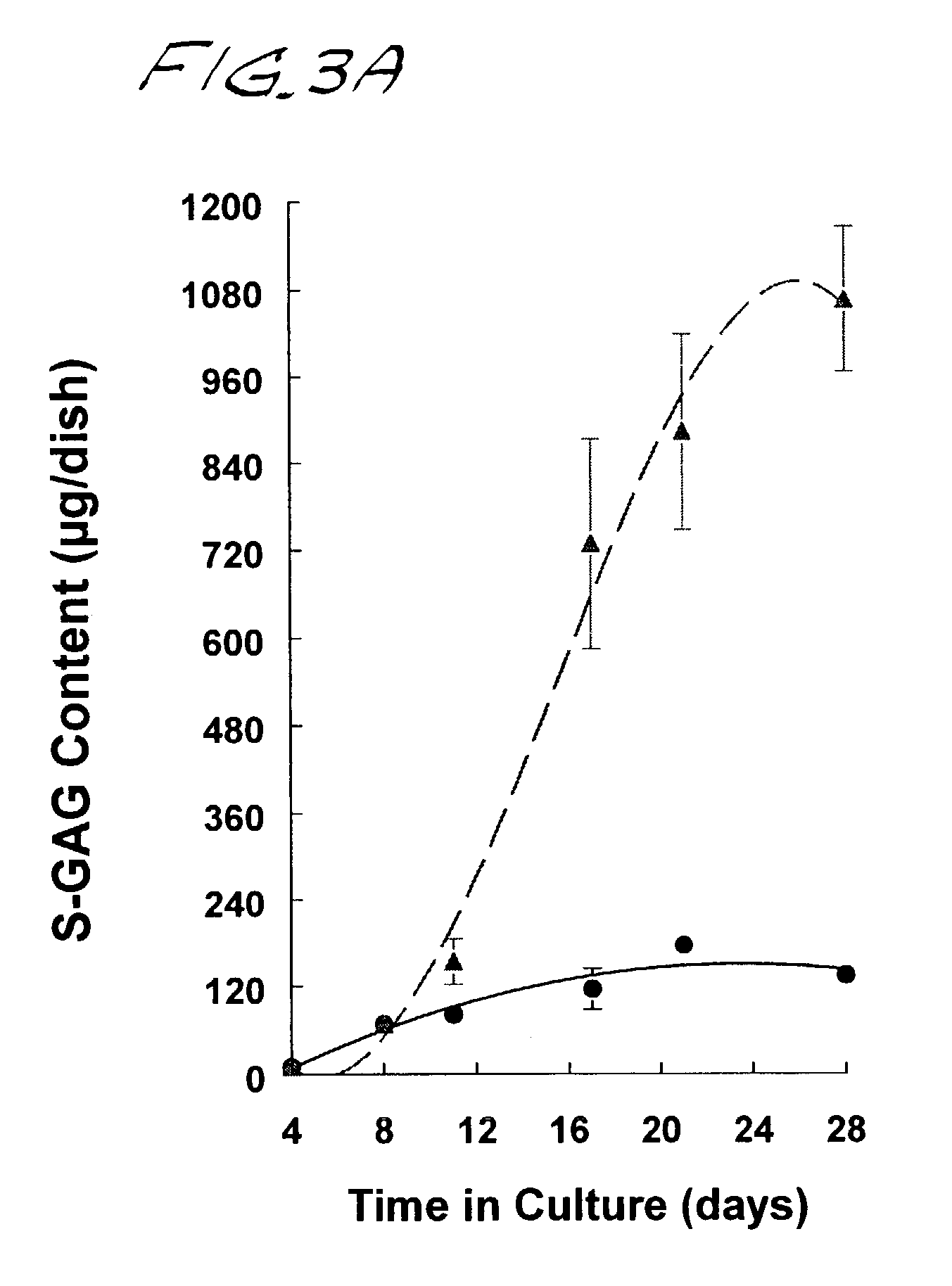
[0137]Neocartilage formation was assessed by calorimetric analyses of sulfated glycosaminoglycan (S-GAG) and hydroxyproline (OH-proline), general measures of proteoglycan and collagen synthesis, respectively. Neocartilage disks were lyophilized and digested 18 hrs at 56° C. in 500 μl of 0.1 M sodium acetate buffer (pH 5.6), containing 5 mM Na4EDTA, 5 mM L-cysteine, and 125 μg / ml papain. The digests were cooled to room temperature for determination of S-GAG, OH-proline, and DNA content.

[0138]Sulfated-GAG and the OH-proline content of papain digested material were determined at the indicated times via microplate colorimetric procedures adapted from Farndale et al., Biochim Biophys Acta 883: 173–177 (1986), and Stegemann and Stalder, Clin Chim Acta 18: 267–273 (1967), respectively. Chondroitin-6-sulfate (Calbiochem) and cis-4-hydroxy-L-proline (Sigma) were used as standard.

[0139]DNA content was estimated by fluorometric analysis using a C...

example 3

Model Systems for Studying Articular Cartilage Disease and Articular Cartilage Response to Natural and Synthetic Compounds

[0148]Neocartilage cultures were established in 24 or 48 well plastic dishes (Corning) as described above (Example 1) and grown to day 30. The hyaline cartilage matrix was then stimulated to undergo autolytic degradation following 30 day activation with increasing concentrations of a variety of cytokine and / or inflammatory agents, including IL-1, Il-6, IL-17, TNF-α, LPS, and phorbol ester. Culture media were collected every 48 hours and subsequently changed by the addition of fresh media±inflammatory stimuli.

[0149]Cultures were terminated and the biochemical composition of the neocartilage matrix that remained was analyzed as described previously. Parallel samples were processed for light and transmission electron microscopy for histological evaluation of extracellular matrix components.

[0150]Conditioned media were thawed and identification of chondrocyte-derived...


PUM

PropertyMeasurementUnit
thickaaaaaaaaaa
diameteraaaaaaaaaa
thicknessaaaaaaaaaa

Abstract

Disclosed are neocartilage compositions characterized by having multiple layers of cells, said cells being surrounded by a substantially continuous insoluble glycosaminoglycan and collagen-enriched hyaline extra-cellular matrix, and which neocartilage phospholipids are advantageously enriched in anti-inflammatory n-9 fatty acids, particularly 20:3 n-9 eicosatrienoic or Mead acid.Also disclosed are methods of growing neocartilage in substantially serum-free growth media and methods of producing a conditioned growth media containing compounds effective to enhance neocartilage formation.The neocartilage compositions are useful as implants and as replacement tissue for damaged or defective cartilage and as a model system for studying articular cartilage disease and response to natural and synthetic compounds.

Description

[0001]This application is a continuation of application Ser. No. 09 / 467,520, filed Dec. 21, 1999, which is a divisional of application Ser. No. 09 / 054,913, filed Apr. 3, 1998, which issued as U.S. Pat. No. 6,235,316 B1 on May 22, 2001, and which claims priority from U.S. Provisional Application Ser. No. 60 / 043369, filed Apr. 4, 1997, all of which are incorporated herein by reference.BACKGROUND OF THE INVENTION[0002]This invention relates generally to novel neocartilage compositions useful as implants and as replacement tissue for damaged or defective cartilage, as model systems for studying articular cartilage disease and articular cartilage response to natural and synthetic compounds and for the isolation of cartilage derived substances useful in the biotechnology industry.[0003]More specifically, the invention concerns neocartilage, particularly human neocartilage, having multiple layers of cells surrounded by a substantially continuous insoluble glycosaminoglycan and collagen-enr...

Claims


Application Information

Patent Timeline
Patent Type & Authority Patents(United States)
IPC IPC(8): C12N5/00A61F2/08A61K35/12A61K35/32A61L27/38C12N5/077
CPCA61L27/3612A61L27/3633A61L27/3654A61L27/3695A61L27/3817A61L27/3852A61L27/3895C12N5/0655A61K35/32A61K35/12C12N2533/90C12N2500/25C12N2500/38C12N2500/99C12N2501/01C12N2501/23C12N2501/25C12N2501/70C12N2501/91C12N2501/998C12N2502/1317C12N2503/00A61L2430/06A61P19/00C12N2500/90
Inventor ADKISSON, HUSTON D.
Owner ZIMMER INC
Learn More