
N-terminal and C-terminal markers in nascent proteins

a technology of c-terminal and nascent proteins, applied in the field of non-radioactive markers, can solve the problems of time-consuming use of radioactive labeling, health risks in the laboratory, and many drawbacks of radioactive labeling methods

Inactive Publication Date: 2006-09-05
AMBERGEN INC
View PDF1 Cites 17 Cited by
  • Summary
  • Abstract
  • Description
  • Claims
  • Application Information

AI Technical Summary

Benefits of technology

The present invention provides methods for labeling, detecting, and isolating nascent proteins in a translation system without using radioactive amino acids or other radioactive labels. The methods involve misaminoacylating tRNA molecules with fluorescent markers and introducing them into a translation system. The resulting nascent proteins containing the fluorescent markers can be detected, isolated, and quantitated. The invention also contemplates the use of BODIPY markers and the introduction of initiator or suppressor tRNA molecules. Kits containing misaminoacylated tRNA and components of a protein synthesis system are also provided. Overall, the invention provides improved methods for studying the translation process and the function and structure of nascent proteins.

Problems solved by technology

Radioactive methods suffer from many drawbacks related to the utilization of radioactively labeled amino acids.
Handling radioactive compounds in the laboratory always involves a health risk and requires special laboratory safety procedures, facilities and detailed record keeping as well as special training of laboratory personnel.
Disposal of radioactive waste is also of increasing concern both because of the potential risk to the public and the lack of radioactive waste disposal sites.
In addition, the use of radioactive labeling is time consuming, in some cases requiring as much as several days for detection of the radioactive label.
The long time needed for such experiments is a key consideration and can seriously impede research productivity.
While faster methods of radioactive detection are available, they are expensive and often require complex image enhancement devices.
The use of radioactive labeled amino acids also does not allow for a simple and rapid means to monitor the production of nascent proteins inside a cell-free extract without prior separation of nascent from preexisting proteins.
However, a separation step does not allow for the optimization of cell-free activity.
Radioactive labeling methods also do not provide a means of isolating nascent proteins in a form which can be further utilized.
The presence of radioactivity compromises this utility for further biochemical or biophysical procedures in the laboratory and in animals.
This is clear in the case of in vitro expression when proteins cannot be readily produced in vivo because the protein has properties which are toxic to the cell.
Recent research has met with some success, but these methods have had numerous drawbacks.
Radioactive labeling methods also do not provide a simple and rapid means of detecting changes in the sequence of a nascent protein which can indicate the presence of potential disease causing mutations in the DNA which code for these proteins or fragments of these proteins.
Current methods of analysis at the protein level rely on the use of gel electrophoresis and radioactive detection which are slow and not amenable to high throughput analysis and automation.
However, this requires large regions of DNA to be sequenced, which is time-consuming and expensive.
Radioactive labeling methods also do not provide a simple and rapid means of studying the interaction of nascent proteins with other molecules including compounds which might be have importance as potential drugs.
Furthermore, site-specific incorporation of non-native amino acids is not suitable in general for detection of nascent proteins in a cellular or cell-free protein synthesis system due to the necessity of incorporating non-sense codons into the coding regions of the template DNA or the mRNA.
This method is of limited use because it requires that the protein be folded into a native form and also for antibodies to have been previously produced against the nascent protein or a known protein which is fused to the unknown nascent protein.
Such procedures are time consuming and again require identification and characterization of the protein.
These types of post-aminoacylation modifications, although useful, do not provide a general means of incorporating non-native amino acids into the nascent proteins.
The disadvantage is that only those non-native amino acids that are derivatives of normal amino acids can be incorporated and only a few amino acid residues have side chains amenable to chemical modification.
This factor can lower the efficiency of incorporation of the non-native amino acid linked to the tRNA.
Non-specific, post-aminoacylation modifications of tRNA structure could also compromise its participation in protein synthesis.
This approach is of limited use since most proteins do not have special properties with which they can be easily detected.
In many cases, however, the expressed protein may not have been previously characterized or even identified, and thus, its characteristic properties are unknown.

Method used


Image

  • N-terminal and C-terminal markers in nascent proteins
  • N-terminal and C-terminal markers in nascent proteins
  • N-terminal and C-terminal markers in nascent proteins

Examples


example 1

Preparation of Markers

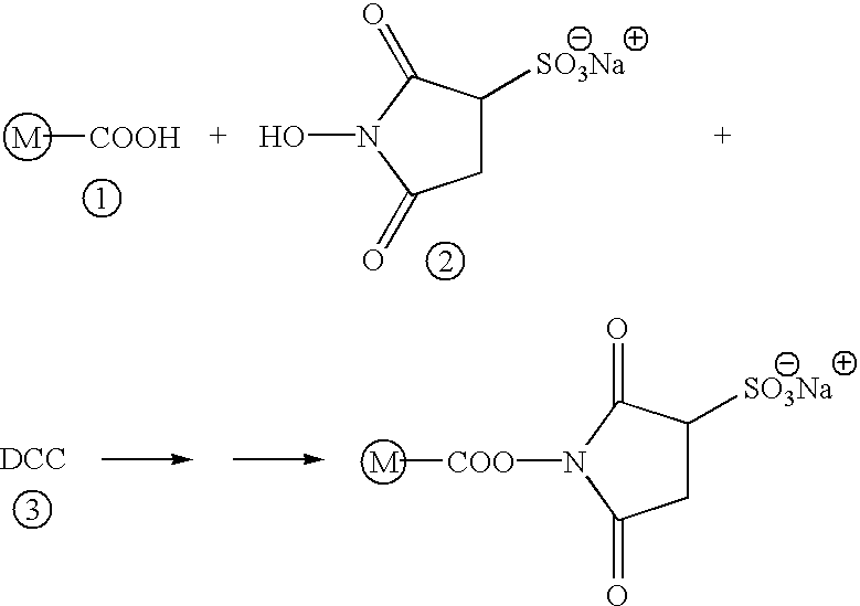
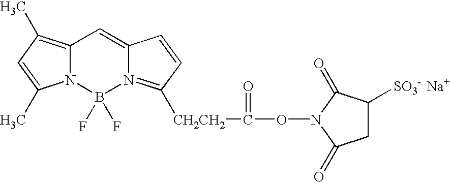
[0232]Synthesis of Coumarin Amino Acid: 4-(Bromomethyl)-7-methoxy coumarin (FIG. 15, compound 1; 6.18 mmole) and diethylacetamidomalonate (FIG. 15, compound 2; 6.18 mmole) were added to a solution of sodium ethoxide in absolute ethanol and the mixture refluxed for 4 hours. The intermediate obtained (FIG. 15, compound 3) after neutralization of the reaction mixture and chloroform extraction was further purified by crystallization from methanolic solution. This intermediate was dissolved in a mixture of acetone and HCl (1:1) and refluxed for one hour. The reaction mixture was evaporated to dryness, and the amino acid hydrochloride precipitated using acetone. This hydrochloride was converted to free amino acid (FIG. 15, compound 4) by dissolving in 50% ethanol and adding pyridine to pH 4-5. The proton (1H) NMR spectrum of the free amino acid was as follows: (m.p. 274-276° C., decomp.) —OCH3 (δ 3.85 s, 3H), —CH2— (δ 3.5 d, 2H), α-CH— (δ 2.9 t, 1H), CH—CO (δ 6.25 s,...

example 2

Misaminoacylation of tRNA

[0235]The general strategy used for generating misaminoacylated tRNA is shown in FIG. 16 and involved truncation of tRNA molecules, dinucleotide synthesis (FIG. 17), aminoacylation of the dinucleotide (FIG. 18) and ligase mediated coupling.

[0236]a) Truncated tRNA molecules were generated by periodate degradation in the presence of lysine and alkaline phosphatase basically as described by Neu and Heppel (J. Biol. Chem. 239:2927-34, 1964). Briefly, 4 mmoles of uncharged E. coli tRNALYS molecules (Sigma Chemical; St. Louis, Mo.) were truncated with two successive treatments of 50 mM sodium metaperiodate and 0.5 M lysine, pH 9.0, at 60° C. for 30 minutes in a total volume of 50 μl. Reaction conditions were always above 50° C. and utilized a 10-fold excess of metaperiodate. Excess periodate was destroyed treatment with 5 μl of 1M glycerol. The pH of the solution was adjusted to 8.5 by adding 15 μl of Tris-HCl to a final concentration of 0.1 M. The reaction volume...

example 3

Preparation of Extract and Template

[0243]Preparation of extract: Wheat germ embryo extract was prepared by floatation of wheat germs to enrich for embryos using a mixture of cyclohexane and carbon tetrachloride (1:6), followed by drying overnight (about 14 hours). Floated wheat germ embryos (5 g) were ground in a mortar with 5 grams of powdered glass to obtain a fine powder. Extraction medium (Buffer I: 10 mM trisacetate buffer, pH 7.6, 1 nM magnesium acetate, 90 mM potassium acetate, and 1 mM DTT) was added to small portions until a smooth paste was obtained. The homogenate containing disrupted embryos and 25 ml of extraction medium was centrifuged twice at 23,000×g. The extract was applied to a Sephadex G-25 fine column and eluted in Buffer II (10 mM trisacetate buffer, pH 7.6, 3 mM magnesium acetate, 50 mM potassium acetate, and 1 mM DTT). A bright yellow band migrating in void volume and was collected (S-23) as one ml fractions which were frozen in liquid nitrogen.

[0244]Preparat...


PUM

PropertyMeasurementUnit
temperatureaaaaaaaaaa
pHaaaaaaaaaa
temperatureaaaaaaaaaa

Abstract

This invention relates to non-radioactive markers that facilitate the detection and analysis of nascent proteins translated within cellular or cell-free translation systems. Nascent proteins containing these markers can be rapidly and efficiently detected, isolated and analyzed without the handling and disposal problems associated with radioactive reagents. Methods are described for incorporating N-terminal, C-terminal and (optionally) affinity markers into a nascent protein

Description

[0001]This is a continuation of application Ser. No. 09 / 382,950, filed Aug. 25, 1999, now U.S. Pat. No. 6,303,337.FIELD OF THE INVENTION[0002]This invention relates to non-radioactive markers that facilitate the detection and analysis of nascent proteins translated within cellular or cell-free translation systems. Nascent proteins containing these markers can be rapidly and efficiently detected, isolated and analyzed without the handling and disposal problems associated with radioactive reagents.BACKGROUND OF THE INVENTION[0003]Cells contain organelles, macromolecules and a wide variety of small molecules. Except for water, the vast majority of the molecules and macromolecules can be classified as lipids, carbohydrates, proteins or nucleic acids. Proteins are the most abundant cellular components and facilitate many of the key cellular processes. They include enzymes, antibodies, hormones, transport molecules and components for the cytoskeleton of the cell.[0004]Proteins are compose...

Claims


Application Information

Patent Timeline
Patent Type & Authority Patents(United States)
IPC IPC(8): C12Q1/68C07K16/40C12P21/02G01N33/68
CPCC07K16/40G01N33/6803G01N33/582C12P21/02
Inventor ROTHSCHILD, KENNETH J.GITE, SADANANDOLEJNIK, JERZY
Owner AMBERGEN INC
Learn More