Method for separating particles in free flow electrophoresis
a free flow electrophoresis and particle technology, applied in the direction of electrolysis components, material analysis by electric/magnetic means, semi-permeable membranes, etc., can solve the problems of limited specificity, large difficulty in diagnostic practice and research, and precisely defined sample preparations
- Summary
- Abstract
- Description
- Claims
- Application Information
AI Technical Summary
Benefits of technology
Problems solved by technology
Method used
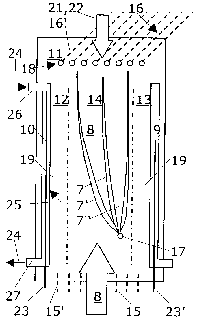
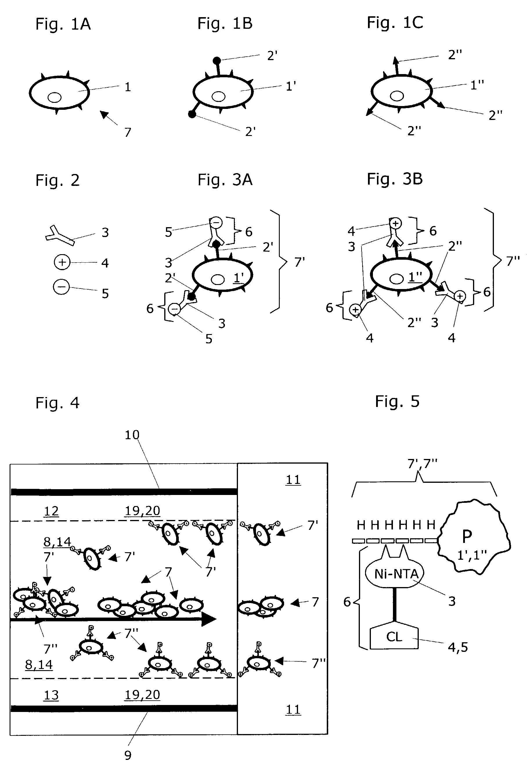
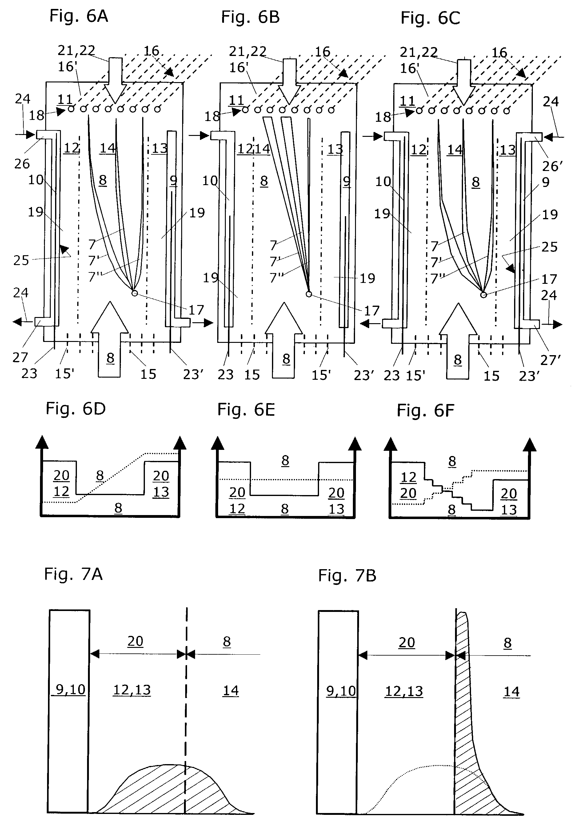
Image

Examples
example a
Purification of Circulating Tumor Cells from Whole Blood
[0054]A summary of the valency of disseminated tumor cells can be found in: Bockmann B, Grill H J, Giesing M. Molecular characterization of minimal residual cancer cells in patients with solid tumors. Biomol. Eng. 2001; 17: 95–111.
[0055]The membrane protein epithelial glycoprotein (EGP) which is specific to epithelial cells is used as a cellular surface marker. This EGP is also expressed from epithelial tumor cells (Moldenhauer G, Momburg F, Möller P, Schwartz R, Hämmerling G J. Epithelium-specific surface glycoprotein of Mr 34,000 is a widely distributed carcinoma marker. Br J Cancer 1987;56:714–21). The literature provides good descriptions of monoclonal antibodies with the designation Ber-EP4 which act against EGP cell surface epitopes (Latza U, Niedobitek G, Schwarting R, Nekarda H, Stein H. “Ber-EP4: new monoclonal antibody which distinguishes epithelia from mesothelia”, J Clin Pathol 1990;43: 213–219).
[0056]This monoclona...
example b
Isolation of Peroxisomes
[0066]Peroxisomes are cell organelles which engage in oxidative reactions with molecular oxygen. They generate peroxide oxygen for oxidative purposes.
[0067]Highly enriched peroxisomes can be isolated, for instance from rat liver using a complex density gradient centrifuge according to the method reported by Lüers et al. (Lüers G H, Hartig R, Mohr H, Hausmann M, Fahimi H D, Cremer C, Völkl A. “Immuno-isolation of highly purified peroxisomes using magnetic beads and continuous immunomagnetic sorting”, Electrophoresis 1998;19: 1205–1210):[0068]1. Homogenization of rat liver using known methods.[0069]2. Centrifugation of homogenized material at 1,900×g to remove cell nuclei, cell debris, mitochondria and cells which are still intact.[0070]3. Centrifugation of the remaining material at 25,000×g.[0071]4. The resulting pellet contains the so-called “light microsomal fraction” (incl. peroxisomes, microsomes and some mitochondria with lysosomes), thereby constituting ...
example c
Isolation of Recombined Proteins
[0077]For the expression of recombinant proteins there are vectors which can attach a chain of 5–10 histidines onto the protein to be expressed at the 5′ or 3′ end of a structure (His-tags, cf. FIG. 5). These additional histidines are used for later purification of the expressed proteins. Other vectors contain, for instance glutathione S-transferase as an additional element. These vectors are suitable for the expression of recombinant proteins, e.g. in E. coli, Baculovirus-infected bacteria and mammalian cells. The corresponding methods for producing recombinant proteins are known to those skilled in the art.
[0078]The currently available options for purifying recombinant expressed proteins are based primarily on affinity chromatography methods. These methods are relatively easy to use and scalable, depending on the quantity of the proteins to be purified. However, affinity chromatography purification of recombinant proteins has several significant dis...
PUM
| Property | Measurement | Unit |
|---|---|---|
| volumes | aaaaa | aaaaa |
| pH | aaaaa | aaaaa |
| temperature | aaaaa | aaaaa |
Abstract
Description
Claims
Application Information
 - R&D
- Intellectual Property
- Life Sciences
- Materials
- Tech Scout
- Unparalleled Data Quality
- Higher Quality Content
- 60% Fewer Hallucinations
Browse by: Latest US Patents, China's latest patents, Technical Efficacy Thesaurus, Application Domain, Technology Topic, Popular Technical Reports.
© 2025 PatSnap. All rights reserved.Legal|Privacy policy|Modern Slavery Act Transparency Statement|Sitemap|About US| Contact US: help@patsnap.com