Method for chondrocyte expansion with phenotype retention

a chondrocyte and phenotype technology, applied in the field of chondrocyte expansion with phenotype retention, can solve the problems of cartilage matrix breakdown, degenerative arthritis, loss of quality of life,

Active Publication Date: 2007-09-25
ZIMMER INC +1
View PDF19 Cites 49 Cited by
  • Summary
  • Abstract
  • Description
  • Claims
  • Application Information

AI Technical Summary

Problems solved by technology

Millions of people in the United States alone are afflicted with painful inflammatory or degenerative arthritis which limits normal joint function and results in loss of quality of life.
The primary cause of degenerative arthritis is breakdown of the cartilage matrix.
Loss of joint articular cartilage due to traumatic injury or disease ultimately results in joint stiffening caused by “bone on bone movement” and painful exposure of nerve endings in subchondral bone.
Damage to articular cartilage results in lesions of the joint surface, and progressive degeneration of these lesions often leads to symptomatic joint pain, disability and reduced or disturbed functionality.
Lack of a blood supply in large part restricts the tissue's ability to recruit chondroprogenitor cells that can act to repair articular cartilage defects.
However, these experimental therapies have not produced long-lasting repair of hyaline cartilage (Buckwatler et al., 1990; Hunziker, 2002).
A key limitation of this method is the relatively small number of donor cells that can be obtained at biopsy, and chondrocytes derived from adult articular cartilage appear to have a limited ability to produce cartilage matrix after expansion.
Loss of chondrocyte phenotype during serial expansion in vitro poses a key limitation to the commercialization of orthobiologic approaches to articular cartilage repair.
However, many of such cultured chondrocytes still produce type I collagen and small proteoglycans, indicating an “incompletely” restored cartilage phenotype.
Furthermore, the potential for carry over of residual materials derived from the three-dimensional hydrogels can complicate the regulatory path.

Method used


Image

  • Method for chondrocyte expansion with phenotype retention
  • Method for chondrocyte expansion with phenotype retention
  • Method for chondrocyte expansion with phenotype retention

Examples


example 1

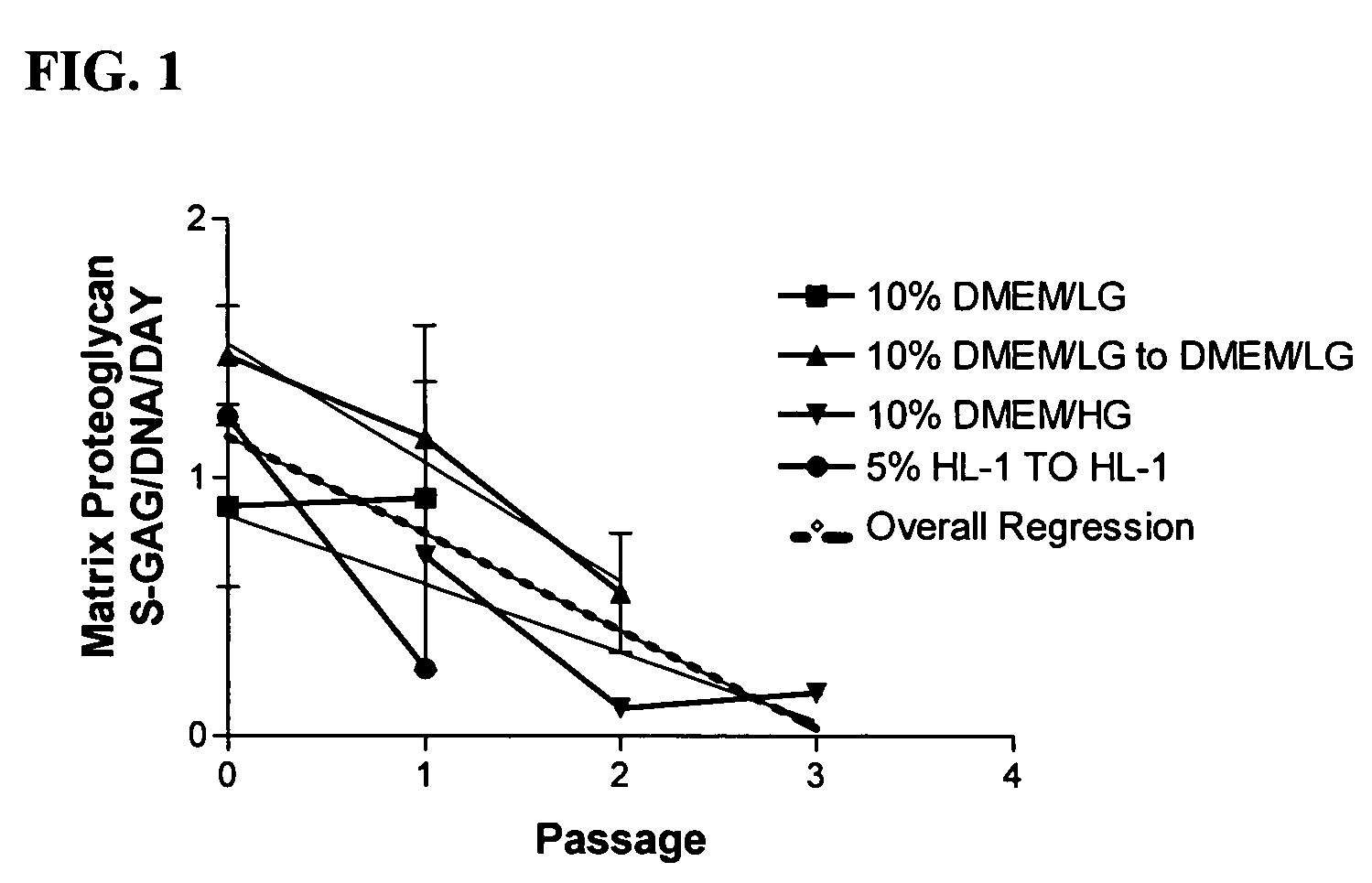
Loss of Chondrocyte Phenotype by Serial Expansion in Serum Containing Media

[0103]Chondrocytes, regardless of tissue origin, rapidly lose the ability to synthesize cartilage specific macromolecules with serial expansion in vitro (Homicz et al., 2002; Mandl et al., 2002). Although juvenile chondrocytes are thought to better retain the ability to synthesize matrix macromolecules than adult articular chondrocytes, the following experiment was performed to determine the effect of serial expansion on chondrocyte matrix synthesis using chondrocytes derived from immature articular cartilage. A variety of media containing serum were evaluated to demonstrate the deleterious effects of serum on chondrocyte differentiation potential.

[0104]Sixteen (16) different donor cell populations were isolated from cadaveric articular cartilage ranging in age from new-born to three years. Four different basal medium formulations containing the indicated amount of fetal bovine serum were used: 10% DMEM / LG, 1...

example 2

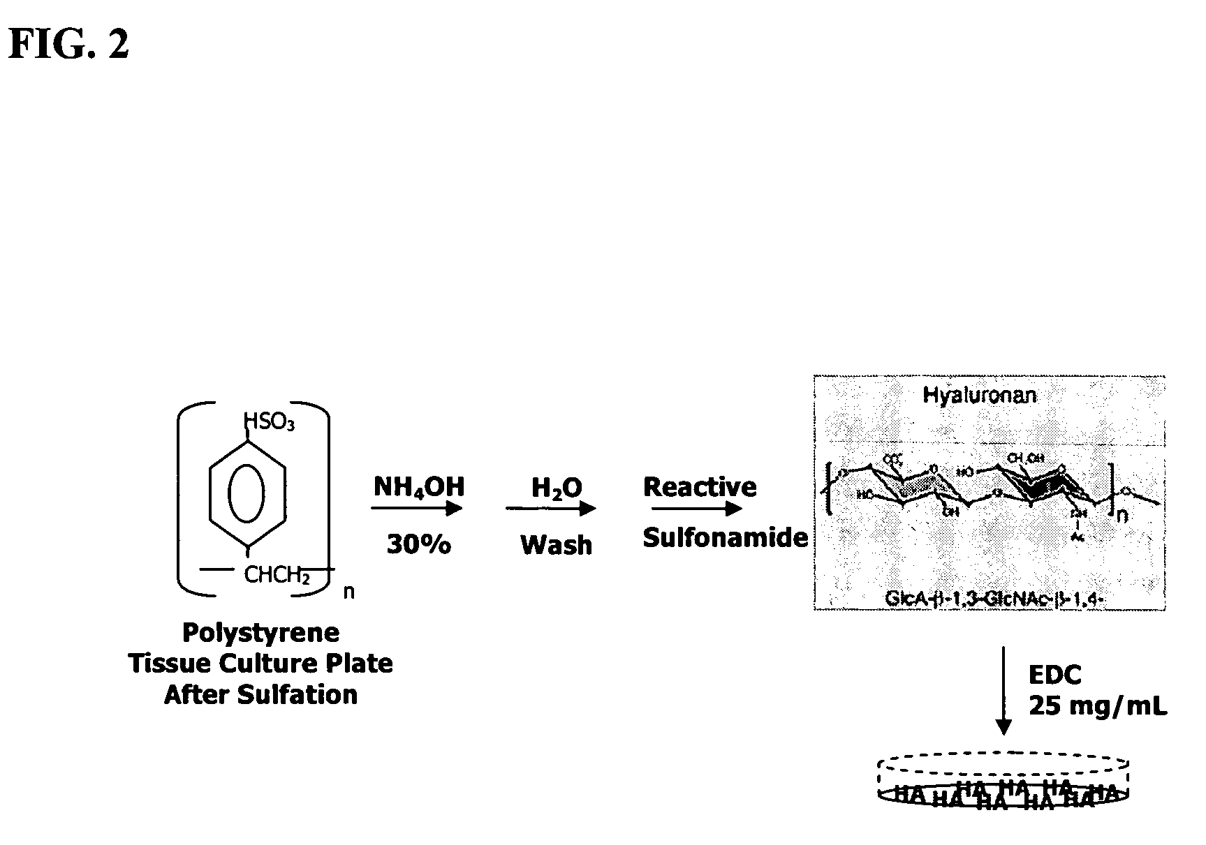
Method for Covalent Immobilization of Sodium Hyaluronate on Polystyrene Surfaces

[0107]To improve the efficiency with which chondrocytes can be expanded in vitro without loss of native chondrocyte phenotype, juvenile chondrocytes were grown on a modified substratum prepared via covalent attachment of high molecular weight sodium hylauronate to polystyrene. It was hypothesized that immobilized sodium hyaluronate, in contrast to unmodified tissue culture plastic, would provide a microenvironment that could more closely mimic that of native articular cartilage. The following method for chemical modification of polystyrene with sodium hylauronate is a modification of that originally described by Turley and Roth (Turely and Roth, 1979).

[0108]In FIG. 2, the virgin polystyrene plates were treated with sulfuric acid at 37° C. for two hours. After removing the sulfuric acid and extensive washing of the plates in deionized distilled water, the reactive sulfonamide was created by adding aqueous...

example 3

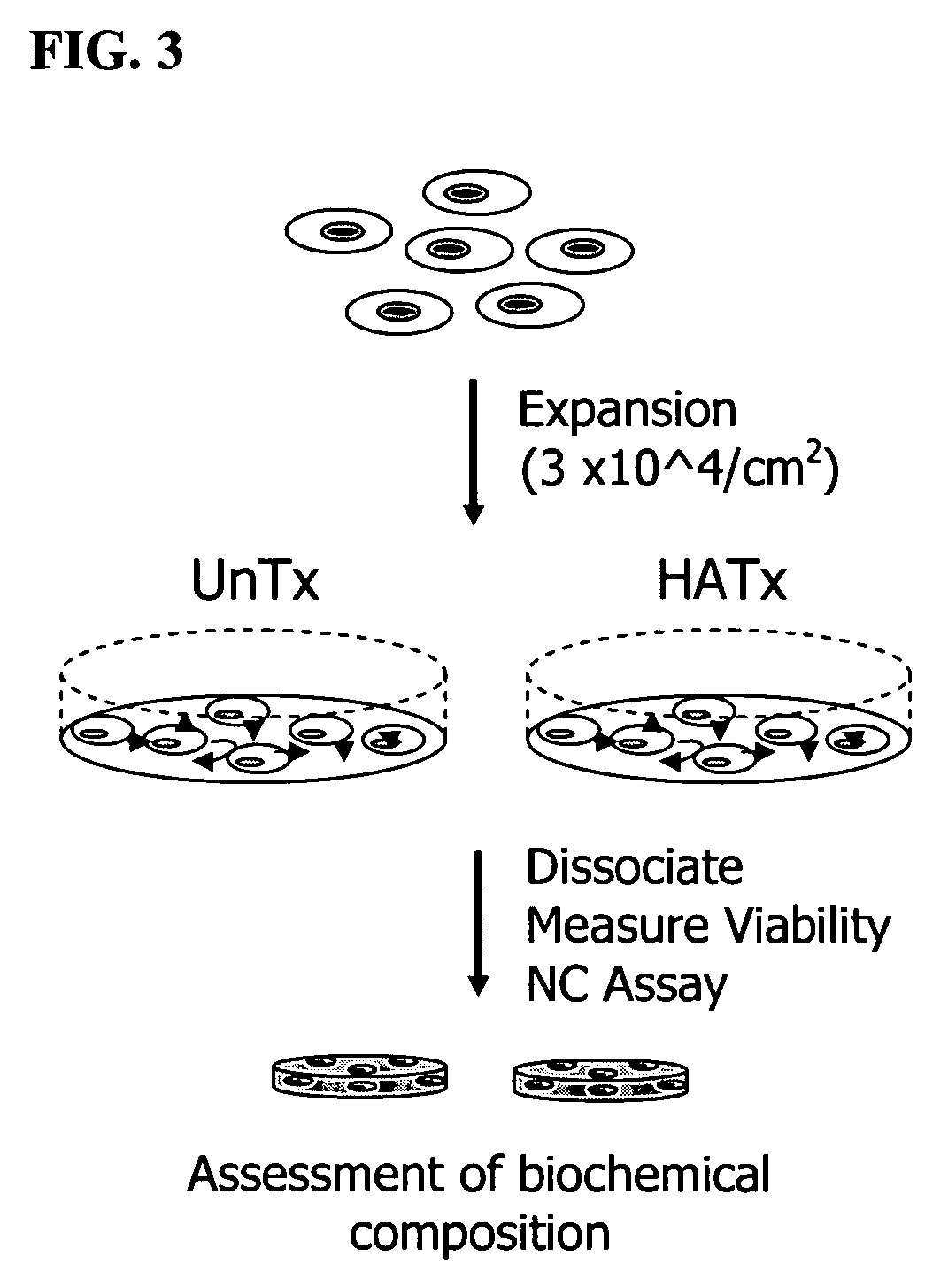
Chondrocyte Phenotype Retained by Serial Expansion on HA-coated Plastic in Serum-free Medium

[0109]Chondrocytes derived from a six week-old subject were taken through two passages using plates that were modified by the method illustrated in Example 2. Differentiation potential of the expanded chondrocytes was assessed after enzymatic dissociation and estimation of total cell number and viability. A proprietary serum-free differentiation medium, developed at Isto Technologies, Inc, was used to stimulate chondrocyte differentiation. In contrast to other methods, such as that described by Martin et al. (U.S. Pat. No. 6,582,960), this medium contains neither TGFβ nor dexamethasone to enhance chondrogenic differentiation potential, and the only protein component in this formulation is recombinant human insulin (Serologicals Corporation, Milford, Mass.).

[0110]FIG. 3 shows a schematic representation of the process of cytokine-mediated chondrocyte expansion in which covalent attachment of so...


PUM

PropertyMeasurementUnit
molecular weightaaaaaaaaaa
temperatureaaaaaaaaaa
concentrationsaaaaaaaaaa

Abstract

The present invention provides a method that maintains chondrocyte phenotype during serial expansion by culturing a population of chondrocytes in a defined serum-free culture medium containing cytokines and on a substrate that is modified by covalent attachment of hyaluronic acid. The underlying principle is to maintain native chondrocyte phenotype by growing the dissociated chondrocytes on a substrate modified by covalent attachment of hyaluronic acid to retain native chondrocyte morphology and function. Chondrocyte expanded in this manner can be used in various medical applications to repair cartilaginous tissues that have been injured by trauma or disease. This substratum provides a microenvironment that more closely mimics that of native articular cartilage, thereby promoting chondrogenesis in a predictable manner.

Description

STATEMENT REGARDING FEDERALLY SPONSORED RESEARCH OR DEVELOPMENT[0001]Research relating to the invention was supported in part by a NIST sponsored ATP Award 70NANB1H3027 The Government has certain rights to paid up, royalty-free non-exclusive license of the invention.CROSS-REFERENCE TO RELATED APPLICATIONS[0002]Not applicable.REFERENCE TO A SEQUENCE LISTING[0003]The Sequence Listing, which is a part of the present disclosure, includes a text file comprising primer nucleotide and / or amino acid sequences of the present invention on a compact disc. The subject matter of the Sequence Listing is incorporated herein by reference in its entirety.BACKGROUND OF THE INVENTION[0004]Millions of people in the United States alone are afflicted with painful inflammatory or degenerative arthritis which limits normal joint function and results in loss of quality of life. The primary cause of degenerative arthritis is breakdown of the cartilage matrix. Cartilage is a smooth, flexible connective tissue...

Claims


Application Information

Patent Timeline
Patent Type & Authority Patents(United States)
IPC IPC(8): C12N5/00C12N5/077
CPCC12N5/0655C12N2500/99C12N2501/115C12N2501/15C12N2500/90C12N2533/80
Inventor ADKISSON, HUSTON DAVISMILLIMAN, CURT L.
Owner ZIMMER INC
Why Eureka
Try Eureka