Cell delivery patch for myocardial tissue engineering

Active Publication Date: 2008-07-08
THE TRUSTEES OF THE UNIV OF PENNSYLVANIA
View PDF6 Cites 33 Cited by
  • Summary
  • Abstract
  • Description
  • Claims
  • Application Information

AI Technical Summary

Problems solved by technology

Both of these procedures carry significant risks with undetermined long-term effects.
With respect to adults, despite tremendous advances in the treatment of acute myocardial infarction, post-infarction congestive heart failure remains a difficult clinical problem.
Coronary occlusion with consequent regional ischemia leads to loss of cardiomyocytes and progressive replacement of muscle by collagenous tissue resulting in a myocardial scar.
Thus, left ventricular dilatation and heart failure can occur even after a myocardial infarction of moderate size.
While novel therapeutic strategies such as biventricular pacing, mitral valve repair and ventricular remodeling surgery may improve the quality of life in a portion of patients with end-stage heart failure, heart transplantation is still the only available treatment that significantly lengthens life expectancy.
Transplantation, however, is limited by a chronic shortage of donor hearts.
Treatment by transplantation is also limited by the problem of rejection.
Advances in immunosuppression have significantly improved the treatment of acute rejection but chronic rejection in the form of cardiac allograft vasculopathy remains a leading cause of death in transplant recipients.
This new paradigm of allorecognition presents yet another major barrier to successful, long-term, solid organ transplantation.
Complete correction of cardiac anomalies, however, is limited by the inability to engineer functional ventricular tissue and to enlarge the ventricular cavity.
Manipulation of the native heart for tissue engineering purposes is possible in a large animal model under complete cardiopulmonary bypass, but no comparable technology has been available for rodent models (Shinoka T, Ma P X, Shum-Tim D, et al., Tissue-engineered heart valves, Autologous valve leaflet replacement study in a lamb model.
Cardiac tissue engineering in a murine model has been limited by the difficulty of achieving cardiopulmonary bypass utilizing standard techniques.
This technology, however, does not address the absence of myocardium r

Method used


Image

  • Cell delivery patch for myocardial tissue engineering
  • Cell delivery patch for myocardial tissue engineering
  • Cell delivery patch for myocardial tissue engineering

Examples


example 1

[0095]Construction of Biocompatible Scaffolding for Ventricular Replacement

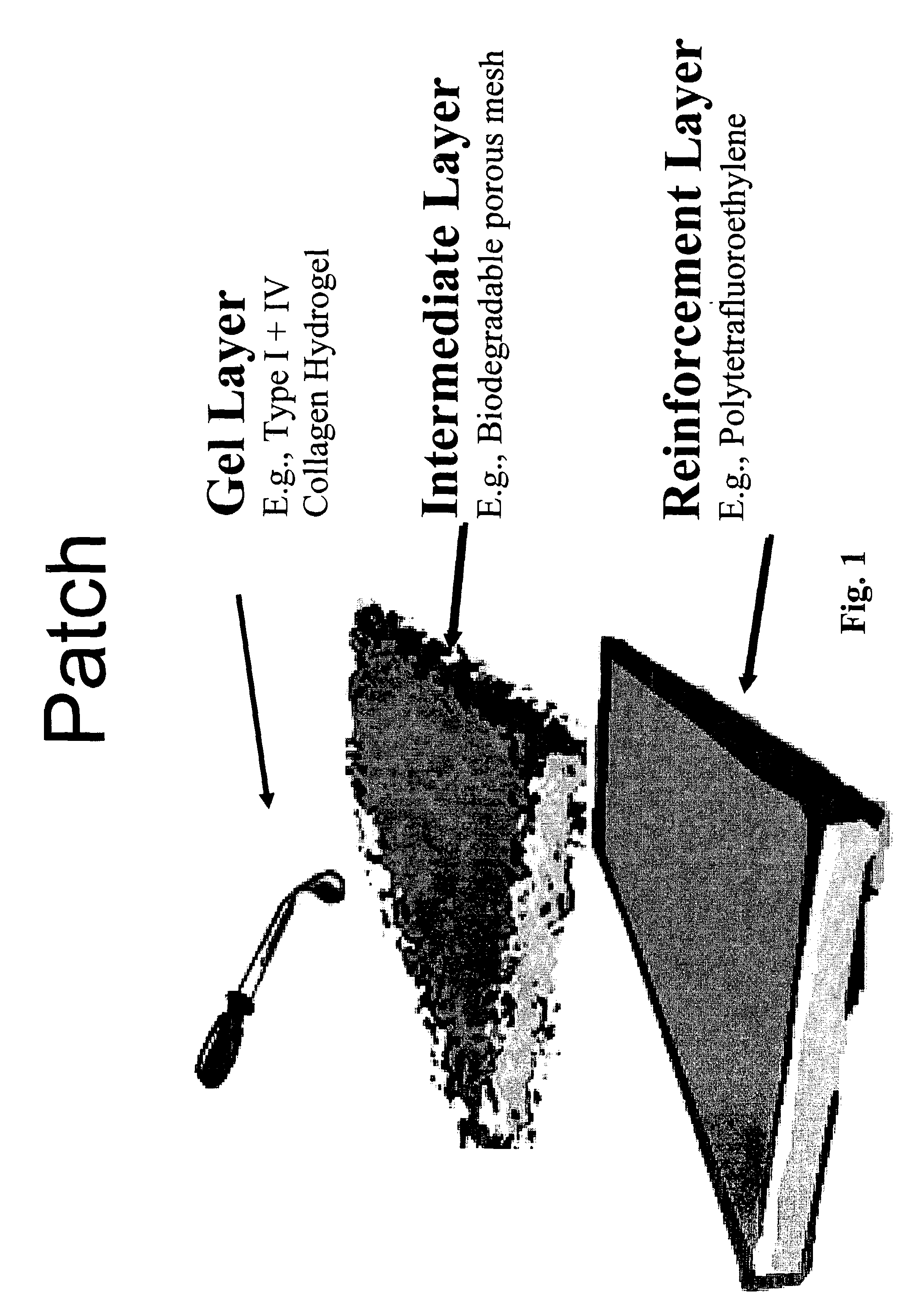
[0096]FIG. 1 depicts a patch and a method of making same according to one aspect of the invention.

[0097]Collagen hydrogels may be utilized as the basis for ventricular reconstruction of the present invention. Such collagen hydrogels have been investigated for the formation of three dimensional cardiac tissue in vitro (Zimmermann W H, Fink C, Kralisch D, Remmers U, Weil J, Eschenhagen T. Three-dimensional engineered heart tissue from neonatal rat cardiac myocytes. Biotechnology &Bioengineering 2000; 68: 106-114). The liquid hydrogel was made by combining 66 μl of type I rat tail collagen (4.6 mg / ml)(BD Biosciences, Bedford, Mass.) with 66 μl of 2× Dulbecco's Modified Eagle's Medium (supplemented with 20% horse serum, 200 μg / ml streptomycin and 200 U / ml penicillin G), 9 μl of 0.1 M NaOH, 25 μl of Matrigelt® basement membrane matrix (growth factor reduced, phenol red free) (Collaborative Biomedical, Bedford, Mas...

example 2

[0102]Donor Operation and Scaffolding Implantation

[0103]For all except liver cell studies, animals utilized for transplantation consisted of male Lewis (LEW / SsHsd) rats weighing 200-225 g at the time of experimentation (Harlan, Indianapolis, In). All experimental protocols were reviewed and approved by the Institutional Animal Care and Use Committee of The University of Pennsylvania, and followed guidelines set forth in the National Institutes of Health Guide for the Care and Use of Laboratory Animals. Techniques to obtain the donor heart were used as described (Asfour B, Hare J M, Kohl T, et al. A simple new model of physiologically working heterotopic rat heart transplantation provides hemodynamic performance equivalent to that of an orthotopic heart, Journal of Heart &Lung Transplantation 1999; 18: 927-936). After induction of anesthesia with enflurane, a midline sternotomy was performed and extended caudally to an abdominal incision, exposing the inferior vena cava (IVC) and aor...

example 3

[0116]Isolation and Expansion of Mesenchymal Stem Cells

[0117]Bone marrow stroma was isolated and enriched for mesenchymal stem cells according to established protocols (Wakitani S, Saito T, Caplan A I. Myogenic cells derived from rat bone marrow mesenchymal stem cells exposed to 5-azacytidine, Muscle &Nerve 1995; 18: 1417-1426). Femora and tibiae of male Lewis (LEW / SsHsd) rats weighing 100-150 g were collected and after carefully removing the adherent soft tissue, the bone marrow plugs were flushed out and disaggregated into a single cell suspension by sequential passage through a 22-gauge and 26-gauge needle. This suspension was seeded onto plastic tissue culture plates and maintained in Dulbecco's Modified Eagle's Medium supplemented with 10% fetal calf serum specially selected to support expansion of mesenchymal stem cells (Stem Cell Technologies, Vancouver, Canada) and 1% penicillin / streptomycin. Three days later the medium was changed and the non-adherent cells discarded. Adher...


PUM

PropertyMeasurementUnit
Shapeaaaaaaaaaa
Biocompatibilityaaaaaaaaaa
Biodegradabilityaaaaaaaaaa

Abstract

A patch for cardiac tissue engineering includes a gel layer supported by an intermediate layer, which is attached to a reinforcement layer. These patches may be implanted in a heart to treat pediatric congenital malformations of the heart as well as adult ischemic myopathies. The gel layer may include cells such as, for example, stem cells; the intermediate layer may be biodegradable porous mesh and the reinforcement layer may be polytetrafluoroethylene. Included are methods for making patches according to the invention and for tissue engineering using patches of the invention.

Description

CROSS-REFERENCE TO RELATED APPLICATIONS[0001]The present patent application claims the benefit of the filing dates of U.S. Provisional Patent Application Ser. No. 60 / 360,820, filed Feb. 28, 2002 and U.S. Provisional Patent Application Ser. No. 60 / 408,448, filed Sep. 5, 2002, both of which are hereby incorporated by reference in their entireties.GOVERNMENT SUPPORT[0002]The present invention was supported by the U.S. National Institutes of Health under Grant No. NIH NRSA F32-HL10251. The government may have certain rights in the invention.FIELD OF THE INVENTION[0003]The present invention is directed to a cell delivery patch used for cardiac tissue engineering, as well as to a method for cardiac tissue engineering using such a patch.BACKGROUND OF THE INVENTION[0004]Congenital heart defects are the most common form of birth anomalies, affecting 6 to 8 patients per 1000 births, and are the leading non-infectious cause of death in children. Hypoplastic left heart syndrome is the most deva...

Claims


Application Information

Patent Timeline
IPC IPC(8): A61K9/00
CPCA61K47/34A61K9/0024
Inventor KRUPNICK, ALEXANDERKREISEL, DANIELROSENGARD, BRUCE R.
Owner THE TRUSTEES OF THE UNIV OF PENNSYLVANIA
Why Eureka
Try Eureka