
Cytotoxicity mediation of cells evidencing surface expression of CD63

a cytotoxicity and surface expression technology, applied in the field of cancer diagnosis and treatment, can solve the problems of difficult to investigate the role of this family of proteins in the modulation of signal transduction pathways, lack of strong and consistent data that would definitively demonstrate, and difficulty in elucidating the mechanisms that lead to tumor progression

Inactive Publication Date: 2008-10-07
F HOFFMANN LA ROCHE & CO AG
View PDF43 Cites 4 Cited by
  • Summary
  • Abstract
  • Description
  • Claims
  • Application Information

AI Technical Summary

Benefits of technology

The patent describes a method for creating individualized anti-cancer antibodies that can be used to treat cancer. These antibodies are generated by immorting cells from a patient's tumor and can target molecules and pathways not previously shown to be essential for the growth and survival of malignant tissue. These antibodies can be used in staging and diagnosis of cancer, and can be produced using the methods outlined in the patent. The antibodies can also be used to treat tumor metastasis and can be produced using phage display libraries and screening methods. The patent also describes the use of these antibodies in a mouse model of breast cancer. Overall, this patent provides a new way to develop personalized cancer treatments that can improve the effectiveness and target specific molecules and pathways in cancer cells.

Problems solved by technology

As a result of these, apparently conflicting, results, there is lack of strong and consistent data that would definitively demonstrate the association of CD63 with cancer.
As a result it has been very difficult to investigate the role of this family of proteins in the modulation of signal transduction pathways.
Elucidation of the mechanisms that lead to tumor progression is a very difficult and complex endeavor frequently marked by apparently contradictory observations and, as a result, it rare that those observations successfully translate into effective therapies.
At least 30 percent of these patients will fail the first line therapy, thus leading to further rounds of treatment and the increased probability of treatment failure, metastases, and ultimately, death.
Chemotherapy and radiation treatment cannot be tailored to the patient, and surgery by itself, in most cases, is inadequate for producing cures.
However, it is now widely recognized that no single monoclonal antibody can serve in all instances of cancer, and that monoclonal antibodies can be deployed, as a class, as targeted cancer treatments.
At the present time, the cancer patient usually has few options of treatment.
However, to the particular individual, these improved statistics do not necessarily correlate with an improvement in their personal situation.
Historically, the use of polyclonal antibodies has been used with limited success in the treatment of human cancers.
Furthermore, there was a lack of reproducibility and no additional benefit compared to chemotherapy.
Solid tumors such as breast cancers, melanomas and renal cell carcinomas have also been treated with human blood, chimpanzee serum, human plasma and horse serum with correspondingly unpredictable and ineffective results.
However, treatment with Herceptin and Taxol® led to a higher incidence of cardiotoxicity in comparison to Taxol® treatment alone (13 versus 1 percent respectively).
Therefore, there is still a large unmet need for patients with breast cancer.
Even those who can benefit from Herceptin treatment would still require chemotherapy and consequently would still have to deal with, at least to some degree, the side effects of this kind of treatment.
To date, Phase III clinical trials of 17-1A have not demonstrated improved efficacy as adjuvant therapy for stage III colon cancer.
In addition, treatment in both Switzerland and the US is only approved for patients as a second line therapy.
There also continues to be poor results for lung, brain, ovarian, pancreatic, prostate, and stomach cancer.
Preclinically, there has been some limited success in the use of monoclonal antibodies for melanoma.
Very few of these antibodies have reached clinical trials and to date none have been approved or demonstrated favorable results in Phase III clinical trials.
The discovery of new drugs to treat disease is hindered by the lack of identification of relevant targets, among the products of 30,000 known genes, that unambiguously contribute to disease pathogenesis.
Firstly, the choice of appropriate targets for therapeutic or diagnostic antibody binding is limited by the paucity of knowledge surrounding tissue specific carcinogenic processes and the resulting simplistic methods, such as selection by overexpression, by which these targets are identified.
Secondly, the assumption that the drug molecule that binds to the receptor with the greatest affinity usually has the highest probability for initiating or inhibiting a signal, may not always be the case.
Despite some progress with the treatment of breast and colon cancer, the identification and development of efficacious antibody therapies, either as single agents or co-treatments, has been inadequate for all types of cancer.

Method used


Image

  • Cytotoxicity mediation of cells evidencing surface expression of CD63
  • Cytotoxicity mediation of cells evidencing surface expression of CD63
  • Cytotoxicity mediation of cells evidencing surface expression of CD63

Examples


example 1

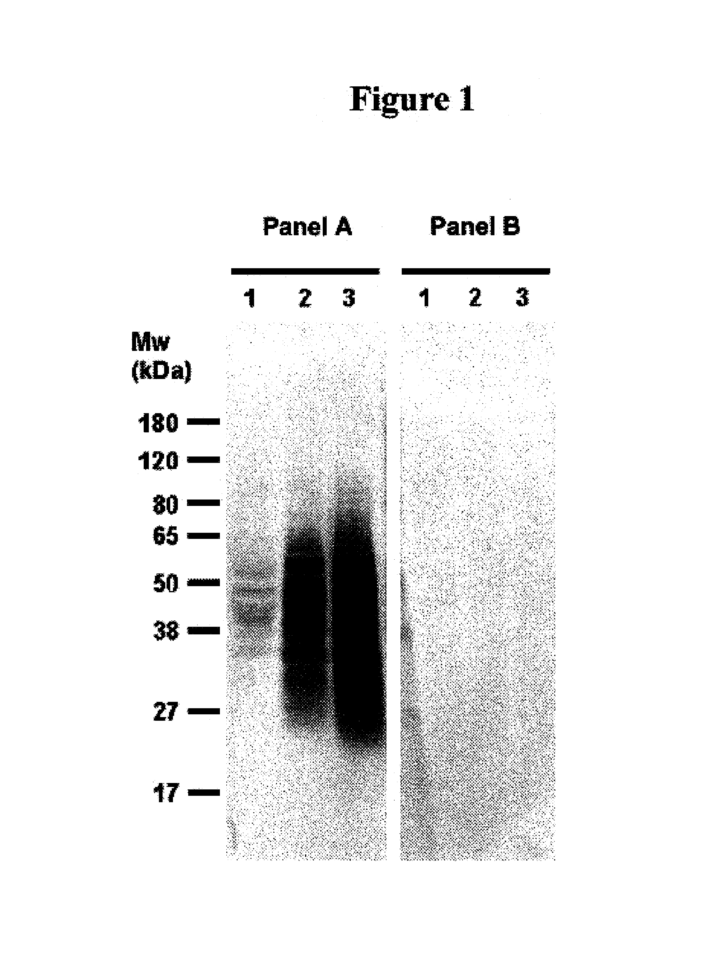
Identification of 7BD-33-11A Binding proteins by Western Immunoblotting

[0133]As outlined and discussed in Ser. No. 10 / 810,751, the contents of are herein incorporated by reference, to identify the antigen(s) recognized by the antibody 7BD-33-11A, cell membrane preparations were subjected to sodium dodecylsulphate polyacrylamide gel electrophoresis (SDS-PAGE), and transferred to membranes. The latter were probed with the antibody 7BD-33-11A to visualize the proteins detected by this antibody.

1.0. Whole Cell Lysate and Total Membrane Fraction Preparation

1.1. Whole Cell Lysate Preparation

[0134]Previous work by FACS demonstrated binding of antibody 7BD-33-11A to the breast cancer cell line MDA-MB-231 (MB-231). As a result total cell membrane preparations and whole cell lysates obtained from this cell line were used for the antigen identification and characterization. Total cell lysate from MB-231 cells was prepared as follows: MB-231 cell pellet (1.5 g) was resuspended in 2 mL lysis buf...

example 2

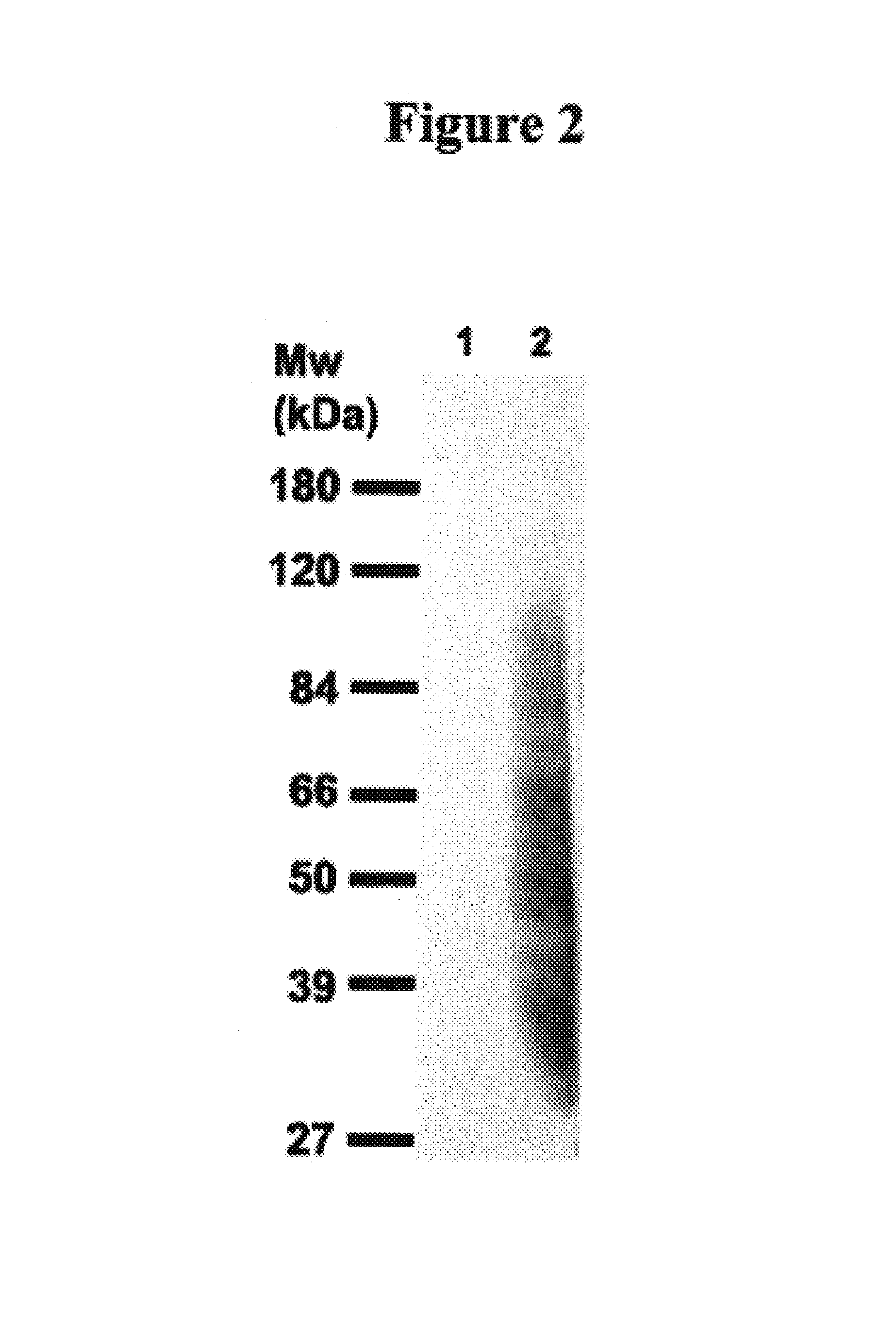
Identification of Antigens Bound by 7BD-33-11A

[0138]The data detailed in this example have been described previously in Ser. No. 10 / 810,751, the contents of which are herein incorporated by reference.

1.0 Immunoprecipitation of Antigens from MB-231 Total Membrane Fraction

[0139]Total membrane extracts (5 mg total protein) were diluted to a 1 mg / mL final protein concentration with the appropriate volume of 1× lysis buffer (50 mM Tris pH 7.4, 150 mM NaCl, 1% Triton X-100, 0.02% NaN3, 2 mM sodium orthovanadate, 50 mM sodium fluoride, and protease inhibitor cocktail (Roche Diagnostics, Manheim, Germany)), and with the appropriate volume of 2×RIPA buffer (50 mM Tris pH 7.4, 150 mM NaCl, 1.0% sodium cholate, 0.2% SDS, 1% Triton X-100 and 0.02% NaN3), in order to obtain a final 1×RIPA buffer concentration. The extracts were pre-cleared for 2 hr with protein G-Sepharose beads (Amersham Biosciences, Uppsala, Sweden) at 4° C. Total membrane extracts were removed and stock BSA (10 mg / mL) was add...

example 3

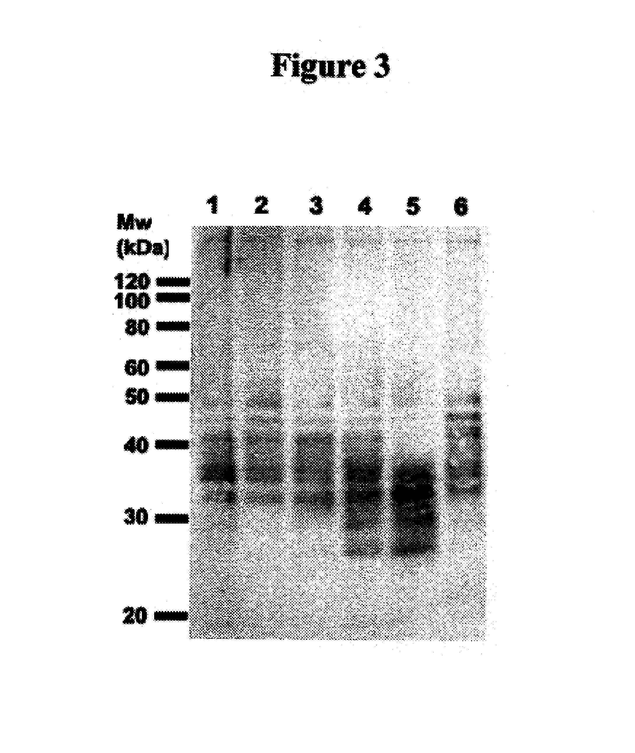
Identification of Antigens Bound by 1A245.6 and H460-22-1

1.0 Immunoprecipitation of Antigens from MB-231 Total Membrane Fraction

[0149]Total membrane extracts (1 mg total protein) were diluted to a 1 mg / mL final protein concentration with the appropriate volume of 1× lysis buffer (50 mM Tris pH 7.4, 150 mM NaCl, 1% Triton X-100, 0.02% NaN3, 2 mM sodium orthovanadate, 50 mM NaF, and protease inhibitor cocktail), and with the appropriate volume of 2× RIPA buffer (50 mM Tris pH 7.4, 150 mM NaCl, 1.0% NaCholate, 0.2% SDS, 1% Triton X-100 and 0.02% NaN3), in order to obtain a final 1× RIPA buffer concentration. The extracts were pre-cleared for 2 hr with protein G-Sepharose beads (Amersham Biosciences; Uppsala, Sweden) at 4° C. Total membrane extracts were removed and stock BSA (10 mg / mL) was added to a 0.5 mg / mL final BSA concentration. While extracts were being pre-cleared, antibody-conjugated protein G-Sepharose beads (60 μg of antibody chemically cross-linked to 30 μl of protein G Sep...


PUM

PropertyMeasurementUnit
median timeaaaaaaaaaa

Abstract

This invention relates to the diagnosis and treatment of cancerous diseases, particularly to the mediation of cytotoxicity of tumor cells; and most particularly to the use of cancerous disease modifying antibodies (CDMAB), optionally in combination with one or more chemotherapeutic agents, as a means for initiating the cytotoxic response. The invention further relates to binding assays which utilize the CDMABs of the instant invention.

Description

REFERENCE TO RELATED APPLICATIONS[0001]This application claims priority of U.S. Provisional Patent Application No. 60 / 642,057, filed Jan. 3, 2005 and is related to U.S. patent application Ser. No. 10 / 810,751, filed Mar. 26, 2004, U.S. patent application Ser. No. 10 / 603,006, filed Jun. 23, 2003, U.S. patent application Ser. No. 10 / 348,231, filed Jan. 21, 2003, now U.S. Pat. No. 7,009,040, issued Mar. 7, 2006 (including divisional application Ser. No. 10 / 891,866, filed Jul. 15, 2004, now U.S. Pat. No. 7,186,808, issued Mar. 6, 2007), the contents of each of which are herein incorporated by reference.FIELD OF THE INVENTION[0002]This invention relates to the diagnosis and treatment of cancerous diseases, particularly to the mediation of cytotoxicity of tumor cells; and most particularly to the use of cancerous disease modifying antibodies (CDMAB), optionally in combination with one or more chemotherapeutic agents, as a means for initiating the cytotoxic response. The invention further r...

Claims


Application Information

Patent Timeline
Patent Type & Authority Patents(United States)
IPC IPC(8): A61K39/395C07K16/00
CPCC07K16/2896G01N33/5743A61K2039/505C07K2317/92G01N2333/70596A61P35/00
Inventor YOUNG, DAVID S. F.HAHN, SUSAN E.FINDLAY, HELEN P.DA CRUZ, LUIS A. G.
Owner F HOFFMANN LA ROCHE & CO AG
Learn More