

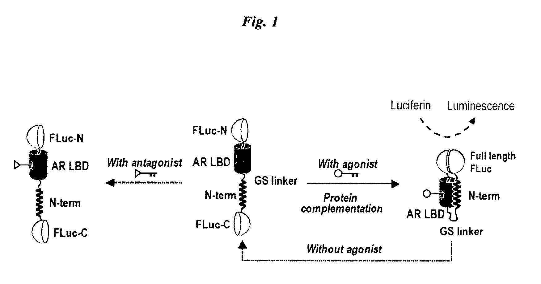
Single molecule-format bioluminescent probe
a single molecule, bioluminescent probe technology, applied in the direction of instruments, immunoglobulins, peptides, etc., can solve the problems of inability to provide a representative readout, inconvenient and accurate probe actions, inconvenient use and cost efficiency, etc., to achieve convenient and accurate introduction, convenient
- Summary
- Abstract
- Description
- Claims
- Application Information
AI Technical Summary
Benefits of technology
Problems solved by technology
Method used
Image

Examples
example 1
Probe Having Firefly Luciferase (FLuc) as a Luminescent Enzyme
(1) Methods
(1-1) Plasmid Construction
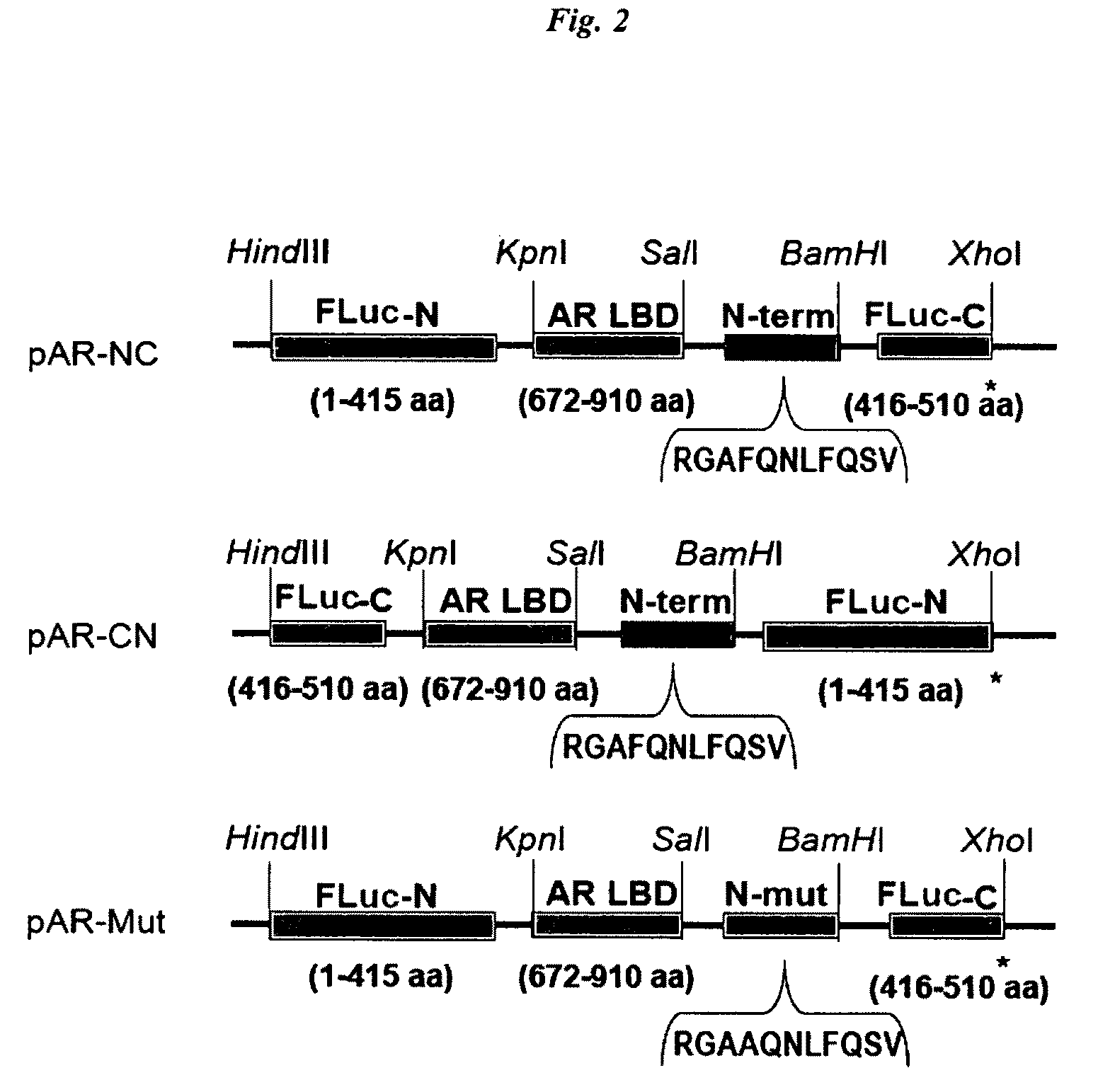
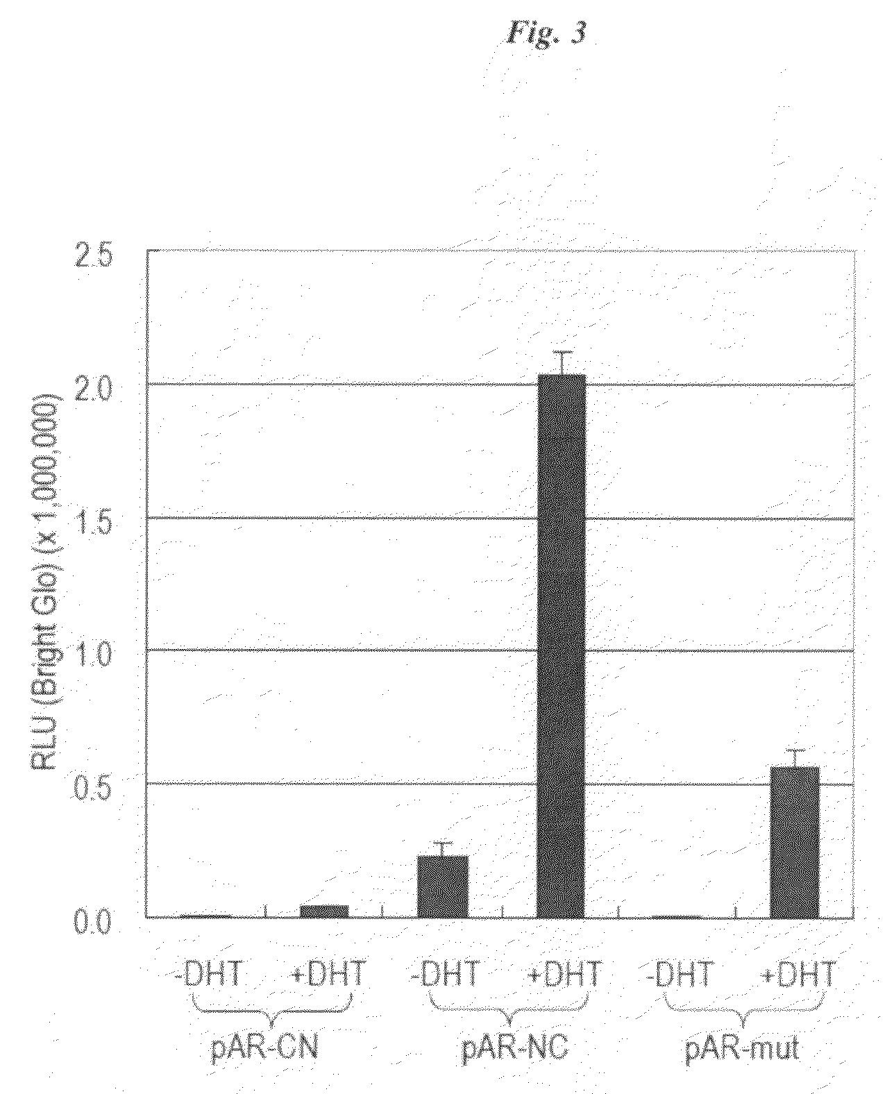
[0090]The cDNAs of N-terminal (Fluc-N; 1-415 AA) and C-terminal (Fluc-C; 416-510 AA) domains of split FLuc were amplified to introduce each unique restriction site at both ends of the domains using adequate primers and a template plasmid carrying the full-length cDNA of Fluc. The cDNA-encoding AR LBD (672-910 AA) was modified by PCR to introduce adequate restriction sites at the both ends of the domains. DNA oligomers encoding an AR N-terminal motif (11 AA; SEQ ID NO: 1; 20RGAFQNLFQSV30) and its alanine mutant (11 AA; SEQ ID NO: 30 20RGAAQNLFQSV30) were custom-synthesized by Exigen (Tokyo, Japan). The amplified fragments of each site were subcloned into the corresponding restriction enzyme-digested pcDNA 3.1 (+) vector backbone (Invitrogen). The constructed plasmids were sequenced to ensure fidelity with a BigDye Terminator Cycle Sequencing kit and a genetic analyzer ABI Prism310.
(1-2)...
example 2
Single Molecule-Format Luminescent Probe with a Click Beetle Luciferase (CBLuc)
[0113]Probe-expression plasmids were constructed by subcloning chimeric DNAs of FIG. 11 into pcDNA 3.1(+) vector. Each of these plasmids was named as pSimbe (Single Molecule-format probe using a click Beetle). They have an AR LBD and various LBD-interacting peptide, i.e., pSimbe-FQ has ‘FQNLF’ motif (SEQ ID NO: 28) (20RGAFQNLFQSV30; SEQ ID NO: 1) derived from human AR NTD, pSimbe-LXP has forward sequence of ‘LXXLL’ motif (686 KHKILHRLLQDSS698; SEQ ID NO: 2) and pSimbe-LXA has a reverse sequence of ‘LXXLL’ motif (698SSDQLLRHLIKHK686; SEQ ID NO: 3) from Xenopus TIF2. The amino acid sequences of N-terminal (CBLuc-N) and C-terminal (CBLuc-C) of CBluc are as in FIG. 11, respectively. The combination of [A] and [a] corresponds to Plasmid [1], and Plasmids [2], [3] to [10] are provided in a corresponding manner. FIG. 12 shows a full-length CBLuc amino acid sequence in which the arrows indicate the cleavage sites...
PUM
| Property | Measurement | Unit |
|---|---|---|
| short time kinetics | aaaaa | aaaaa |
| pH | aaaaa | aaaaa |
| half-time | aaaaa | aaaaa |
Abstract
Description
Claims
Application Information