Method of isolation of nucleic acids
a nucleic acid and cell technology, applied in the field of nucleic acid cell isolation, can solve the problems of impeded many molecular biology reactions and techniques, time-consuming and laborious, and dna contamination of rna preparations, so as to improve the nucleic acid isolation process, reduce the volume or reagents added in the initial process step, and reduce the effect of proteinase treatmen
- Summary
- Abstract
- Description
- Claims
- Application Information
AI Technical Summary
Benefits of technology
Problems solved by technology
Method used
Image
Examples
example 1
Isolation of Genomic DNA from 200 μl Human Whole Blood
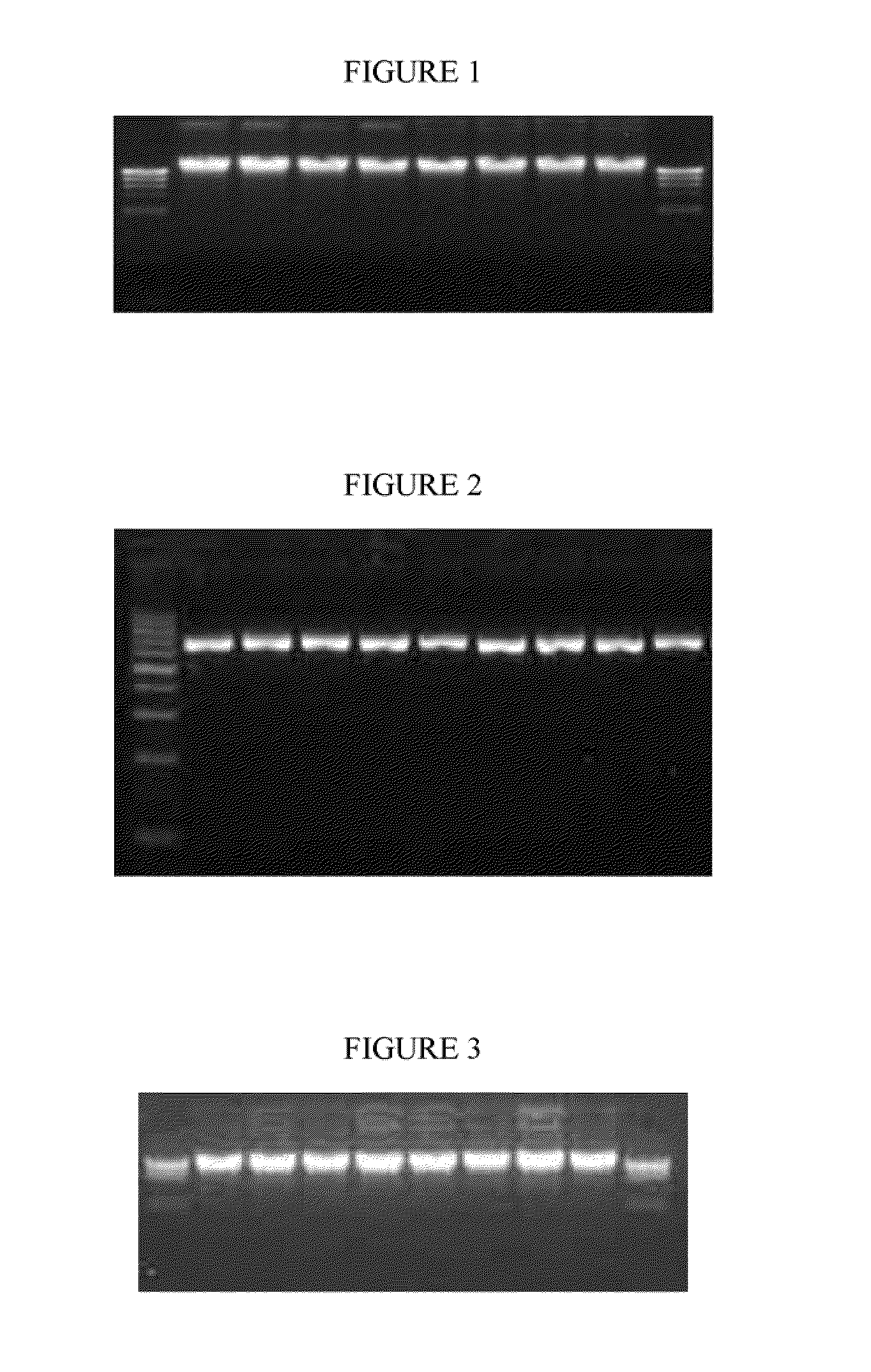
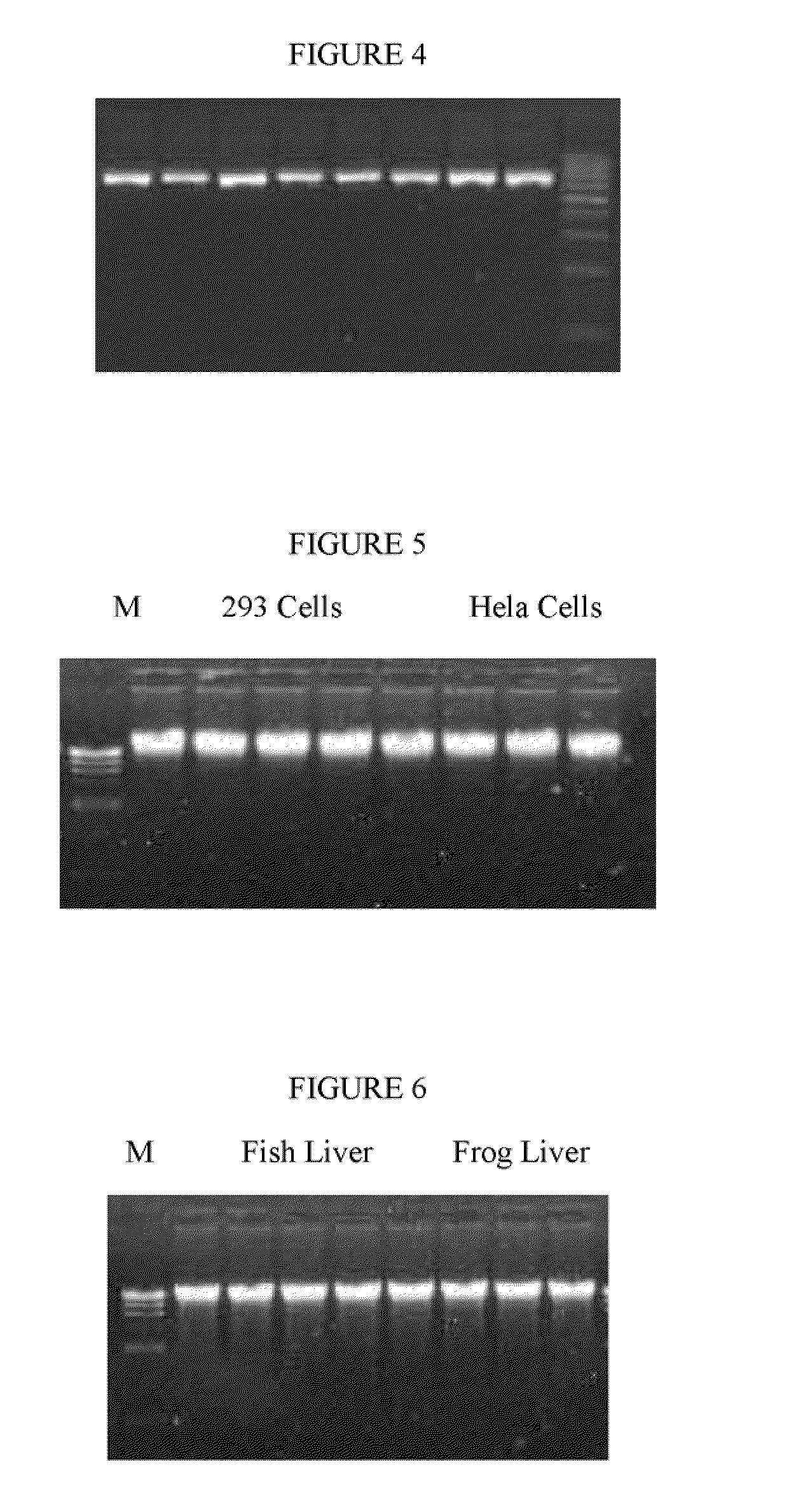
[0056]DNA Isolation: 0.2 ml whole human blood was mixed with 500 μl of cell membrane lysis buffer (1% TRITON X-100, 40 mM sodium citrate, 3 mM MgCl2) and 10 μl beads (50 mg / ml) in a microcentrifuge tube. The mixture was inverted 10 times and incubated at room temperature for 3 minutes, then the tube was placed in a magnet stand (Omega Bio-Tek, Atlanta, Ga.) for 7 minutes. The cell membrane supernatant was carefully removed. 300 μl of nuclear membrane lysis buffer (3.5 M Guanidine Hydrochloride, 20 mM Tris-HCl pH 7.4,) and 10 ul proteinase K (20 mg / ml) was added and incubated at 65° C. for 10 minutes. Then 200 μl of 100% isopropanol was added and the incubation continued for 5 minutes at room temperature. The beads were attracted to the tube wall with the magnet and the supernatant removed. The complex was washed twice with 500 μl 70% EtOH. All of the ethanol was removed and 50 μl water added. To remove residual ethanol the tubes ...
example 2
Isolation of Genomic DNA from 3 ml Human Whole Blood
[0058]DNA isolation: 3 ml whole human blood was mixed with 2 ml of cell membrane lysis buffer (0.5% TRITON X-100, 40 mM sodium citrate, 3 mM MgCl2) and 200 μl beads (50 mg / ml) in a 15 ml centrifuge tube. The mixture was inverted 10 times and incubated at room temperature for 3 minutes, then the tube was placed in a magnet stand (Omega Bio-Tek, Atlanta, Ga.) for 7 minutes. The cell membrane supernatant was carefully removed. 3 ml of nuclear membrane lysis buffer (3.5 M Guanidine Hydrochloride, 20 mM Tris-HCl pH 7.4,) and 10 ul proteinase K (20 mg / ml) was added and incubated at 65° C. for 10 minutes. Then 2 ml of 100% isopropanol was added and the incubation continued for 5 minutes at room temperature. The beads were attracted to the tube wall with the magnet and the supernatant removed. The complex was washed twice with 3 ml 70% EtOH. All of the ethanol was removed and 500 μl water added. To remove residual ethanol the tubes were in...
example 3
Isolation of Genomic DNA from Cultured Cells
[0060]Cell membrane lysis buffer: 0.01 M phophate buffered isotonic saline (146 mM) with calcium (1.0 mM CaCl2) and magnesium (0.5 mM MgSO4.7 H2O) which contained 0.6% NP40 (v / v) (Accurate Scientific and Chemical Co., Hicksville, N.Y.) and 0.2% bovine serum albumin (BSA) (w / v) (Fraction Five GIBCO).
[0061]DNA isolation: 106 Cultured HEK 293 cells (Clontech, Mountain View, Calif. 94043) and Hela cells (ATCC, Manassas, Va. 20108) were mixed with a cell membrane lysis buffer (0.6% NP40 v / v; 0.01 M phosphate buffer saline (146 mM); 1.0 mM CaCl2 and 0.5 mM MgSO4) in a microcentrifuge tube. The mixture was inverted 20 times and incubated at room temperature for 3 minutes, then the tube was placed in a magnet stand (Omega Bio-Tek, Atlanta, Ga.) for 7 minutes. The cell membrane supernatant was carefully removed. 300 μl of nuclear membrane lysis buffer (3.5 M Guanidine Hydrochloride, 20 mM Tris-HCl pH 7.4,) and 10 ul proteinase K was added and incub...
PUM
| Property | Measurement | Unit |
|---|---|---|
| pH | aaaaa | aaaaa |
| diameter | aaaaa | aaaaa |
| diameter | aaaaa | aaaaa |
Abstract
Description
Claims
Application Information
 - R&D
- Intellectual Property
- Life Sciences
- Materials
- Tech Scout
- Unparalleled Data Quality
- Higher Quality Content
- 60% Fewer Hallucinations
Browse by: Latest US Patents, China's latest patents, Technical Efficacy Thesaurus, Application Domain, Technology Topic, Popular Technical Reports.
© 2025 PatSnap. All rights reserved.Legal|Privacy policy|Modern Slavery Act Transparency Statement|Sitemap|About US| Contact US: help@patsnap.com


