

Polypeptides, cyclic polypeptides and pharmaceutical comprising thereof for non invasive specific imaging of fibrosis
a fibrosis and fibrosis specific imaging technology, applied in the direction of peptide/protein ingredients, drug compositions, peptide sources, etc., can solve the problems of destroying the affected structure, affecting the function of the body, and imposing an enormous health burden
- Summary
- Abstract
- Description
- Claims
- Application Information
AI Technical Summary
Benefits of technology
Problems solved by technology
Method used
Image

Examples
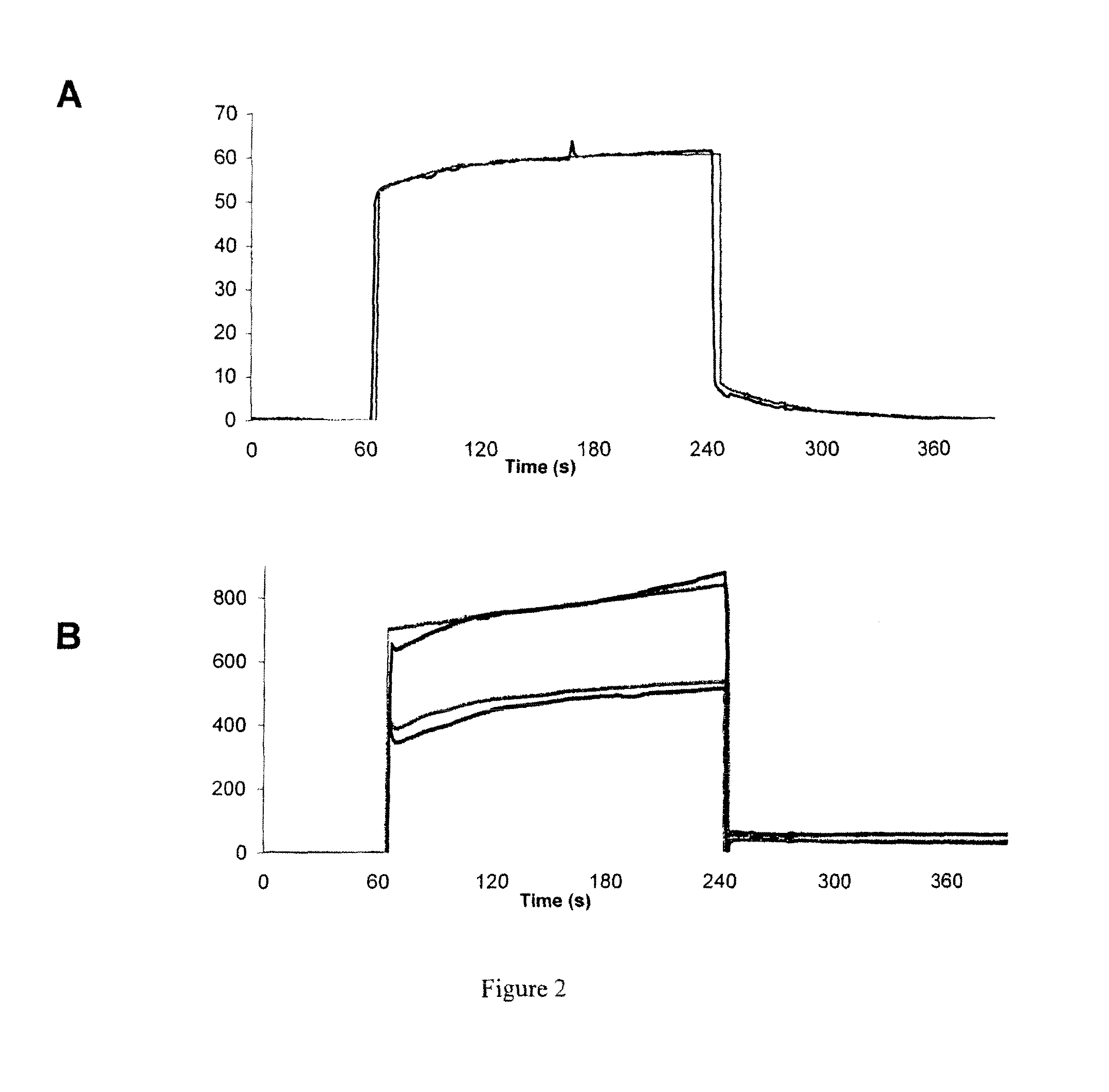
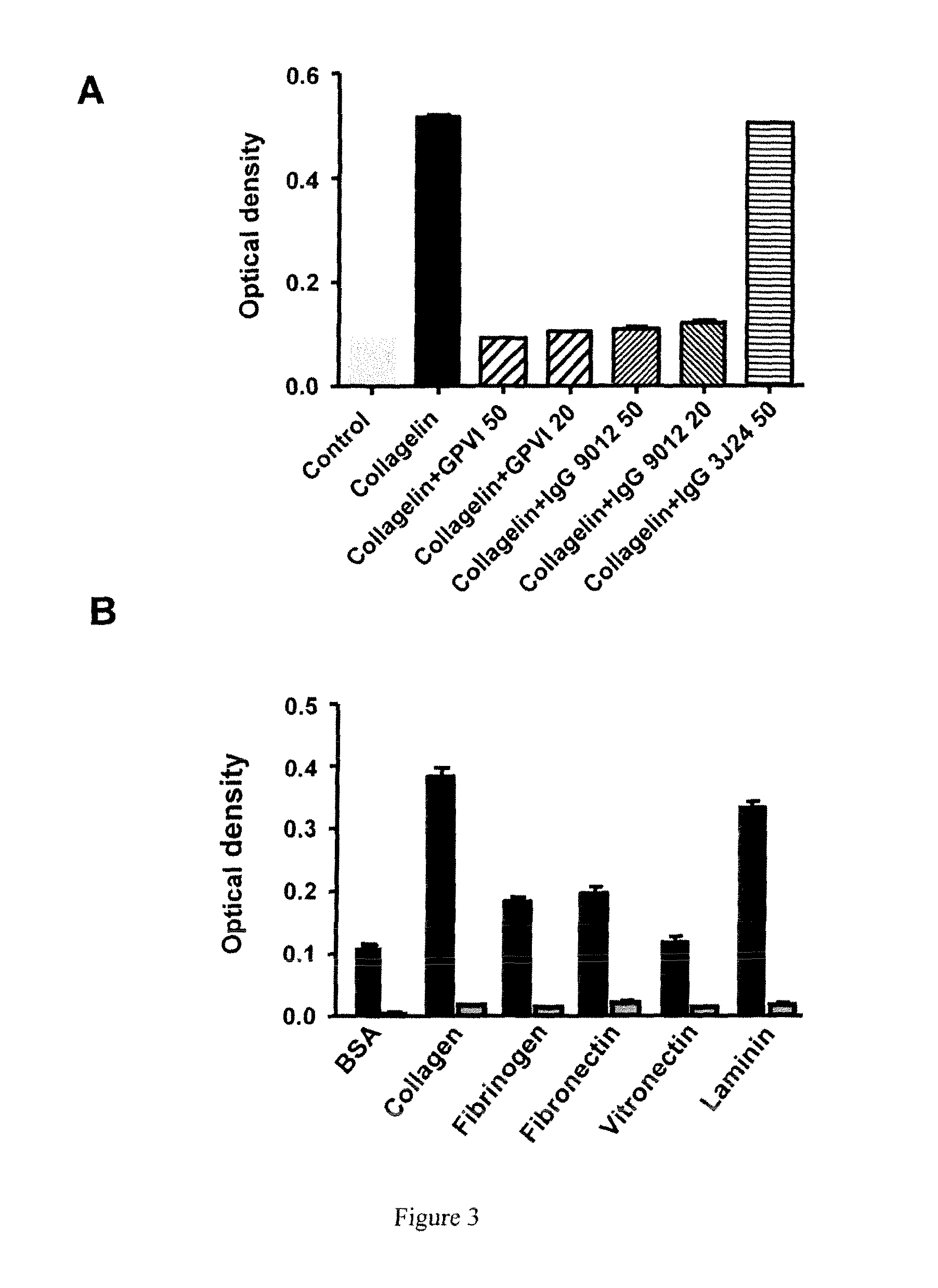
example 1
Identification and Characterization of a Peptidomimetic of Human Platelets Glycoprotein VI with Collagen Binding Activity. Its Use for Molecular Imaging of Fibrosis
[0174]Material & Methods:
[0175]FliTrx™ Peptide Library and Anti-GPVI Antibody:
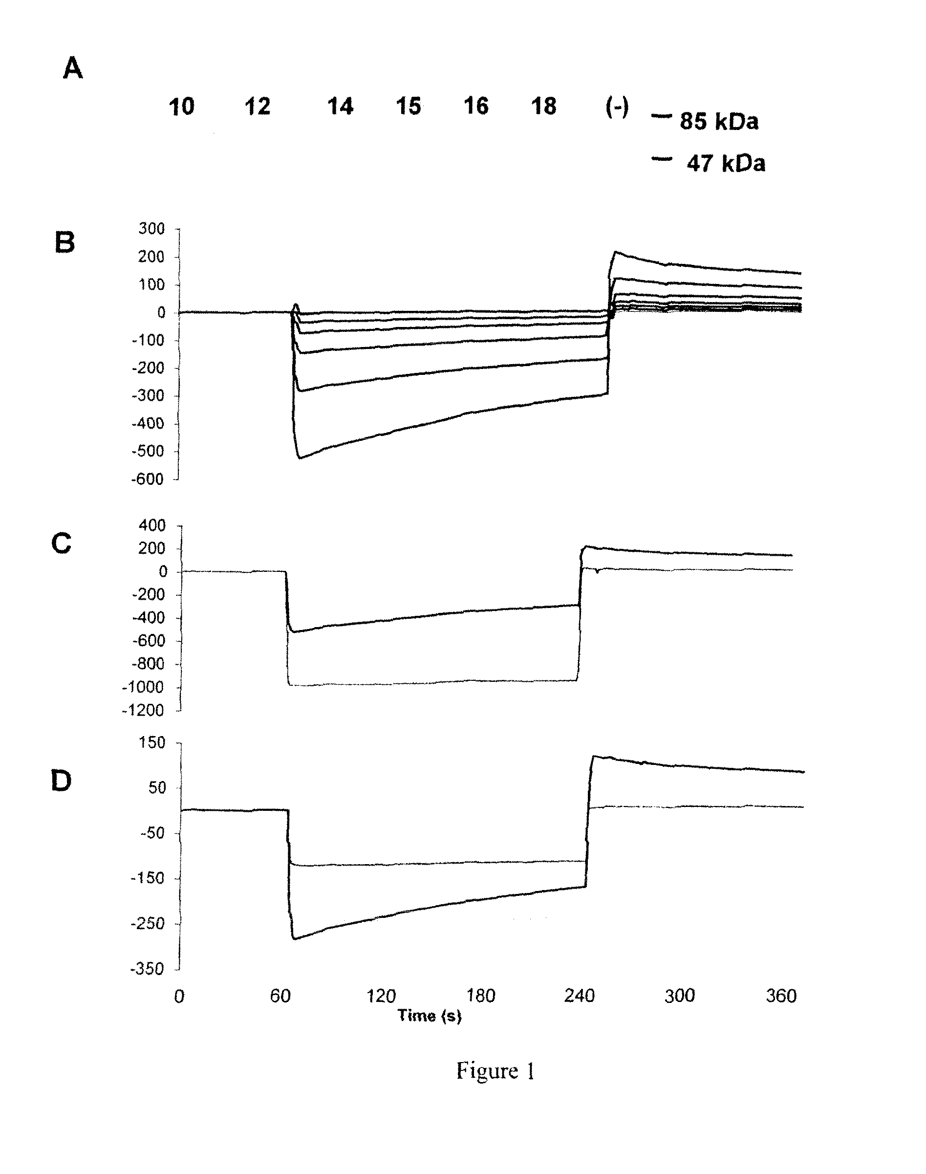
[0176]The FliTrx™ Random Peptide Display Library is an E. coli-based system allowing the screening of peptide interactions. The FliTrx™ Library was constructed in the pFliTrx™ vector (Lu Z. et al. 1995). A diverse library of random dodecapeptides (108) is positioned in the active site loop of the thioredoxin protein (trxA), inside the dispensable region of the bacterial flagellin gene (fliC). The resultant recombinant fusion protein (FLITRX) is exported and assembled into partially functional flagella on the bacterial cell surface. The dodecapeptides are displayed on the cell surface in a conformationally constrained by a disulfide bridge. The FliTrx™ random peptide library, based on the system was obtained from Invitrogen (San Diego, Calif.).
[0...
example 2
Imaging of Pulmonary Fibrosis by Scintigraphy Using 99Mtc-Labeled Collagelin
[0232]Methods:
[0233]Male C57BL / 6J mice, aged 6-7 weeks were kept in accordance with INSERM rules. On day 0, mice were administered 80 μg of bleomycin hydrochloride (Bleomycine Bellon, Aventis, France) intratracheally. Mortality was assessed daily over a 14 day period. Naïve mice were used as controls.
[0234]At day 14 mice received one intravenous injection of 99 mTc-B-collagelin or of 99 mTc-B-Pc (3 MBq). Then planar whole-body scintigraphic imaging (60 min duration) was performed 1 h after tracer injection, using Biospace Lab dedicated small animal gamma camera.
[0235]At the end of the experiment, animals were sacrificed and lung were dissected for gamma counting, autoradiography, and histology (Sirius red coloration).
[0236]Results:
[0237]Scintigraphy:
[0238]Significant 99 mTc-B-collagelin uptake was observed in pulmonary areas of the mice that received bleomycin (lung / muscle background activity ratio: 3.65±0.3...
PUM
| Property | Measurement | Unit |
|---|---|---|
| molecular weight | aaaaa | aaaaa |
| concentration | aaaaa | aaaaa |
| concentration | aaaaa | aaaaa |
Abstract
Description
Claims
Application Information