Methods and apparatus for reducing deleterious effects of x-ray radiation upon implantable medical device circuitry

a technology for implantable medical devices and circuits, applied in the field of methods and apparatus for reducing the negative effects of x-ray radiation impinging on the operation circuitry, can solve the problems of increasing the complexity and sophistication of the imds, unable to perform significantly more complex tasks, and the imds are vastly more sophisticated and complex. achieve the effect of small surface area

Active Publication Date: 2015-12-08
MEDTRONIC INC
View PDF18 Cites 1 Cited by
  • Summary
  • Abstract
  • Description
  • Claims
  • Application Information

AI Technical Summary

Benefits of technology

[0009]In one aspect of the present invention, methods of fabricating an IMD having selective x-ray impervious shielding are provided. These methods include identifying one or more components, circuits and / or circuit paths that are potentially vulnerable to x-ray radiation and then coupling one or more x-ray radiation shields to potentially vulnerable circuitry operatively coupled of an IMD. In one form of the invention one or more shield members having at least one radiation-admitting aperture selectively allows radiation from an x-ray source to impinge upon circuitry or the like disposed below the aperture. During and / or following x-ray exposure the exposed circuitry is operated to determine whether the x-ray radiation modified or damaged any parts thereof. If modification or damage is confirmed then the location of the aperture is used to guide installation of radiation opaque material (e.g., a plate or portion of lead, relatively thicker region of IMD housing, a layer of material disposed to overlay a part of an integrated circuit or integrated circuit packaging, etc.). Of course, the final orientation of the IMD should be considered since as will be described and depicted, x-ray radiation attenuates when it travels through a body. Thus, the shield should be located on the interior of an IMD between the outer cover and the vulnerable circuitry. If the radiation opaque material is not biocompatible (e.g., lead) then the material must be installed within the interior of the IMD housing. Due to the ever decreasing volume and weight of current IMDs, the size and weight of the radiation material needs to be considered as well. Due to the foregoing the inventors have discovered that a lead plate having a relatively small surface area, if properly located within the IMD housing, can essentially eliminate x-ray radiation from impinging upon possibly vulnerable circuitry operatively coupled therein.

Problems solved by technology

Today's state-of-the-art IMDs are vastly more sophisticated and complex than earlier ones, and are capable of performing significantly more complex tasks.
As the functional sophistication and complexity of IMDs have increased over the years, however, they have also been found to be vulnerable to more sophisticated and complex sources of interference.
In particular, conventional IMDs have been found to be vulnerable to electromagnetic interference signals produced for example by magnetic resonance imaging (MRI) devices during a magnetic resonance imaging (MRI) scanning session.
The inventors have discovered that x-ray radiation exposure can negatively affect both the sensing amplifiers (causing so-called over-sensing), the amplifiers used to collect EGMs, the clock monitor circuitry used in the crystal oscillator circuit (used to generate the main timing-clock for certain IMDs) among other components and circuits.
Furthermore, other components or circuits that can be negatively affected include battery status monitors, impedance measuring amplifiers, activity sensor amplifier and the like.
That is, during exposure to x-ray radiation for example during a CT-scanning session or a fluoroscopy-imaged examination, the collected cardiac EGMs may become distorted due to over-sensing and / or modified due to an incorrect clock performance so that an accurate assessment of the cardiac rhythm and / or function becomes more difficult.
In addition, in certain IMDs a full and / or a partial electrical reset (PER) can occur which inevitably temporarily reduces therapy delivery.

Method used


Image

  • Methods and apparatus for reducing deleterious effects of x-ray radiation upon implantable medical device circuitry
  • Methods and apparatus for reducing deleterious effects of x-ray radiation upon implantable medical device circuitry
  • Methods and apparatus for reducing deleterious effects of x-ray radiation upon implantable medical device circuitry

Examples


example

Standard X-Ray

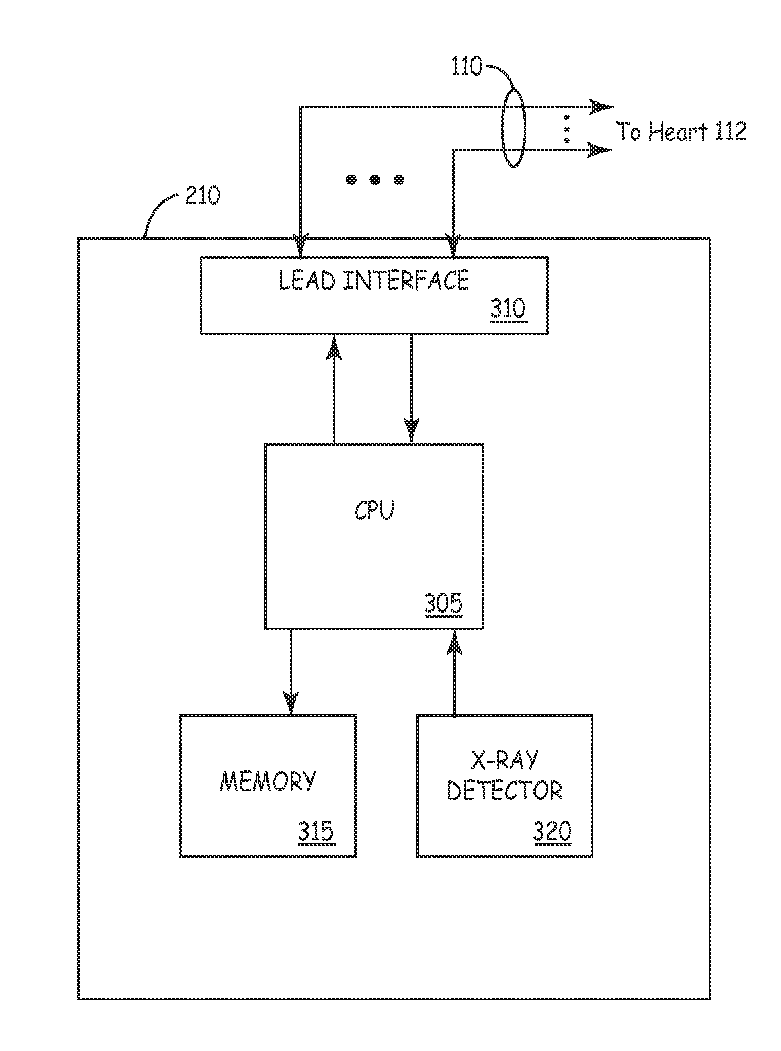
[0061]For this testing, the x-ray source was directed from the topside of a table, so it impinged upon the flip-chip side of the hybrid first. The microprocessor and sense amplifier chips are mounted on the flip-chip side of the hybrid, so the radiation penetrated from the backside of the die. The tube settings ranged from 120 KV & 143 mA for a 1 sec (143 mAs) exposure to 120 KV & 49 mA for the 16 sec (778 mAs) exposure. The response observed at the sense amplifier was an AC-coupled high-pass waveform. The voltage gain to the sense amplifier was approximately 1000. The most likely cause of the large transient voltage is a shift in the offset voltage of one of the previous stages in the sense amplifier. A shift of 8 mV was seen in the voltage reference, and it is assumed that a similar shift is occurring somewhere in the sense amplifier. The sensor output was 120 mV for the 120 KV / 143 mA / 1 sec exposure setting. The IB50 test output was monitored during the exposure by r...


PUM

PropertyMeasurementUnit
threshold energyaaaaaaaaaa
voltageaaaaaaaaaa
sampling clock frequencyaaaaaaaaaa

Abstract

According to the invention methods and structures for determining whether impingement of x-ray radiation upon potentially vulnerable discrete components, circuits and / or circuit pathways of an implantable medical device (IMD) causes modifications or damage thereto. The invention includes structures for deflecting impingement of x-ray radiation from said potentially vulnerable components, circuits and / or circuit pathways. The x-ray radiation can be generated by various imaging modalities or unknown sources. Myriad IMDs can utilize the teaching of the present invention, including for example, a pacemaker, a drug delivery pump, a nerve stimulation device, a muscle stimulation device, an implantable cardioverter-defibrillator, a subcutaneous defibrillator, a deep brain stimulator or the like.

Description

CROSS REFERENCE TO RELATED APPLICATION[0001]The present patent document is related to co-pending non-provisional U.S. patent application Ser. No. 11 / 096,671 entitled, “METHODS AND APPARATUS FOR REDUCING SPURIOUS SIGNALS IN IMPLANTABLE MEDICAL DEVICES CAUSED BY X-RAY RADIATION,” filed on even date herewith; the contents of which are hereby incorporated by reference.FIELD OF THE INVENTION[0002]This invention relates generally to implantable medical devices (IMDs) and, more particularly, to a method and apparatus for reducing negative effects of x-ray radiation impinging upon operative circuitry of active IMDs (e.g., pacemakers, nerve stimulators, therapeutic-substance pumps, physiologic monitors, etc.); in particular, the invention relates to reducing such x-ray radiation impingement during positron emission tomography (PET), computed tomography (CT), fluoroscopy-type and other x-ray imaging scans including newer imaging modalities such as digital subtraction angiography (DSA) and Ang...

Claims


Application Information

Patent Timeline
Patent Type & Authority Patents(United States)
IPC IPC(8): A61N1/05A61L31/18A61N1/37A61N1/375
CPCA61L31/18A61N1/37A61N1/375A61N1/3718H01L2224/49431H01L2924/19105H01L2224/49171H01L2224/48137A61N1/37512
Inventor CLEMENT, WESLEY J.FUJIMOTO, HIROSHIWILKINSON, JEFFREY D.BUYSMAN, JOHN R.BORGERDING, GIRARD B.
Owner MEDTRONIC INC
Why Eureka
Try Eureka