Co-delivery of stimulatory and inhibitory factors to create temporally stable and spatially restricted zones

a technology of stimulatory factors and inhibitory factors, applied in the field of drug delivery, can solve the problems of difficult spatial cue cellular discrimination, achieve the effects of preventing the negative effects of initial bursts, promoting cell-based maintenance of tissue structure and function, and halting or retarding disease progression or age-related tissue changes

Active Publication Date: 2017-07-04
PRESIDENT & FELLOWS OF HARVARD COLLEGE
View PDF159 Cites 3 Cited by
  • Summary
  • Abstract
  • Description
  • Claims
  • Application Information

AI Technical Summary

Benefits of technology

[0013]In one aspect, the stimulatory and inhibitory agents are delivered with polymeric scaffolds in vivo or in vitro. Exemplary scaffold compositions include polylactic acid, polyglycolic acid, poly(lactic-co-glycolic acid) (PLGA) polymers, alginates and alginate derivatives, gelatin, collagen, fibrin, hyaluronic acid, laminin rich gels, agarose, natural and synthetic polysaccharides, polyamino acids, polypeptides, polyesters, polyanhydrides, polyphosphazines, poly(vinyl alcohols), poly(alkylene oxides), poly(allylamines)(PAM), poly(acrylates), modified styrene polymers, pluronic polyols, polyoxamers, poly(uronic acids), poly(vinylpyrrolidone) and copolymers or graft copolymers of any of the above. One preferred scaffold composition includes an arginine-glycine-aspartate (RGD)-modified alginate. The density and mixture of the bioactive agents and inhibitors is controlled by initial doping levels or concentration gradient of the substance, by embedding the bioactive substances in scaffold material with a known leaching rate, by release as the scaffold material degrades, by diffusion from an area of concentration, by interaction of precursor chemicals diffusing into an area, or by production / excretion of compositions by resident support cells. The physical or chemical structure of the scaffold also regulates the diffusion of bioactive agents.
[0018]This approach is exemplified herein in the context of angiogenesis via the administration of VEGF and anti-VEGF, but is also useful in other drug delivery applications in order to mitigate the negative effects of initial bursts, as well as regenerative processes in general. The in vivo and in vitro methods described herein promote regeneration of a tissue or organ immediately adjacent to the bioactive agents, or at some distant site. Alternatively, the methods described herein promote destruction of a tissue (locally or at a distant site). The methods are also useful for disease prevention, e.g., to promote cell-based maintenance of tissue structure and function and to stop or retard disease progression or age-related tissue changes.
[0019]The invention provides in vivo and in vitro methods for inducing angiogenesis by contacting a tissue with an agent that promotes angiogenesis (i.e., growth and development of new blood vessels from pre-existing vessels), and contacting the tissue with an inhibitor of angiogenesis. In one aspect, the inhibitor of angiogenesis is a specific inhibitor of the agent that promotes angiogenesis. Alternatively, the inhibitor of angiogenesis is a general inhibitor of angiogenesis. In one aspect, the agent and agent inhibitor are administered in a 1:1 ratio. In other aspects, the agent / agent inhibitor are administered in a 1:2; 1:5; 1:10; 1:100; 1:1,000; or 1,000:1, 100:1, 10:1, 5:1, or 2:1 ratio. Preferably, the agent and the inhibitor are administered simultaneously. In one aspect, the agent and the inhibitor are incorporated in or on a device comprising a scaffold composition. The agent and the inhibitor are spatially segregated in or on the scaffold composition. Alternatively, the agent and inhibitor are injected directly into the tissue, e.g., an artery. The simultaneous delivery of agents and agent inhibitors maintains the temporal stability of the active agent concentration profile. Preferably, the angiogenesis methods described herein result in heterogeneous distribution of blood vessels.
[0022]Regeneration can be temporally and spatially (directionally) controlled by simultaneously delivering from a single source / location stimulatory and inhibitory agents that are spatially segregated. Specifically, the invention also provides a method of inducing regeneration in a spatially restricted zone by contacting a target site with an agent that promotes regeneration and contacting a target site with an inhibitor of regeneration. In one aspect, the inhibitor of regeneration is a specific inhibitor of the agent that promotes regeneration. In another aspect, the inhibitor of regeneration is a general inhibitor of regeneration. Preferably, the agent and the inhibitor are administered simultaneously. Optionally, the agent and the inhibitor are incorporated in or on a device comprising a scaffold composition. The agent and the inhibitor are spatially segregated in or on the scaffold composition. Alternatively, the agent and inhibitor are injected directly into the tissue, e.g., an artery. The simultaneous delivery of agents and agent inhibitors maintains the temporal stability of the active agent concentration profile. Also provided are methods of temporally and spatially (directionally) controlling regenerative processes by simultaneously delivering from a single source / location agents that act synergistically rather than antagonistically to create spatial patterns / control.
[0024]The invention provides for methods for inducing cell differentiation / specialization in a spatially restricted zone by contacting a cell with a promoter of differentiation and an inhibitor of differentiation. The agent and the inhibitor interact, thereby inducing cell differentiation in a spatially restricted zone at or near a site at which the agent and the inhibitor interact. Optionally, the agent and the inhibitor are administered simultaneously. In one aspect, the agent and the inhibitor are incorporated in or on a device comprising a scaffold composition. The agent and the inhibitor are spatially segregated in or on said scaffold composition. The simultaneous delivery of agents and agent inhibitors maintains the temporal stability of the active agent concentration profile. Suitable combinations of agents to promote differentiation of stem cells into bone cells or tooth cells include BMP4 and anti-BMP4. Suitable combinations of agents to promote differentiation of stem cells into dentin include TGF-β1 and latency associated peptide (LAP).

Problems solved by technology

However, the diffusion / reaction of stimulatory factors alone results in the formation of shallow gradients that make cellular discrimination of spatial cues difficult.

Method used


Image

  • Co-delivery of stimulatory and inhibitory factors to create temporally stable and spatially restricted zones
  • Co-delivery of stimulatory and inhibitory factors to create temporally stable and spatially restricted zones
  • Co-delivery of stimulatory and inhibitory factors to create temporally stable and spatially restricted zones

Examples


example 1

VEGF and Anti-VEGF on Angiogenesis

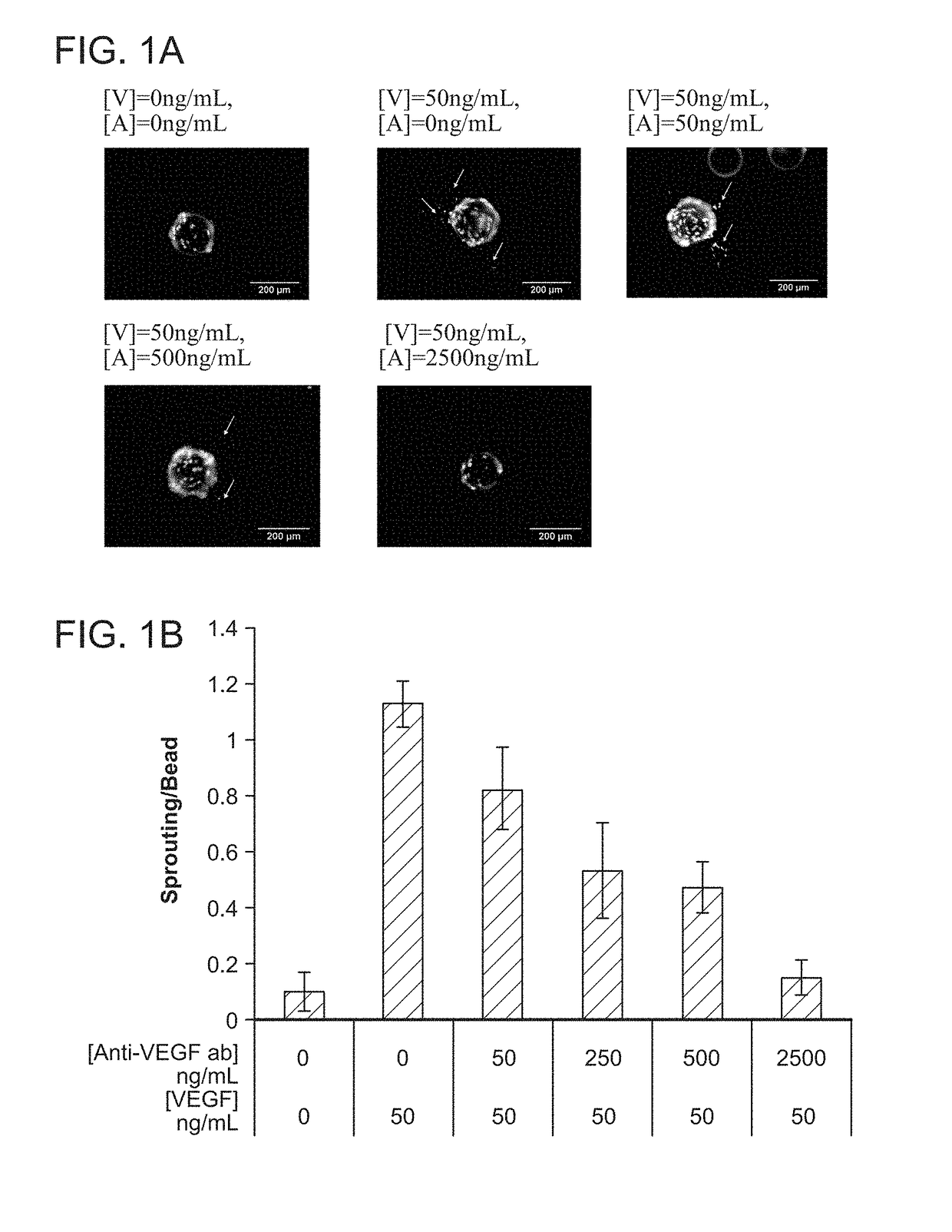
[0119]The relation between VEGF and anti-VEGF concentrations on angiogenesis was first evaluated using a common in vitro sprouting assay, in order to quantitatively determine the appropriate doses of the two factors for subsequent in vivo studies.

Cell Culture and In Vitro Sprouting Assay

[0120]Briefly, dermal human vascular endothelial cells (HMVECs) were purchased from Lonza (CC-2543) and cultured to confluence at 37° C. and 5% CO2 in microvascular endothelial cell growth medium-2 (EGM-2MV) (Lonza) containing all supplements. Angiogenic activity of endothelial cells was assessed using a modification of a widely used in vitro sprouting assay (Nehls, V., and Drenckhahn, D. 1995 Microvasc Res 50, 311-322). Briefly, dextran beads microcarriers (Cytodex 3) with a dry weight of 50 mg were swollen in PBS and autoclaved. The microcarriers were washed in EGM-2MV medium and seeded with 3×10⁶ HMVECs in a spinner flask. The microcarriers were stirred for 2 minu...

example 2

cal Model of Protein Distribution

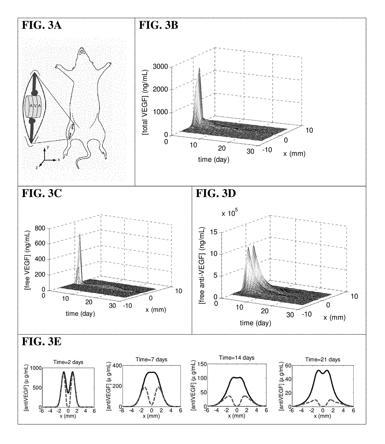
[0126]In order to design appropriate encapsulated doses of VEGF and anti-VEGF to create spatially defined angiogenic regions, mass transport PDEs of the proteins in the scaffolds and the underlying tissues were simulated.

[0127]Briefly, a computational model was generated to depict the concentration profiles of free VEGF anti-VEGF, and VEGF complexed with anti-VEGF. This model accounted for diffusion, release from scaffolds, binding kinetics, and protein degradation. The governing equations of the VEGF and anti-VEGF concentrations inside the scaffold and underlying muscle were:

[0128]∂c1∂t=D1⁢∇2⁢c1-k1⁢c1+f1-kon⁢c1⁢c2+koff⁢c3∂c2∂t=D2⁢∇2⁢c2-k2⁢c2+f2-kon⁢c1⁢c2+koff⁢c3∂c3∂t=D3⁢∇2⁢c3+kon⁢c1⁢c2-koff⁢c3whereci=concentrationci⁡(x,y,z,t=0)=0;∀i,x,y,zfi={release⁢⁢function,inside⁢⁢scaffold0,inside⁢⁢muscle⁢⁢i={1free⁢⁢VEGF2free⁢⁢antiVEGF3VEGF⁢-⁢antiVEGF⁢⁢complex⁢⁢D1=7×10⁻⁷⁢⁢cm2s=Effective⁢⁢interstitial⁢⁢diffusion⁢⁢coefficient⁢⁢of⁢⁢VEGF165⁢⁢D2=3.2× 10-9⁢⁢cm2s=  Effe...

example 3

Regulated Angiogenesis In Vivo

[0135]To test the ability of this system to provide spatial control over angiogenesis, scaffolds were subsequently implanted into the ischemic hindlimbs of SCID mice.

Mouse Model of Hindlimb Ischemia

[0136]Scaffolds were implanted in 6-week-old SCID mice (Taconic, Hudson, N.Y.) that had undergone unilateral ligation of hindlimb blood vessels to create a severe model of hindlimb ischemia (Sun et al., 2005 Pharm Res 22, 1110-1116). The SCID model was chosen because it offered a stable loss of perfusion over weeks and the angiogenic effects from inflammation were reduced. Briefly, animals were anesthetized by IP injection of a 7:1 mixture of ketamine and xylazine. The targeted hindlimb was shaved and sterilized with ethanol prior to making an incision through the dermis. Ligation sites were made on the external iliac artery and vein, and on the femoral artery and vein using 5-0 Ethilon (Ethicon, Somerville, N.J.). The vessels were severed between the ligatio...


PUM

PropertyMeasurementUnit
timeaaaaaaaaaa
timeaaaaaaaaaa
timeaaaaaaaaaa

Abstract

The invention provides methods and compositions for local manipulation of regenerative processes via exogenous factor delivery.

Description

RELATED APPLICATIONS[0001]This application is a national stage application, filed under 35 U.S.C. §371, of International Application No. PCT / US2011 / 042051, filed Jun. 27, 2011, which claims the benefit of U.S. Provisional Patent Application No. 61 / 358,499, filed on Jun. 25, 2010, the contents of which are incorporated herein by reference in their entireties.STATEMENT AS TO FEDERALLY SPONSORED RESEARCH[0002]This invention was made with Government support under National Institutes of Health awards R01HL069957 and R37DE013033. The Government has certain rights in the inventionFIELD OF THE INVENTION[0003]This invention relates generally to the field of drug delivery.BACKGROUND OF THE INVENTION[0004]Nature frequently utilizes opposing factors to create a stable activator gradient to robustly control pattern formation. Specifically, during developmental processes, tight spatial regulation often results from the combined action of stimulatory and inhibitory factors. In such reaction scheme...

Claims


Application Information

Patent Timeline
Patent Type & Authority Patents(United States)
IPC IPC(8): A61K38/18A61K31/33A61K35/28A61K35/54A61P19/02A61K9/00A61F2/30A61K45/06A61K35/545A61K31/16A61K31/335A61K31/381A61K31/4192A61K31/454A61K35/12A61K39/395
CPCA61K38/1841A61K38/185A61K38/1866A61K38/1875A61K31/381A61K31/16A61K31/454A61K31/335A61K31/4192A61K9/0087A61F2/30756A61K35/12A61K35/28A61K35/545A61K38/1808A61K39/3955A61K45/06A61K2300/00A61F2002/30766A61P19/02A61P9/00
Inventor MOONEY, DAVID J.YUEN, WILLIAM W.ARANY, PRAVEEN
Owner PRESIDENT & FELLOWS OF HARVARD COLLEGE
Why Eureka
Try Eureka