
Biodegradable in-situ forming implants and methods of producing the same

a biodegradable, insitu forming technology, applied in the direction of prosthesis, surgical adhesives, dentistry, etc., can solve the problems of reluctance of patients to accept such implants or drug-delivery systems, important limitations of their use, and inability to meet the demand for biodegradable implants

Inactive Publication Date: 2002-12-31
ATRIX LAB
View PDF69 Cites 111 Cited by
  • Summary
  • Abstract
  • Description
  • Claims
  • Application Information

AI Technical Summary

Benefits of technology

In both the thermoplastic and the thermosetting systems, the advantages of liquid application are achieved. For example, the polymer may be injected via syringe and needle into a body while it is in liquid form and then left in-situ to form a solid biodegradable implant structure. The need to form an incision is eliminated, and the implant will assume the shape of its cavity. Furthermore, a drug-delivery vehicle may be provided by adding a biologically active agent to the liquid prior to injection. Once the implant is formed, it will release the agent to the body and then biodegrade. The term "biologically active agent" means a drug or some other substance capable of producing an effect on a body.
It is a further object of the present invention to provide implants having a range of properties from soft and elastomeric to hard and rigid, so as to be usable with both soft and hard tissue.

Problems solved by technology

Although these two classes of biodegradable polymers have many useful biomedical applications, there are several important limitations to their use in the body where body is defined as that of humans, animals, birds, fish, and reptiles.
These incisions are often larger than desired by the medical profession and lead to a reluctance of the patients to accept such an implant or drug-delivery system.
Although these small particles can be injected into the body with a syringe, they do not always satisfy the demand for a biodegradable implant.
When inserted into certain body cavities such as the mouth, a periodontal pocket, the eye, or the vagina where there is considerable fluid flow, these small particles, microspheres, or microcapsules are poorly retained because of their small size and discontinuous nature.
In addition, microspheres or microcapsules prepared from these polymers and containing drugs for release into the body are sometimes difficult to produce on a large scale, and their storage and injection characteristics present problems.
Furthermore, one other major limitation of the microcapsule or small-particle system is their lack of reversibility without extensive surgical intervention.
That is, if there are complications after they have been injected, it is considerably more difficult to remove them from the body than with solid implants.

Method used


Image

  • Biodegradable in-situ forming implants and methods of producing the same
  • Biodegradable in-situ forming implants and methods of producing the same

Examples


example 1

Poly(DL-lactic acid) was prepared by the simple poly-condensation of lactic acid. No catalysts were used, and the reaction times were varied to produce polymers with different theoretical molecular weights. These polymers were designated as DL-PLA oligomers. A quantity of the solid oligomer was dissolved in NMP to give a 68:32 ratio of polymer to solvent. Sanguinarine chloride(SaCl), a benzophenanthridine alkaloid with antimicrobial activity especially toward periodontal pathogens, was added to the polymer solution to give a 2% by weight dispersion of the drug in the total mixture. The dispersion of drug and polymer solution was then injected into a dialysis tube (diameter of 1.5 mm) with a sterile disposable syringe without a needle. Each end of the 6-in. length of dialysis tubing was tied with a knot to prevent loss of the drug / polymer mass, and the tube with the injected material was placed in a pH 7 Sorenson's buffer receiving fluid maintained at 37.degree. C. Upon immersion in ...

example 2

Ethoxydihydrosanguinarine(SaEt), the ethanol ester of sanguinarine, was added to the same DL-PLA oligomer / NMP solution described in Example 1. SaEt dissolved in the polymer solution to give a homogeneous solution of drug and polymer. Approximately 250 .mu.L of the solution was added to receiving fluid and the release of drug measured as described in Example 1. The release of SaEt was slower than that for SaCl as expected because of its lower water solubility. After the first day, approximately 45% was released, 52% after 2 days, 60% after 5 days, 70% after 9 days, and 80% after 14 days.

example 3

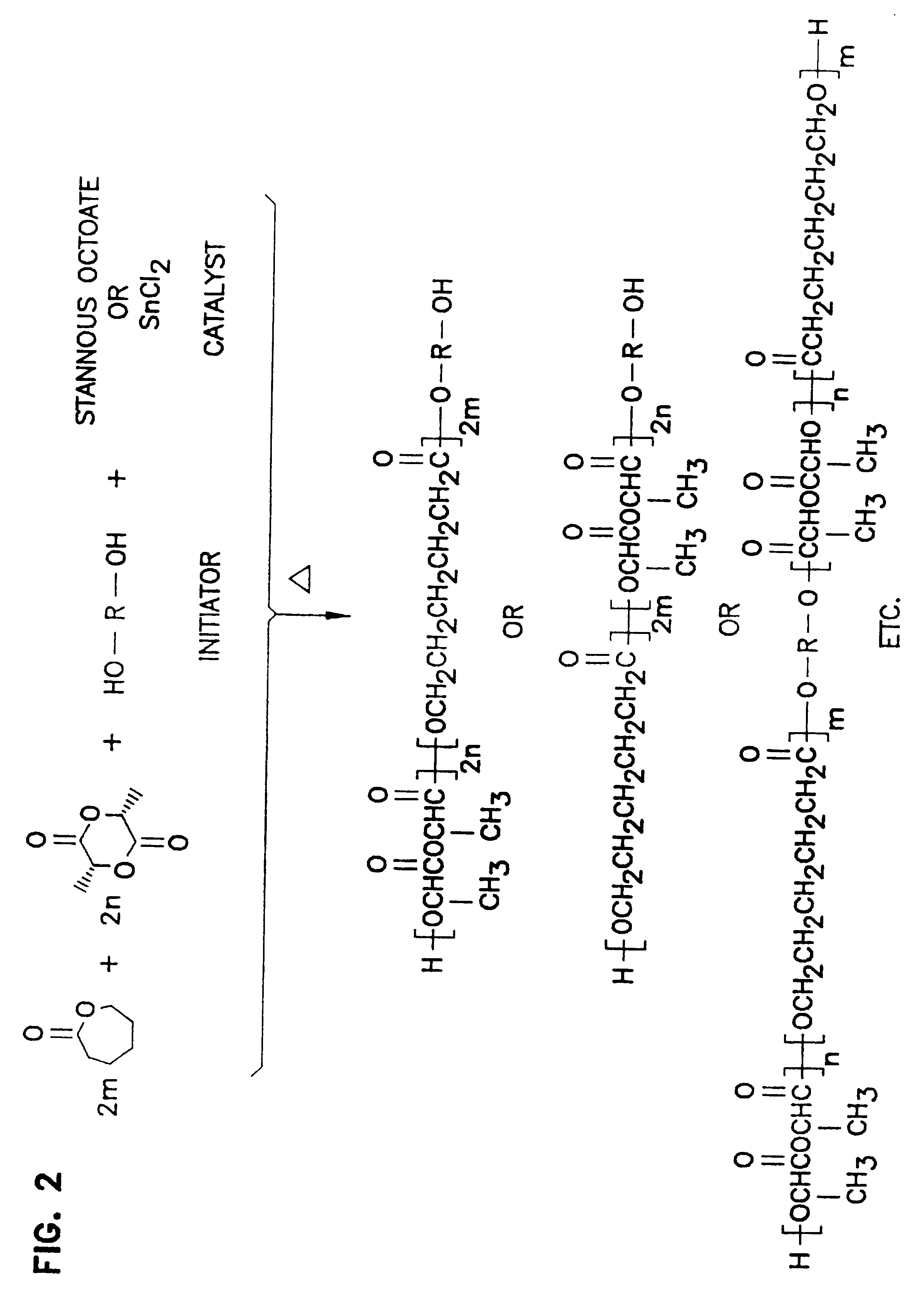
Poly(DL-lactide) with an inherent viscosity of 0.08 dL / g and a theoretical molecular weight of 2,000 was prepared by the ring-opening polymerization of DL-lactide using lauryl alcohol as the initiator and stannous chloride as the catalyst. This polymer was then dissolved in NMP to give a 40% by weight polymer solution. SaCl was dispersed in the solution of this polymer in NMP to give a 1.5% by weight dispersion of the drug in the solution and the release rate determined as described in Example 1. The release rate of the drug from this higher molecular-weight polymer was slower than from the DL-PLA oligomer. After the first day, approximately 32% was released, 40% after 2 days, 45% after 5 days, and 50% after 15 days.


PUM

PropertyMeasurementUnit
diameteraaaaaaaaaa
pHaaaaaaaaaa
pHaaaaaaaaaa

Abstract

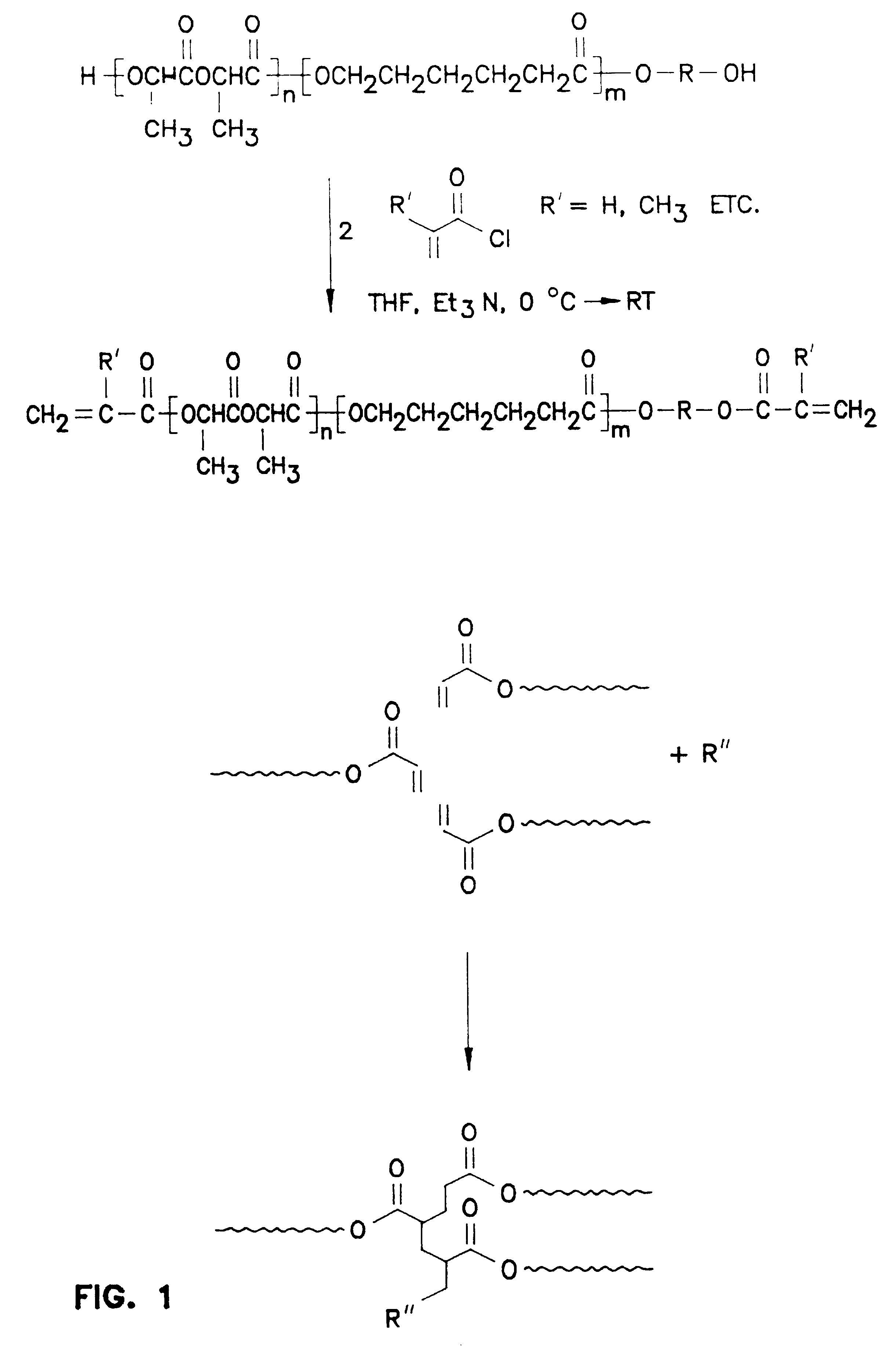
A biodegradable polymer is provided for use in providing syringeable, in-situ forming, solid biodegradable implants for animals. The polymer is placed into the animal in liquid form and cures to form the implant in-situ. A thermoplastic system to form said implant comprises the steps of dissolving a non-reactive polymer in biocompatible solvent to form a liquid, placing the liquid within the animal, and allowing the solvent to dissipate to produce the implant. An alternative, thermosetting system comprises mixing together effective amounts of a liquid acrylic ester terminated, biodegradable prepolymer and a curing agent, placing the liquid mixture within an animal and allowing the prepolymer to cure to form the implant. Both systems provide a syringeable, solid biodegradable delivery system by the addition of an effective level of biologically active agent to the liquid before injection into the body.

Description

BACKGROUND OF THE INVENTIONThe present invention relates to a method and composition for producing biodegradable polymers, and more particularly to the use of such polymers for providing syringeable, in-situ forming, solid, biodegradable implants.Biodegradable polymers have been used for many years in medical applications. These include sutures, surgical clips, staples, implants, and drug delivery systems. The majority of these biodegradable polymers have been thermoplastic materials based upon glycolide, lactide, .epsilon.-caprolactone, and copolymers thereof. Typical examples are the polyglycolide sutures described in U.S.-Pat. No. 3,297,033 to Schmitt, the poly(L-lactide-co-glycolide) sutures described in U.S. Pat. No. 3,636,956 to Schneider, the poly(L-lactide-co-glycolide) surgical clips and staples described in U.S. Pat. No. 4,523,591 to Kaplan et al., and the drug-delivery systems described in U.S. Pat. No. 3,773,919 to Boswell et al., U.S. Pat. No. 3,887,699 to Yolles, U.S. ...

Claims


Application Information

Patent Timeline
Patent Type & Authority Patents(United States)
IPC IPC(8): A61K9/00A61L15/62A61L15/16A61L15/44A61L27/50A61L26/00A61L27/00A61L24/04A61L24/00A61L27/18A61F2/30A61F2/02
CPCA61F2002/30062A61F2002/30583A61F2210/0004A61F2210/0085A61K9/0024A61L15/44A61L15/62A61L24/0042A61L24/046A61L26/009A61L27/18A61L27/50A61L2300/252A61L2300/404A61L2300/406C08L75/04C08L67/04
Inventor DUNN, RICHARD L.ENGLISH, JAMES P.COWSAR, DONALD R.VANDERBILT, DAVID P.
Owner ATRIX LAB
Learn More