Method and reagent kit for rare-earth element metal chelating making quantitative proteome

A rare earth element and metal chelation technology, applied in the direction of measuring devices, instruments, scientific instruments, etc., to achieve the effect of convenient use, wide practicability, and simple operation

Inactive Publication Date: 2007-09-26
INST OF RADIATION MEDICINE ACAD OF MILITARY MEDICAL SCI OF THE PLA
View PDF0 Cites 3 Cited by
  • Summary
  • Abstract
  • Description
  • Claims
  • Application Information

AI Technical Summary

Problems solved by technology

The inventors found that it is feasible to use rare earth elements whose price is much lower than that of stable isotopes as internal quality standards in quantitative proteomics by combining multidimensional liquid chromatography separation means and multi-stage tandem mass spectrometry, which can be used to solve the problem of proteomics. Relative Quantitative Questions

Method used


Image

  • Method and reagent kit for rare-earth element metal chelating making quantitative proteome
  • Method and reagent kit for rare-earth element metal chelating making quantitative proteome

Examples


Embodiment 1

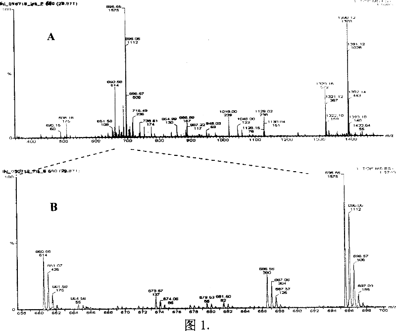
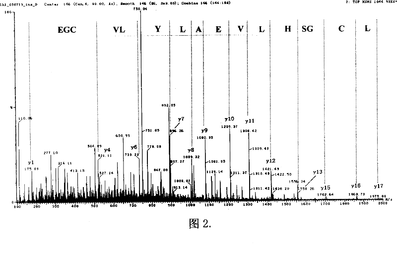
[0032] The relative quantification of embodiment 1 standard protein insulin

[0033] 1.1 Take 0.1 mg of insulin and dissolve it in 100 μL of 50 mM pH 7.0 triethylamine bicarbonate buffer solution. Add 0.2 μL of 100 mM reducing agent tricarboxyethyl phosphine (TCEP), react at 37°C for 4 hours, then cool to room temperature, add 0.5 μL of 100 mM iodoacetamide (IAA), react at room temperature for 1 hour, and then add 10 mM DTT to terminate the reaction. Add 2ug trypsin, digest at 37°C for 2 hours. Then add 2ug of trypsin, and digest at 37°C for 10 hours. The insulin peptide mixture obtained by enzymatic digestion was added to 360 μg of diethylenetriaminepentaacetic acid dianhydride solid powder, vigorously vortexed for 1 min, and then left at room temperature for 1 hour. Then put the reaction solution in a vacuum dryer and dry it completely, add 0.1M ammonium acetate solution to redissolve, and divide it into two parts according to the volume, and add 40 μL of YCl with a concen...

Embodiment 2

[0036] Embodiment 2 Relative Quantification of Standard Protein Horse Myoglobin

[0037] 2.1 Take 0.1 mg of horse myoglobin and dissolve it in 100 μL of 50 mM pH 7.0 triethylamine bicarbonate buffer solution. Myoglobin has no cysteine ​​residues in its amino acid sequence, so a reductive alkylation step is not required. Add 2ug trypsin, digest at 37°C for 2 hours. Then add 2ug of trypsin, and digest at 37°C for 10 hours. The myoglobin peptide mixture obtained by enzymatic digestion was added to 360 μg of diethylenetriaminepentaacetic acid dianhydride solid powder, vigorously vortexed for 1 min, and then left at room temperature for 1 hour. Then put the reaction solution in a vacuum dryer and dry it completely, add 0.1M ammonium acetate solution to redissolve, and divide it into two parts on average, one of which is added with 40 μL of YCl with a concentration of 0.15M 3 , add the same concentration and volume of TbCl to the other 3 aqueous solution. Incubate at 37°C for 2...

Embodiment 3

[0040] The relative quantification of embodiment 3 six kinds of standard protein mixtures

[0041] 3.1 Configure six protein solutions (bovine serum albumin, transferrin, α-lactoglobulin, β-lactoglobulin, myoglobin and lysozyme protein) with a concentration of 0.1mmoL / L for each protein, The buffer solution was 50 mM triethylamine bicarbonate pH 7.0. Take 70 μL of each and mix together. Add 0.8 μL of 500 mM reducing agent tricarboxyethylphosphine (TCEP), react at 37°C for 4 hours, then cool to room temperature, add 1 μL of 1M iodoacetamide (IAA), and react at room temperature for 1 hour in the dark. Add 5ug of trypsin, digest at 37°C for 2 hours. Then add 5ug of trypsin, and digest at 37°C for 10 hours. The myoglobin peptide mixture obtained by enzymatic digestion was added to 360 μg of diethylenetriaminepentaacetic acid dianhydride solid powder, vigorously vortexed for 1 min, and then left at room temperature for 1 hour. Then put the reaction solution into a vacuum dryer ...


PUM


Abstract

This invention provides to rare earth element metal chelation label protein for quantitative protein study method and agent case. This invention method relates to protein restore alkali base and pancreatic enzymes slices, peptide section double function agent decoration, rare metal chelation, multi-diemension liquid phase spectrum isolation and multiple degrees series spectrum quantitative and testing. Through one degree spectrum ion signal intensity content and protein validating through series mass spectrum and database. This invention also describes this method agent case.

Description

technical field [0001] The invention relates to a method and kit for protein quantification in proteomics. Specifically, it relates to a new method and kit for rare earth element metal chelate labeling protein for quantitative proteomics. Background technique [0002] Quantitative proteomics is the comparative analysis of protein content in biological samples (cells, tissues or body fluids, etc.) It is a necessary means and research frontier to study the pathogenic mechanism and pharmacological control mechanism of major diseases. [0003] At present, the relative quantitative methods widely used in proteome research mainly rely on two-dimensional electrophoresis (Twodimensional electrophoresis, 2-DE) staining method and stable isotope-labeled mass spectrometry detection technology. The 2-DE relative quantification method can intuitively reflect the difference in protein expression through the staining intensity, but the existence of the dye is likely to cause interference...

Claims


Application Information

Patent Timeline
Patent Type & Authority Applications(China)
IPC IPC(8): G01N30/02G01N30/06
Inventor 钱小红刘慧玲张养军王京兰
Owner INST OF RADIATION MEDICINE ACAD OF MILITARY MEDICAL SCI OF THE PLA
Why Eureka
Try Eureka