
Fluorescent micro-ball immune chromatography test paper strip for detecting residual animal medicine and preparing method thereof

An immunochromatographic test paper and fluorescent microsphere technology, applied in the field of veterinary drug detection in food safety, can solve the problems of low contrast between color bands and background, limited detection sensitivity, large background interference, etc., to increase stability and fluorescence lifetime. , the effect of expanding the range and variety, and improving the detection sensitivity

Active Publication Date: 2009-05-20
WUXI ZODOLABS BIOTECH
View PDF0 Cites 60 Cited by
  • Summary
  • Abstract
  • Description
  • Claims
  • Application Information

AI Technical Summary

Problems solved by technology

[0005] (1) The colloidal gold labeling process is an electrostatic adsorption process, which is a physical adsorption, so the stability in the liquid phase is poor, often causing the labeled protein molecules to fall off again
[0006] (2) The test result is judged by displaying a single purple-red strip, and the color is single, so it is difficult to realize multiple inspection and joint inspection
[0007] (3) Only when the gold particles gather to a certain amount, the human naked eye can observe the purple-red band, and the color band has little contrast with the background, thus limiting the detection sensitivity
[0008] (4) The matrix effect of different materials is obvious, and the background interference is very large
[0009] (5) The detection sensitivity is low
[0010] (6) Quantitative detection cannot be realized
[0011] At present, there is also a detection method that uses quantum dots to label rapid immunochromatographic test strips, but this method cannot achieve quantitative detection of the test substance.

Method used


Image

  • Fluorescent micro-ball immune chromatography test paper strip for detecting residual animal medicine and preparing method thereof
  • Fluorescent micro-ball immune chromatography test paper strip for detecting residual animal medicine and preparing method thereof
  • Fluorescent micro-ball immune chromatography test paper strip for detecting residual animal medicine and preparing method thereof

Examples


Embodiment 1

[0063] Example 1: Preparation of Fluorescent Microsphere Immunochromatographic Test Strips for Detecting Clenbuterol Hydrochloride Residues in Samples

[0064] 1. Preparation of chromatography test strips

[0065] 1. Preparation of nitrocellulose membrane;

[0066] (1) Preparation of clenbuterol-BSA conjugate (Clen-BSA) artificial antigen;

[0067] The Clen-BSA was coupled by the diazonium method.

[0068] (2) Preparation of detection area and quality control area:

[0069] Clenbuterol-BSA conjugates and anti-mouse antibodies were coated onto nitrocellulose membranes: with 0.01M pH7.2 PBS (phosphate buffered saline containing 5% sucrose and 0.05% Tween-20 (Tween -20)) adjust the concentration of clenbuterol-BSA conjugate to be 0.5mg / ml, the resulting solution is sprayed on the membrane as the detection area, with 0.01M PBS of pH7.2 (which contains 5% sucrose and 0.05% Tween-20 (Tween-20)) regulates the concentration of anti-mouse antibody to be 0.5mg / mL, and the solution o...

Embodiment 3

[0084] Example 3: Preparation of Fluorescent Microsphere Immunochromatographic Test Strips for Detecting Residual Furazolidone in Samples

[0085] 1. Preparation of chromatography test strips

[0086] 1. Preparation of nitrocellulose membrane;

[0087] (1) preparing furazolidone artificial antigen;

[0088] The diazo method was used to couple furazolidone-OVA (chicken ovalbumin) as the coating antigen.

[0089] (2) Preparation of detection area and quality control area: adjust the concentration of furazolidone-OVA conjugate to 1.0 mg / mL with 0.01M PBS pH7.2 (containing 5% sucrose and 0.05% Tween-20), and the obtained The solution was sprayed on the membrane as the detection area, and the concentration of the anti-mouse antibody was adjusted to 1.0 mg / mL with 0.01 MpH7.2 PBS (which contained 5% sucrose and 0.05% Tween-20), and the resulting solution was sprayed on the membrane as the detection area. In the control area, the amount of spray film in the two areas is 0.74μl / cm,...

Embodiment 4

[0100] Example 4: Preparation of fluorescent microsphere immunochromatographic test strips for detecting residual ractopamine hydrochloride and clenbuterol hydrochloride in samples

[0101] 1. Preparation of chromatography test strips

[0102] 1. Preparation of nitrocellulose membrane;

[0103] (1) Prepare clenbuterol (Clen) and ractopamine (Rac) artificial antigens;

[0104] Clen-BSA conjugates were prepared by the diazo method, and Rac-BSA conjugates were prepared by the EDC method

[0105] (2) Preparation of the detection area: adjust the concentration of the clenbuterol-BSA conjugate to 0.5 with 0.01M PBS at pH 7.2

[0106] mg / mL, adjust the concentration of ractopamine-BSA conjugate to 0.8 mg / mL, and then spray the resulting solution on the membrane to form two detection areas, the two areas are separated by 5 mm, after drying overnight at 37 ° C, in Store in a dry environment at room temperature for later use.

[0107] 2. Preparation of fluorescent microsphere pads; ...


PUM

PropertyMeasurementUnit
Diameteraaaaaaaaaa

Abstract

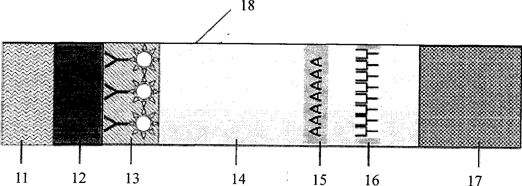
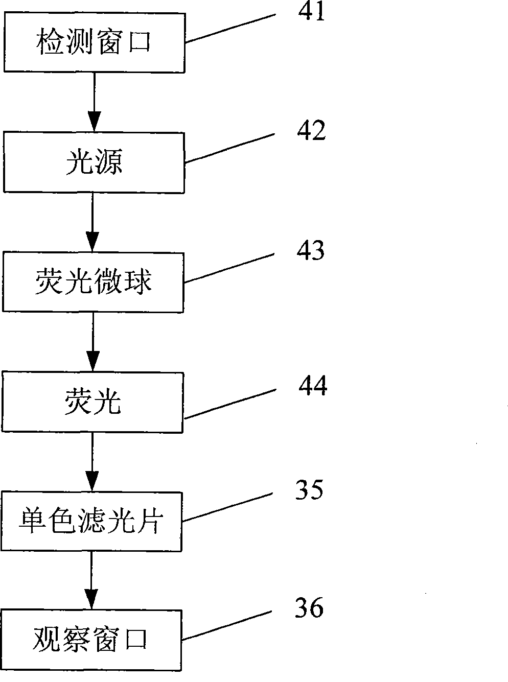
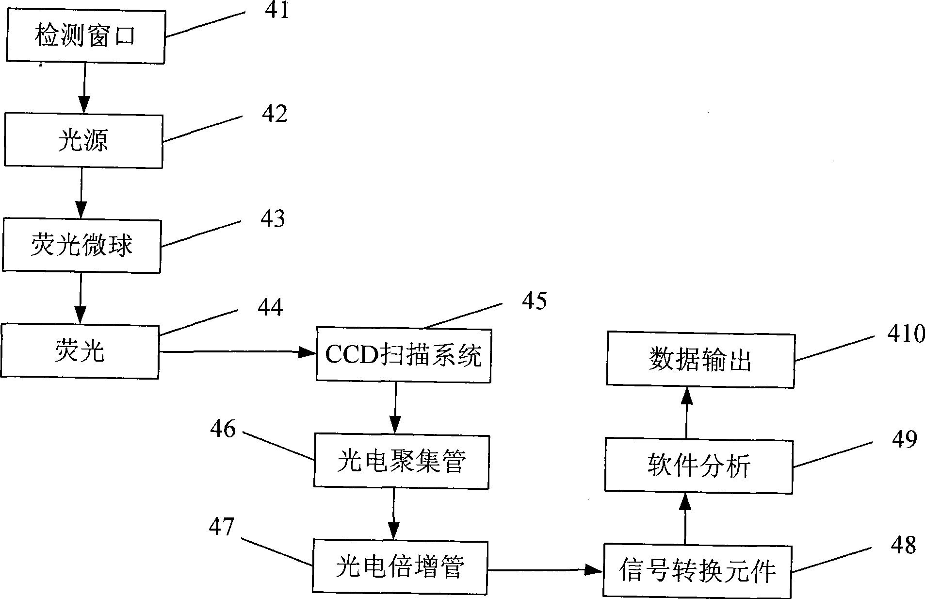
The invention discloses a test paper strip for the fluorescent bead immunochromatography of veterinary medicine remained in a detected sample and a preparation method thereof. A filter paper, a sample pad, a fiberglass membrane, a pyroxylin membrane and water sucking paper are sequentially stuck on a base plate in related joint; fluorescent beads are sprayed on the pyroxylin membrane for marking a veterinary medicine resistant molecule monoantibody; the pyroxylin membrane coated with a veterinary medicine molecule holoantigen is used as a detection region; the pyroxylin membrane coated with an anti-mouse antibody is used as a quality control region; and the test paper strip is prepared through the following steps: (1) the preparation of the pyroxylin membrane; (2) the preparation of a fluorescent bead pad; and (3) the assembling of the test paper strip. In the detecting process, emitted spectrums pass through a proper optical filter device; and all the emitted spectrums are collected through CCD scanning technology, are congregated, are multiplied and are analyzed through a fluorescence analyzer and relevant software to obtain a quantized fluorescence signal, thereby realizing quantitative detection. The test paper strip is mainly used for the qualitative detection and the quantitative detection of all the veterinary medicine classes in food safety detection.

Description

technical field [0001] The invention belongs to the field of veterinary drug detection in food safety, in particular to a fluorescent microsphere immunochromatographic test strip for qualitatively and quantitatively detecting residual veterinary drugs in a sample and a preparation method thereof. Background technique [0002] In food safety testing, technologies such as GC-MS, LC-MS, enzyme-linked immunosorbent assay (ELISA) and colloidal gold-labeled immunochromatographic test strips are mainly used at home and abroad to achieve qualitative and quantitative detection. In food safety testing, in order to achieve high efficiency and quickness, colloidal gold labeling technology or ELISA is often used for preliminary screening, and then positive samples are confirmed by GC-MS or LC-MS. Since the above methods all require expensive and complicated instruments and equipment, the cost is high, and special training is required for operators, and the experimental results cannot be ...

Claims


Application Information

Patent Timeline
IPC IPC(8): G01N33/577G01N33/558G01N33/544G01N33/52
Inventor 魏华熊勇华赖卫华陈雪岚金晶李林陈媛
Owner WUXI ZODOLABS BIOTECH
Learn More