Construction method for dry eye model in vitro

An established method, dry eye technology, applied in the research and prevention of dry eye, simulation field, can solve problems such as lack of histology, and achieve the effect of low price, high repeatability and simple production

Active Publication Date: 2011-08-10
福建和泽生物科技有限公司
View PDF0 Cites 1 Cited by
  • Summary
  • Abstract
  • Description
  • Claims
  • Application Information

AI Technical Summary

Problems solved by technology

[0005] The purpose of the present invention is that there are many influencing factors that affect the production of dry eye animal models, but the pathogenesis research and drug screening method of dry eye through ocular surface epithelial cell culture lacks histological evidence. All disease research models have their corresponding limitations and other problems, and provide a method for establishing an in vitro dry eye model that is not only simple and easy to implement, has good reproducibility, and is less expensive than animal models, but also has many histopathological characteristics similar to dry eye and applications in modeling, research and prevention of dry eye

Method used


Image

  • Construction method for dry eye model in vitro
  • Construction method for dry eye model in vitro
  • Construction method for dry eye model in vitro

Examples


Embodiment 1

[0039] The preparation method of the dry eye model established by conjunctival tissue cultured under the air-liquid interface environment of the present invention, its specific steps are as follows:

[0040] ①The bulbar conjunctiva and subconjunctival tissue of the donated cadaver eye obtained from the eye bank were cut out, washed 3 times with phosphate buffered saline (PBS) containing antibiotics, and placed in epithelial cell culture medium (SHEM medium) added with 3X antibiotics 30min, and then transferred to SHEM medium containing common antibiotic concentrations for later use.

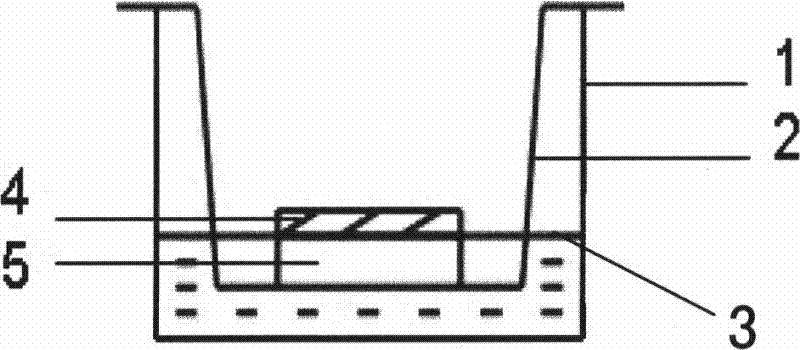
[0041] ②Coat the bottom of the culture dish insert with type I collagen: the collagen concentration is 1mg / ml, and 0.3ml is used in each insert.

[0042] ③ Trim the conjunctival tissue into a 5mm square size, place it in the center of the coated culture dish insert, with the epithelial side facing up.

[0043] ④ Add SHEM medium to the outside of the insert of the culture dish, and keep the liqui...

Embodiment 2

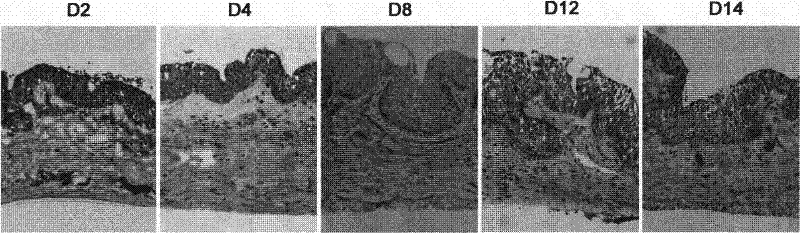
[0047] Conjunctival tissues cultured for 2 to 14 days under the above conditions were frozen and embedded, and frozen sections were performed for histopathological and immunohistochemical staining observations, as follows:

[0048] Main reagents and instruments: mouse anti-human monoclonal anti-keratin K19 antibody, anti-keratin K10 antibody and anti-P63 antibody (Dako, USA), mouse anti-human monoclonal anti-mucin MUC5AC antibody (Abcam, USA), FITC-labeled Goat anti-mouse IgG secondary antibody (Sigma, USA), OCT embedding agent (SAKURA, USA, model OCT4583). Leica CM1850 cryostat (Leica, Germany), Nikon TE2000 inverted fluorescence microscope (Nikon, Japan).

[0049] Hematoxylin-eosin staining: The conjunctival tissue specimens cultured under the above conditions were immediately embedded in OCT at different time points, pre-cooled in liquid nitrogen for 1 min, and stored in a -80°C low-temperature refrigerator. The cryopreserved specimens were made into serial frozen sections...

Embodiment 3

[0054] The preparation method of the dry eye model established by conjunctival tissue cultured under the air-liquid interface environment of the present invention, its specific steps are as follows:

[0055] ① The bulbar conjunctiva and subconjunctival tissue of the donated cadaver eyes obtained from the eye bank were cut out, and after necessary anti-pollution treatment, they were transferred to a medium containing ordinary antibiotic concentrations for later use.

[0056] ②Coat the bottom of the culture dish insert with type I collagen: the collagen concentration is 1mg / ml, and 0.3ml is used in each insert.

[0057] ③Use a trepan drill with a diameter of 7 mm to remove conjunctival tissue pieces of the same size, and place them in the center of the coated culture dish insert with the epithelial side facing upward.

[0058] ④ Add the culture medium to the outside of the insert of the culture dish, and keep the liquid level at the junction of the epithelium and stroma of the c...


PUM

PropertyMeasurementUnit
diameteraaaaaaaaaa

Abstract

A method for constructing a xerophthalmia model in vitro relates to a xerophthalmia model. A method for constructing a xerophthalmia model in vitro with easy operation, good repeatability, lower cost than animal model and a plurality of similar histopathological characteristics similar to xerophthalmia and application thereof in simulating, studying and preventing xerophthalmia are provided. The conjunctiva tissue containing conjunctival epithelium and conjunctival submucosa is cut and placed in culture media; the conjunctiva tissue is trimmed into a conjunctiva tissue block which is placed on a culture vessel inserter coated by type I collagen, with conjunctival epithelium being upward; the culture media are added into the culture vessel outside the inserter to cause the gas-liquid interface of the culture media to be at the junction of the conjunctival epithelium and conjunctival submucosa of the conjunctiva tissue block; and the conjunctiva tissue block is placed into a culture tank to be cultured. The xerophthalmia model in vitro can be used for such researches as xerophthalmia squamous epithelium metaplasia, ocular surface epithelial barrier breakdown, ocular surface epithelial mucoprotein change, and function of the participation and atopsis of inflammatory mediator in xerophthalmia diseases in the generation and development of xerophthalmia.

Description

technical field [0001] The invention relates to a dry eye model, in particular to a method for preparing an in vitro dry eye model by means of conjunctival tissue culture and its application in simulation, research and prevention and treatment of dry eye. Background technique [0002] Dry eye is a kind of disease caused by abnormal tear quality, quantity and dynamics caused by any reason, resulting in tear film instability and / or abnormal ocular surface, accompanied by ocular symptoms. According to the elements of maintaining a stable tear film, dry eye can be divided into: evaporative dry eye (mainly lipid layer abnormality), water deficiency dry eye ( syndrome and non- Syndrome), mucin abnormal dry eye (mainly damaged ocular surface epithelial cells), abnormal tear dynamics dry eye and mixed dry eye ([1] Liu Zuo. Diagnosis of dry eye. Chinese Journal of Ophthalmology, 2002 , 38(5):318-320). Clinical surveys show that dry eye is currently the most common ocular surface...

Claims


Application Information

Patent Timeline
Patent Type & Authority Patents(China)
IPC IPC(8): C12N5/08C12N5/071
Inventor 刘祖国林辉李炜瞿杨洛娃
Owner 福建和泽生物科技有限公司
Why Eureka
Try Eureka