Cytoactive detection method based on capillary zone electrophoresis mode

A technology of zonal electrophoresis and cell activity, which is applied in the determination/inspection of microorganisms, biochemical equipment and methods, color/spectral characteristic measurement, etc. It can solve the problems of high experimental cost, human error, expensive equipment, etc., and achieve storage The effect of long time, avoiding errors, high accuracy and sensitivity

Inactive Publication Date: 2010-07-21
BEIJING INSTITUTE OF TECHNOLOGYGY
View PDF0 Cites 4 Cited by
  • Summary
  • Abstract
  • Description
  • Claims
  • Application Information

AI Technical Summary

Problems solved by technology

[0008] The object of the present invention is to provide a method for detecting cell activity by capillary zone electrophoresis combined with a diode array detector, to overcome the human error existing in the existing method, expensive instruments and equipment, high experimental cost, and not suitable for high-throughput analysis. Accurate, fast, simple, low-cost and qualitative and quantitative detection of cell viability

Method used


Image

  • Cytoactive detection method based on capillary zone electrophoresis mode
  • Cytoactive detection method based on capillary zone electrophoresis mode

Examples


Embodiment 1

[0030] To culture 6 groups of Hela cells, you can use six-well plates or culture flasks with the same specifications. One group was used as the control group without induction agent, and the other five groups were added with methylmercury at concentrations of 2, 4, 6, 8 and 10 μg / ml respectively. at 37°C, 5% CO 2 The normal cell samples and the test cell samples induced by different concentrations of methylmercury were obtained by culturing for 12 hours under the condition.

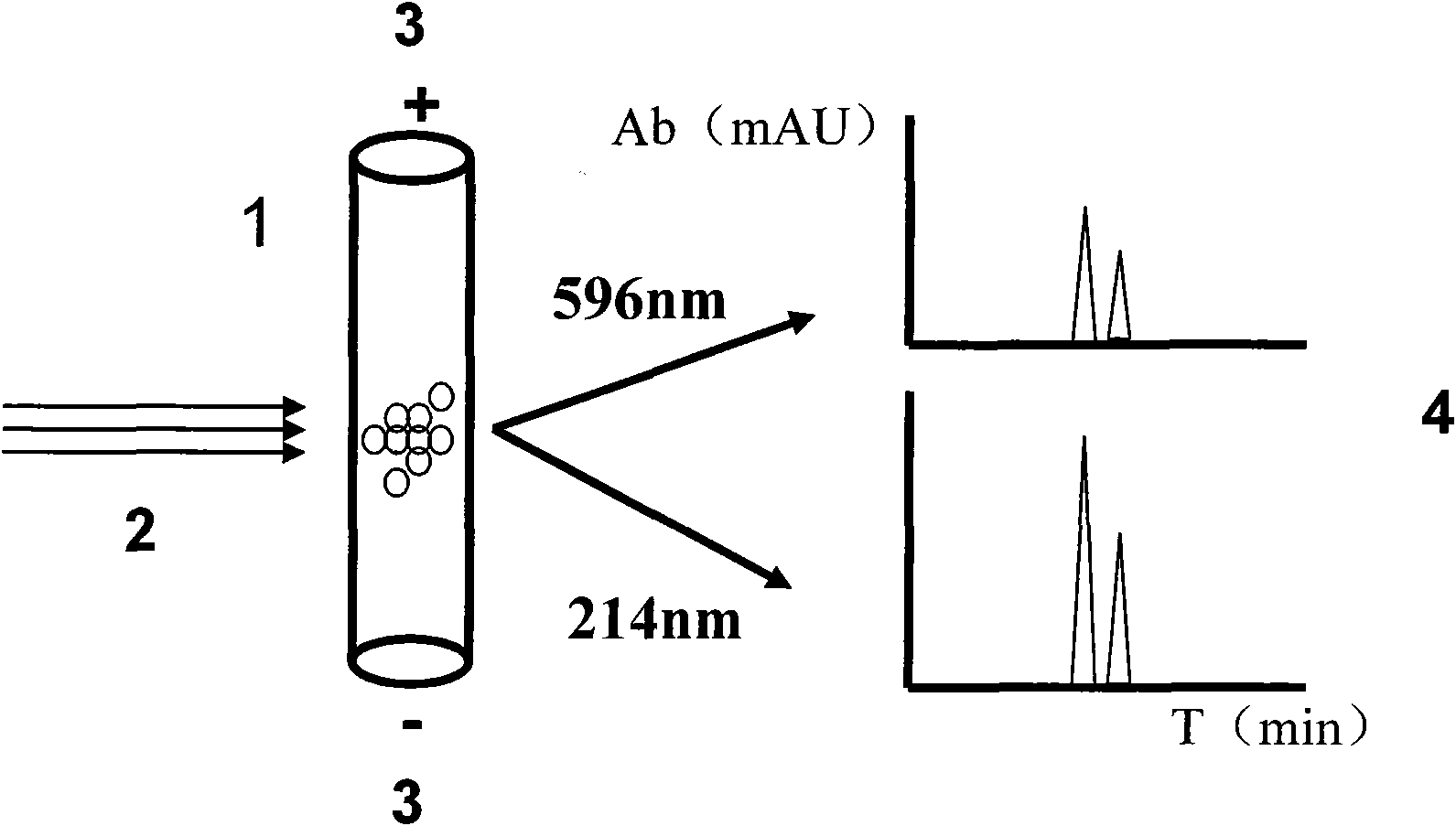
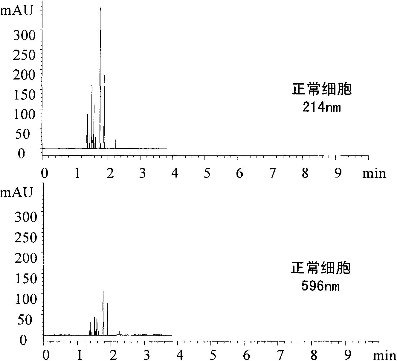
[0031] Step 1. Pretreatment is performed on both the normal cell sample and the cell sample to be tested. That is, normal cell samples and cell samples to be tested were stained with 0.2 mg / mL Jiana Green for 10 min, centrifuged, and the supernatant was removed, and then phosphate buffered saline (PBS) was added to wash the cells until the residual Jiana Green Dye wash.

[0032] Afterwards, 200 μL of 4% formaldehyde solution was added to the washed cell pellet, thereby immobilizing the cells, and the i...

Embodiment 2

[0041] To culture 6 groups of Hela cells, you can use six-well plates or culture flasks with the same specifications. One group was used as the control group without induction agent, and the other five groups were added with methylmercury at concentrations of 2, 4, 6, 8 and 10 μg / ml respectively. at 37°C, 5% CO 2 The normal cell samples and the test cell samples induced by different concentrations of methylmercury were obtained by culturing for 12 hours under the condition.

[0042] Step 1. Pretreatment is performed on both the normal cell sample and the cell sample to be tested. That is, the cell samples to be tested were stained with 10 μg / mL Rhodamine 123 at 37°C for 10 min, centrifuged, and the supernatant was removed, and then the cells were washed with phosphate buffered saline (PBS) until the residual Rhodamine 123 was washed away.

[0043] Afterwards, 200 μL of 4% paraformaldehyde was added to the washed cell pellet to immobilize the cells, and the immobilization pro...

Embodiment 3

[0050] To culture 6 groups of Hela cells, you can use six-well plates or culture flasks with the same specifications. One group was used as the control group without induction agent, and the other five groups were added with methylmercury at concentrations of 2, 4, 6, 8 and 10 μg / ml respectively. at 37°C, 5% CO 2 The normal cell samples and the test cell samples induced by different concentrations of methylmercury were obtained by culturing for 16 hours under the condition.

[0051] Step 1. Pretreatment is performed on both the normal cell sample and the cell sample to be tested. That is, stain the cell sample to be tested with 0.1 mg / mL neutral red at 37°C for 10 min, remove the supernatant after centrifugation, and then add phosphate buffered saline (PBS) to wash the cells until the residual neutral red Red dye wash.

[0052] Afterwards, 200 μL of 4% formaldehyde solution was added to the washed cell pellet, thereby immobilizing the cells, and the immobilization process w...


PUM

PropertyMeasurementUnit
lengthaaaaaaaaaa
lengthaaaaaaaaaa

Abstract

The invention discloses a cytoactive detection method based on a capillary zone electrophoresis mode, belonging to the technical field of biological cytoactive detection. Firstly, cells to be detected are dyed and fixed to prepare cell suspension with certain concentration; capillary zone electrophoresis (CZE) carries out visible absorption detection of single-wavelength or multiple-wavelength ultraviolet to cells, detected peak area summation represents the light absorption value of cells under the detection wavelength, and a product of the ultraviolet and visible double-wavelength absorption value represents cytoactive. The method of the invention is accurate, quick and simple, has low cost, can qualitatively and quantificationally detect cytoactive, can detect cytoactive and analyze toxicant cytotoxicity and can be applied in relevant fields, such as cytobiology, cytotoxicology, medicine research and development, toxicant cytotoxicity evaluation and the like.

Description

technical field [0001] The invention relates to a cell activity detection method based on capillary zone electrophoresis, and belongs to the technical field of biological cell activity detection. Background technique [0002] Cell viability and toxicity detection is widely used in cell biology, cytotoxicology, drug development and other fields, and is an important technology and means in biology and medicine. In recent years, according to the changes of cell characteristics in each state in the process of cell death, a variety of cell viability detection methods have been established. Conventional cell viability detection methods mainly include cell counting method, trypan blue exclusion method, neutral red staining method, tetrathiazolium blue (MTT) colorimetric method, lactate dehydrogenase (LDH) leakage rate detection and other methods. In the process of cell viability detection, instruments such as microscope, microplate reader and flow cytometer play an important role....

Claims


Application Information

Patent Timeline
Patent Type & Authority Applications(China)
IPC IPC(8): G01N21/33C12Q1/02
Inventor 屈锋丁金美张璐江伦郝帅胡美龄
Owner BEIJING INSTITUTE OF TECHNOLOGYGY
Why Eureka
Try Eureka