
Tissue engineered artificial optic nerve conduit and preparation method thereof

A technology of tissue engineering and optic nerve, which is applied in the field of biomedical engineering, can solve the problems of virus transfection, biological safety, limited application prospects, short half-life of nutritional factors, and limited duration, etc., to achieve small foreign body reactions, promote axon regeneration, and avoid The effect of injurious manipulation

Active Publication Date: 2012-09-12
EYE & ENT HOSPITAL SHANGHAI MEDICAL SCHOOL FUDAN UNIV +1
View PDF0 Cites 2 Cited by
  • Summary
  • Abstract
  • Description
  • Claims
  • Application Information

AI Technical Summary

Problems solved by technology

[0006] Most of the studies on artificial optic nerve disclosed in the prior art are to directly fill the nutrient factors into the hollow catheter braided with absorbable material filaments, but because the half-life of the nutrient factors is generally short, especially the only one currently proven to promote optic nerve regeneration The ciliary neurotrophic factor CNTF has a very short half-life and a limited duration in the eye. Direct protein filling cannot provide lasting nutritional support for nerve axon regeneration, which is undoubtedly a major bottleneck affecting the effect of artificial nerves.
[0007] Some studies have attempted to transfect neurotrophic factors into Schwann cells by virus transfection, but the biosafety of virus transfection also limits its clinical application prospects

Method used


Image

  • Tissue engineered artificial optic nerve conduit and preparation method thereof
  • Tissue engineered artificial optic nerve conduit and preparation method thereof
  • Tissue engineered artificial optic nerve conduit and preparation method thereof

Examples


Embodiment 1

[0056] Example 1 Preparation of tissue-engineered artificial optic nerve (filled with common Schwann cells)

[0057] The Schwann cells cultured in vitro were divided into 5×10 6 Cell / ml density was inoculated into 6-well plates, 10% FBS / DMEM culture medium was added, and when the confluence of cell growth reached 90%, it was digested with trypsin and transplanted.

[0058] The PLGA filament is woven into a hollow catheter as required, and the surface of the catheter is coated with a layer of chitosan polymer, and ethylene oxide is fumigated for sterilization. The surgical 4-0 PGA absorbable suture was disassembled into PGA filaments under a microscope, and the PGA filaments (with a diameter of 10 μm) were evenly filled into the PLGA hollow catheter as the internal scaffold of the artificial optic nerve. Then put the catheter into 75% alcohol to sterilize for half an hour, then rinse it with PBS three times, dry it naturally in an ultra-clean bench, and then fill it with cells...

Embodiment 2

[0060] Example 2 Preparation of tissue-engineered artificial optic nerve (filled with transgenic Schwann cells)

[0061] The Schwann cells cultured in vitro were divided into 5×10 6 Inoculate the cell / ml density into a 6-well plate, add 10% FBS / DMEM culture medium, when the confluence of cell growth reaches 90%, suck out the culture medium, wash 3 times with PBS solution, and then add 100ml D-Hank's solution to each well , which contains 12.5μg CNTF plasmid, after standing for 10 minutes, add 900ml D-Hank's solution, and then Petri Pulser TM Gently insert the PP35-2P electrode into the six-hole plate, immerse part of the electrode in the liquid surface, gently place it on the cell surface, and connect the electroporation instrument (ECM-830, BTX) for electric shock. The electric shock parameters are voltage 192v, time 20ms, interval 500ms, A square wave pulsed 5 times. After the electric shock is completed, stand still for 5 minutes, then aspirate the electrotransfer soluti...

Embodiment 3

[0064] Embodiment 3 animal experiments

[0065] Experimental animals: 16 SD rats (200-250 g) at 8-10 weeks, purchased from the Institute of Zoology, Chinese Academy of Sciences, and kept in a clean animal room in accordance with the requirements of the Experimental Animal Ethics Association of Fudan University.

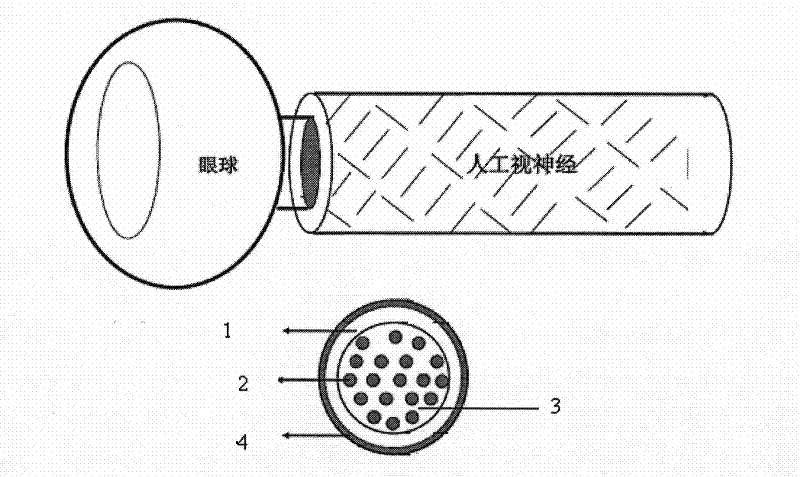
[0066] Experimental method: Under general anesthesia, the optic nerve of the left eye of the rat was separated, the optic nerve sheath was incised, the axon was cut off at 0.5 mm behind the optic nerve head, and the axon at the distal stump was about 3 mm long, and the sheath was avoided during the process. One end of the artificial optic nerve with a length of 11 mm was sutured to the sheath at the proximal end of the optic nerve with 10-0 suture, and the other end was sutured under the scalp with 8-0 suture ( Figure 6 ), check the fundus after the operation, and confirm that there is no damage to the blood vessels on the optic nerve sheath to affect the blood suppl...


PUM

PropertyMeasurementUnit
diameteraaaaaaaaaa

Abstract

The invention belongs to the field of biomedical engineering, and relates to a tissue engineered artificial optic nerve conduit and a preparation method thereof. The tissue engineered artificial optic nerve conduit comprises a hollow conduit woven from polylactic-polyglycolic acid filaments (PLGA), polyglycolic acid (PGA) filaments and transgenic schwann cell suspension, wherein chitosan polymer is coated on the surface of the conduit; an external bracket of artificial optic nerve is constructed by the PLGA hollow conduit; and the PGA filaments are filled in an internal bracket in which the artificial optic nerve is constructed. The conduit can effectively promote regeneration of damaged optic nerve axon, and has the advantages of persistently providing nerve nutritional support, guiding the regenerated nerve axon and the like; the conduit has high tissue compatibility and safety; the specifications of the transfected gene and the conduit can be adjusted according to requirements; andthe conduit and the method can be used for experimental research of optic nerve regeneration and expected to promote regeneration and restoration of damaged optic nerve in clinical use.

Description

technical field [0001] The invention belongs to the field of biomedical engineering, and in particular relates to a tissue-engineered artificial optic nerve catheter and a preparation method thereof. Background technique [0002] Optic nerve disease is a large category of common diseases in ophthalmology clinics. Due to the poor repair ability of the optic nerve after injury, it often causes irreversible consequences, leading to severe vision loss and loss. The clinical treatment methods are limited and the treatment effect is poor, which is a major clinical problem at present. With the development of neuroscience and material science, various tissue-engineered artificial nerve grafts have been applied to promote the regeneration of peripheral nerves and central nerves. The tissue-engineered catheters used to promote the repair or regeneration of optic nerve injuries are artificial optic nerves. research has received more and more attention. [0003] The so-called artifici...

Claims


Application Information

Patent Timeline
Patent Type & Authority Patents(China)
IPC IPC(8): A61B17/00A61L31/14A61L31/12
Inventor 莫晓芬方媛郯志清孙兴怀王艳张佩华郭文毅
Owner EYE & ENT HOSPITAL SHANGHAI MEDICAL SCHOOL FUDAN UNIV
Learn More