
Cell DNA damage detection kit and detection method thereof

A DNA damage and detection kit technology, applied in biochemical equipment and methods, microbial determination/inspection, etc., can solve the problems of long experimental period, limited detection samples, cumbersome production process, etc., to improve sensitivity and image quality, The effect of reducing fluorescence attenuation and improving fluorescence brightness

Inactive Publication Date: 2011-08-10
XIAMEN UNIV
View PDF0 Cites 7 Cited by
  • Summary
  • Abstract
  • Description
  • Claims
  • Application Information

AI Technical Summary

Problems solved by technology

The traditional laboratory method of single-cell gel electrophoresis has cumbersome steps, requires a separate configuration of lysis reagents, and needs to process the sample DNA in advance. The experiment period is long, and the samples are limited each time. gel, the production process is cumbersome, and it is easy to remove the film in the later stage of electrophoresis and washing, resulting in experimental failure, which affects the promotion and application of single-cell gel electrophoresis

Method used


Image

  • Cell DNA damage detection kit and detection method thereof
  • Cell DNA damage detection kit and detection method thereof
  • Cell DNA damage detection kit and detection method thereof

Examples


Embodiment 1

[0032] The making of embodiment 1 gel plate

[0033] The gel plate used in the present invention does not need to use glass slides, and the plate cover of the 96-well culture plate commonly used in the laboratory can be used instead of glass slides for single-cell gel electrophoresis. Every depression on the 96-well plate cover Both can be used as sample wells. Take a 96-well plate cover, use a sharp paper knife to cut off the raised part around it, and cut it into a gel plate of appropriate size according to the number of samples, for example, it can be cut into gel plates containing 12, 24, 36, 48, 64, 96 A concave gel plate, each sample well can be scratched, so that the agarose gel is not easy to degumming.

Embodiment 2

[0034] Example 2 Preparation of Cell DNA Damage Detection Kit

[0035] The configuration of the reagent solution adopts a conventional method, and the kit for DNA damage detection of 400 samples includes:

[0036] 1) 1 g of low melting point agarose.

[0037] 2) 150ml solution A and 150ml solution B. The formula of solution A is: 2.0~3.0mol / L NaCl, 0.1~0.2mol / LEDTA-Na 2 , 5 ~ 100mmol / L Tris, mass fraction 0.5% ~ 1.5% sodium sarcosinate, the rest is deionized water. The preferred formulation of solution A is: 2.5mol / L NaCl, 0.1mol / L EDTA-Na 2 , 10mmol / L Tris (pH=10.0), the mass fraction is 1.0% sodium sarcosinate, and the rest is deionized water.

[0038] The formula of the solution B is as follows: the volume fraction is 1.0%-2.0% Triton-100, 5.0%-15.0% dimethyl sulfoxide (Dimethyl sulfoxide, DMSO), and the rest is deionized water. The preferred formula of solution B is: the volume fraction is 1% Triton-100 and 10% dimethyl sulfoxide, and the balance is deionized water. ...

Embodiment 3

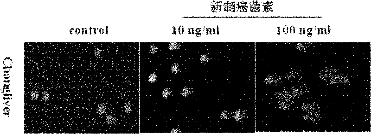
[0042] 1) Preparation of liver cancer SMMC7721 cell samples: Liver cancer SMMC7721 cells were cultured in DMEM culture medium with 10% newborn calf serum, 100u / ml penicillin, and 100u / ml streptomycin at 37°C in CO 2 Incubator cultivation. Pour off the culture medium, wash twice with PBS, digest with 0.25% trypsin, inoculate the cells in a 6-well plate for 24 hours to 80% confluence according to the needs of the experiment, and then set 12.5 μmol / L, 25 μmol / L L, 50 μmol / L 3 concentrations of DNA double-strand break reagent etoposide were used for 12 hours, and a blank control group was set up. At the end of the action time, wash twice with PBS, collect the cells by centrifugation at 1000r / m for 5min, wash once with PBS, and dilute with DMEM culture medium to form a cell suspension for use.

[0043] 2) Glue dispensing: take 20 μl of the liver cancer cell SMMC7721 cell suspension to be tested and 60 μl of 0.8% agarose gel preheated at 37° C. In the sample well of the rubber pla...


PUM


Abstract

The invention provides a cell DNA damage detection kit and a detection method thereof, relating to the cell DNA damage detection technique. The cell DNA damage detection kit comprises low melting-point agarose, a cell lysis solution, a cell despiralization solution and a DNA staining agent. The detection method comprises the following steps of: evenly mixing the cell DNA damage sample suspension with the low melting-point agarose gel, spotting the mixed liquid into sample holes on a gel board; immersing the gel board into the cell lysis solution for lysis in a dark place; immersing the gel board after lysis into the cell despiralization solution for despiralization, performing electrophoresis after despiralization; and dropwise adding the DNA staining agent for staining after electrophoresis, and analyzing the detection result under a fluorescence microscope. According to the detection method, the board cover of a cell culture board with 96 holes is used as the gel board to substitute for a glass slide for performing single cell gel electrophoresis, and compared with the traditional process in which 2 or 3 layers of gel are laid on a sample glass slide for performing single cell gel electrophoresis, two steps of gel laying and spotting are combined into one step, so that the sample preparation process is simple and easy to operate, the gel can not fall off and the success rate is high.

Description

technical field [0001] The invention relates to cell DNA damage detection technology, in particular to a cell DNA damage detection kit and a detection method thereof. Background technique [0002] DNA is an important biomacromolecule that has the function of transmitting genetic information in cells, and is an important research object of biochemistry and molecular biology. The DNA molecule is composed of two antiparallel polynucleotide chains through the principle of base pairing to form a double helix structure, and the genetic information carried by it is determined by the type, quantity and arrangement order of the bases on the polynucleotide chain. The base sequence is hidden deep inside the DNA double helix, and the double helix structure composed of two polynucleotide chains forms a stable DNA molecule and enables it to transmit genetic information correctly. [0003] There are many factors in the living environment of organisms, such as physical factors such as ioni...

Claims


Application Information

Patent Timeline
IPC IPC(8): C12Q1/68
Inventor 宋刚陈以旺王伟伟胡天惠
Owner XIAMEN UNIV
Learn More