Preparation method for acellular biological tissue material heparin coating

A biological tissue and decellularization technology, applied in the direction of coating, packaging item types, special packaging items, etc., can solve the problems of low heparin binding, promotion of monocyte adhesion, easy tissue calcification, etc., and achieve low immunogenicity. , beneficial to tissue reconstruction and host endothelial cell regeneration, good biocompatibility

Active Publication Date: 2012-05-30
吴忠仕 +1
View PDF3 Cites 3 Cited by
  • Summary
  • Abstract
  • Description
  • Claims
  • Application Information

AI Technical Summary

Problems solved by technology

At present, there are many methods for immobilizing heparin on the surface of biological tissue materials at home and abroad: (1) Using glutaraldehyde as a cross-linking agent to covalently cross-link heparin with the surface of biological tissue materials, the disadvantage is that the binding amount of heparin is small , and the cross-linking agent glutaraldehyde has many defects in the treatment of biological tissue materials (the existence of aldehyde groups, the tissue is easy to calcify after cross-linking; cytotoxicity, slow release in the body is not conducive to the proliferation and growth of host endothelial cells on the surface of the material) , which limits its application; (2) use EDC/NSH as a cross-linking agent to immobilize heparin on the surface of biological tissue materials in the form of covalent bonding, but its disadvan

Method used


Image

  • Preparation method for acellular biological tissue material heparin coating
  • Preparation method for acellular biological tissue material heparin coating
  • Preparation method for acellular biological tissue material heparin coating

Examples


Embodiment 1

[0037] Preparation of heparin-coated bovine jugular vein (BJV) material and determination of heparin binding

[0038] Methods: (1) 20 local BJVs were collected, and each root was divided into two sections, one section was used for decellularized photooxidized BJV (DP-BJV), and the other section was used for heparin-coated BJV after decellularized photooxidized (LBL-BJV). Decellularized photooxidation is prepared by CN100443064C patented method: 300-500 kg of healthy buffaloes are taken, the jugular vein is taken out within 30 minutes of warm ischemia time, and the fascia tissue is removed under aseptic conditions. Soak in PBS (pH 7.4) solution of 0.5% Trinaton X-100 for 48 hours, then soak in PBS solution of 0.025% trypsin and 0.02% EDTA for 30 minutes, rinse with PBS for 3 times, and then put in 20 μg / ml DNase·I and 0.2 mg / ml RNase·A in PBS solution for 24 hours, and washed with PBS solution 3 times. The above treatment was performed on a shaker with a shaking frequency of 7...

Embodiment 2

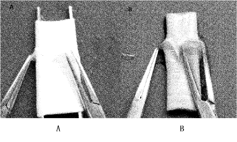
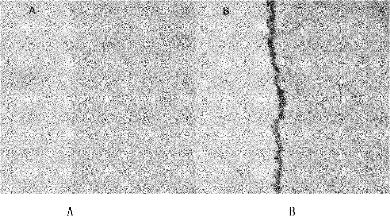
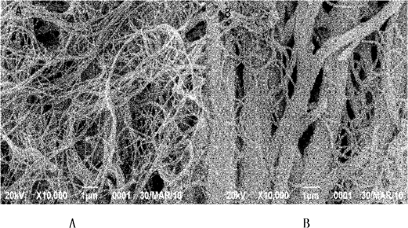
[0046] Electron microscope inspection and toluidine blue staining inspection of heparin-coated BJV materials

[0047] Methods: 10 local BJVs were collected, each root was divided into 2 sections, and randomly divided into the decellularized photooxidized group and the decellularized photooxidized heparin-coated group. All BJVs used in the experiment were cut into vascular slices (1cm×1cm) at a distance of 1cm from the valve sinus. . The decellularized photooxidation method is the same as that in Example 1. Preparation of heparin-coated BJV: immerse the decellularized photooxidized BJV in 5% heparin (containing 2% sodium chloride), and soak for 24 hours at 0-4°C. After rinsing with normal saline for 3 times, soak in 5% dihydroxy iron (Ⅲ) solution for 5 minutes; wash with normal saline for 3 times, 5 minutes each time, soak with 5% heparin (containing 2% sodium chloride) for 5 minutes, wash with normal saline for 3 minutes times, 5 min each time; repeat the above steps 4 times...

Embodiment 3

[0054] Detection of biomechanical properties of heparin-coated bovine jugular vein (BJV) material

[0055] Methods: 20 local BJVs were collected, each BJV was cut into two slices (4cm×1cm) of blood vessel strips (4cm×1cm) at a distance of 1cm from the valve sinus, and divided into decellularized photooxidation group and heparin-coated group. 20 tablets (decellularized photooxidation and heparin coating method are the same as in Example 2). The biomechanical properties of the two groups of blood vessel strips were detected by INSTRON tensile machine.

[0056] Results: The test results are shown in Table 1. The tensile strength of heparin-coated BJV was increased, and the elastic modulus, maximum load, and maximum tensile stress were significantly improved compared with decellularized photooxidized BJV.

[0057] Table 1 Pull test results

[0058]

[0059] There is a statistically significant difference between the two groups (P<0.05).

[0060] Conclusion: After heparin co...


PUM


Abstract

The present invention relates to a preparation method for modifying an acellular biological tissue material by using heparin. According to the present invention, the adopted material is mainly a biological tissue material used for cardiovascular repair and other tissue repair, wherein the material comprises vascular walls, pericardium tissues, cerebral dura maters, intestinal submucosa, and the like of various heterogeneous animals; with the preparation method for modifying the heterogeneous acellular biological tissue material for in vivo tissue reconstruction and regeneration by using the heparin, the biological tissue material coated by using the heparin has good performances of good biological compatibility, low immunogenicity, no cell toxicity, thrombus resistance, calcification resistance, anastomotic stenosis resistance, contribution to tissue reconstruction and host endothelial cell regeneration, and the like.

Description

technical field [0001] The invention relates to a preparation method for modifying decellularized biological tissue materials with heparin. Background technique [0002] Vascular transplantation has been widely used in the fields of cardiovascular diseases, tumors, trauma, organ transplantation reconstruction and microsurgery. Commonly used graft replacement vessels include autologous blood vessels (autologous arteries and autologous veins), allogeneic pipelines and artificial synthetic material pipelines. Autologous vessels and allogeneic conduits are ideal alternatives, but their sources are limited. The autogenous venous blood supply is relatively more, but its blood vessel elasticity is poor, and it is easy to form thrombus and aneurysm. Moreover, some patients have varicose veins and atherosclerosis, which also affects the application of autologous blood vessels. The supply of artificial synthetic materials is abundant, but the low patency rate of small-diameter vascu...

Claims


Application Information

Patent Timeline
IPC IPC(8): A61L27/54A61L27/36A61L27/34A61L33/10
Inventor 陶运明吴忠仕
Owner 吴忠仕
Why Eureka
Try Eureka